ISMRM 25th Annual Meeting & Exhibition • 22-27 April 2017 • Honolulu, HI, USA

Traditional Poster Session: Body: Breast, Chest, Abdomen, Pelvis
1990 -2030 Hepatopancreaticobiliary
2031 -2063 Body Imaging Novel Techniques & Indications
2064 -2103 Body: Cancer
2104 -2135 Breast Cancer
2136 -2164 Lung
 
Hepatopancreaticobiliary
Traditional Poster
Body: Breast, Chest, Abdomen, Pelvis

 
Wednesday, 26 April 2017
Exhibition Hall  08:15 - 10:15


1990.  
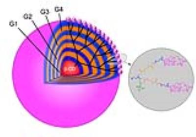
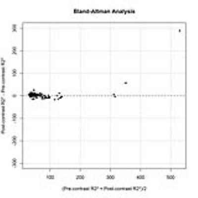
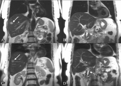
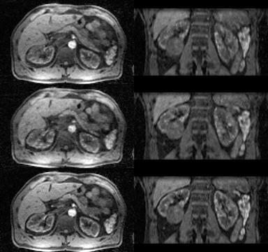
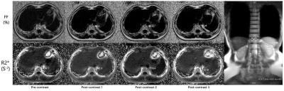
A biodegradable macromolecular MRI contrast agent for enhanced liver metastasis
Xiaoxuan Zhou, Hongjie Hu, Yue Qian, Yuxin Han, Mingzhou Ye, Jianbin Tang, Peipei Pang, Jun Yang
Tumor metastasis accounts for the related mortality, and precise diagnostic imaging of distant metastasis plays a significant role in clinical administration and treatment plan. Enhanced MRI with small-molecule contrast agent (CA) is a favorite imaging modality. However, small-molecule CA has some unsatisfied flaws such as short blood circulation time, low relaxivity and non-specificity. In this study, we synthesized a macromolecular CA with high relaxivity and long blood circulation time, and assessed early liver metastasis with enhanced MRI using this macromolecular CA.


1991.  
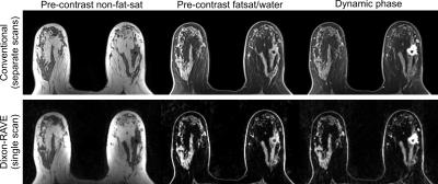
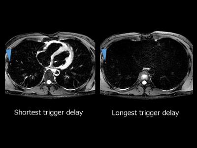
3D contrast enhanced high definition, free-breathing Dixon imaging using radial stack of stars and respiratory gating and tracking: Clinical comparison to state of the art breath hold Dixon imaging
Gabriele Beck, Michael Wyss, Rene Patzwahl, Joachim Hohmann, Christoph Andreas Binkert, Lars van Loon, Hans Peeters
We investigated a motion immune Dixon TFE approach incorporating a 3D radial stack-of-stars acquisition module interleaved with a spiral excitation navigator to gate and track the acquisition. Image quality, sharpness and streaking artifact level and its variability over different patient breathing patterns is efficiently improved to an extent where it is preferred over the state-of-the art breath hold (BH) Dixon scans. Next to that, it allowed scan time reductions of 30% in average. This technique provides motion-free, high resolution hepatic imaging with the benefits of Dixon providing consistent good fat suppression over a large FOV with water, fat, IP and OP diagnostic information all-in-one scan.


1992.  
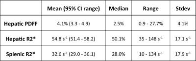
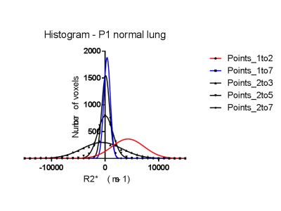
Quantification of Hepatic Fat Fraction and Liver/Spleen R2* in a Healthy Cohort at 3 Tesla
Rosalind Gerson, Chris Bowen, Manjari Murthy, Sharon Clarke
Non-invasive quantification of hepatic fat and iron with MRI has generated increasing clinical interest, particularly given the prevalence of chronic liver disease. Elevated R2* is a biomarker of iron deposition in the liver and spleen. The normative distribution of R2* in these organs at 3T is not well described, but is necessary for confident diagnosis of mild to moderate levels of iron deposition.  Based on measurements in 97 adults selected from the general population, we confirm that ~20% have fatty liver and suggest that a hepatic R2* > 89 s-1 and splenic R2* > 69 s-1 can be considered abnormal.


2009.   
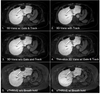
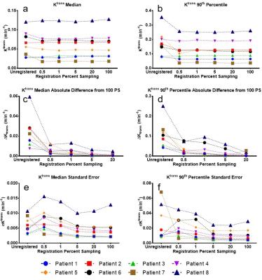
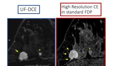
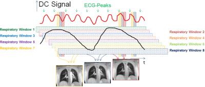
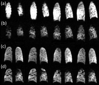
GRASP with Motion Compensation for DCE-MRI of the Abdomen
Koji Fujimoto, Li Feng, Rocardo Otazo, Kai Block, Henry Rusinek, Nicole Wake, Hersh Chandarana
A method to combine information of respiratory states with a non-rigid motion model is proposed to reconstruct motion-compensated 4D images of dynamic-contrast enhanced MRI with high temporal resolution. Abdominal DCE-GRASP MRI was reconstructed by grouping 4 consecutive spokes as one dynamic frame (0.6sec/frame). Respiratory motion was obtained from central k-space data (as in XD-GRASP). A temporal TV constraint was applied separately for each respiratory state. By using an optical-flow algorithm, four sets of motion vectors were obtained. Motion-compensated GRASP reconstruction was performed by including the motion vectors and showed improved image quality and reduced motion blurring. 


1994.   
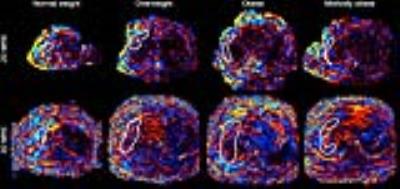
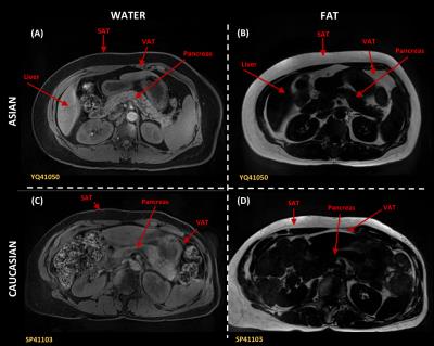
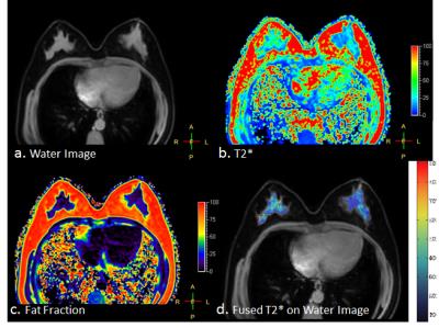
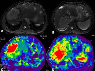
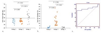
Correlation between incidental fat deposition in the liver and pancreas in asymptomatic patients
Mounes Aliyari Ghasabeh, Manijeh Zarghampour, Li pan, Pegah Khoshpouri, Farnaz Najmi Varzaneh, Nannan Shao, Ankur Pandy, Pallavi Pandy, Danial Fouladi, Ihab R Kamel
Liver steatosis is the most common parenchymal liver disease in Western Countries and it may progress to steatohepatis, fibrosis and cirrhosis. Also, fat deposition in liver and pancreas can cause diabetes by increasing resistance to insulin. Magnetic Resonance Spectroscopy (MRS) has been shown to strongly correlate with histology in liver fat quantification. However, MRS has some limitations such as breathing artifact and difficulties in avoiding vessels or bile ducts within the voxel. So, it is desirable to utilize a novel and robust imaging technique that can screen for the presence of fat in the liver and pancreas.


1999.  
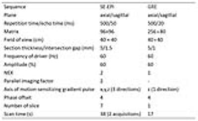
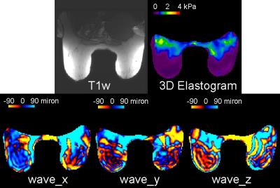
Simultaneous liver and spleen 2D MRE and 3D MRE acquisitions: Preliminary results
Paul Kennedy , Kevin Glaser, Curtis Johnson, Bradley Bolster Jr. , Jalpan Jani, Kashif Khokhar, Richard Ehman, Bachir Taouli
We present initial results of 2D MR elastography (MRE) and 3D MRE acquired in liver and spleen using a dual driver configuration in 13 subjects, 5 healthy and 8 with liver disease. 2D MRE showed a trend to higher stiffness in healthy subjects however in cirrhotic subjects liver stiffness was generally higher with 3D MRE. 3D MRE showed significantly higher liver stiffness in cirrhotic subjects compared to healthy subjects, with spleen stiffness also increased but not reaching significance. Coefficient of variation with single and dual drivers was 5% for liver and spleen in 2D and 3D MRE. 


1995.  
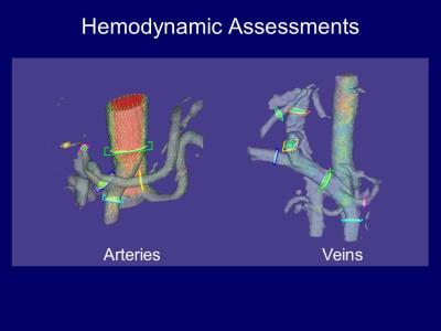
Hemodynamic Assessments of Hepatic Vasculatures using 4D-PCA and MRFD
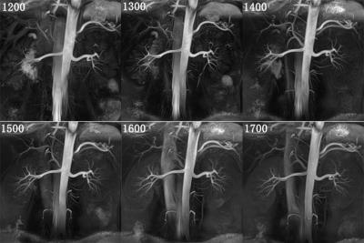
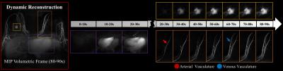
Takeshi Yoshikawa, Yoshiharu Ohno, Katsusuke Kyotani, Kouya Nishiyama, Shinichiro Seki, Yuji Kishida
We introduced new assessment method of liver hemodynamics using 4D-PCA and new flow analytic technique including wall shear stresses. We found 4D-PCA and MRFD enables detailed hemodynamic assessment and has the potential to be used for liver disease assessments.


1996.  
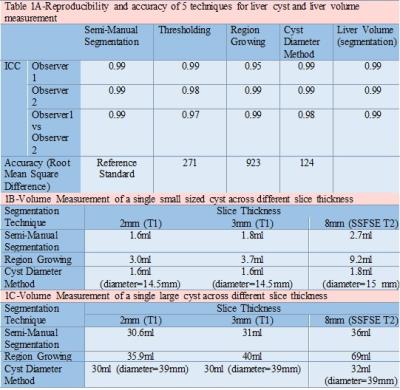
MRI of Liver Cysts in Autosomal Dominant Polycystic Kidney Disease: Effect of Genotype
Zerwa Farooq, Ashkan Heshmatzadeh Behzadi, Jon Blumenfeld, Martin Prince
ADPKD patients (n=25) undergoing abdominal MRI were analyzed 2 times each by 2 separate reviewers using a) thresholding; b) region growing c) cyst diameter d) semi-manual segmentation to determine liver cyst volume. Using the most reproducible technique, 75 additional ADPKD patients were studied correlating their liver parameters with genotype, gender and age. Semi-manual segmentation was the best technique for measuring cyst volume with intraclass correlation coefficient of 0.99. PKD1 mutation was found to be associated with higher total liver volume and liver cyst volume across different age groups and gender compared to PKD2 mutation, suggesting greater hepatic involvement with PKD1. 


1997.   
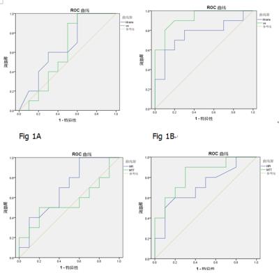
Feasibility and grading performance of Quantitative Susceptibility Mapping for Hepatic Iron quantification
Huimin Lin, Hongjiang Wei, Xu Yan, Caixia Fu, Stephan Kannengiesse, Chunlei Liu, Fuhua Yan
This study aimed to evaluate the performance of Quantitative Susceptibility Mapping (QSM) on estimating and grading the liver iron concentration (LIC), using Ferriscan-R2 values as reference. Thirty-three patients suspected of hepatic iron overload were included in this study. The results showed a significant positive correlation between QSM and Ferriscan-based LIC (r=0.924). ROC analysis revealed that QSM could accurately grade LIC for low and moderate iron overload patients, indicating that high-quality QSM maps may allow to reliably estimate and grade iron deposition in the liver.


1998.  
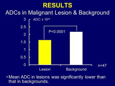
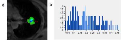
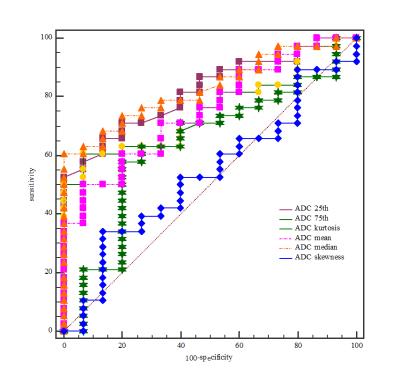
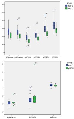
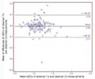
Characterization of Primary Liver Cancers with DWI Histogram Analysis
Sara Lewis, Steven Peti, Stefanie Hectors, Michael King, Juan Putra, Swan Thung, Bachir Taouli
ADC measurement using DWI have shown promise for characterizing focal liver lesions. In this study, we assessed the ability of advanced ADC histogram parameters to distinguish the histological diagnosis and the grade of primary malignant liver cancers. We found that both ADC mean and ADC percentiles could distinguish between tumor types; ADC percentiles were also predictive of tumor grade for HCC and ICC. Advanced ADC histogram analysis may be useful for accurate tumor diagnosis and prediction of tumor grade.


2000.   
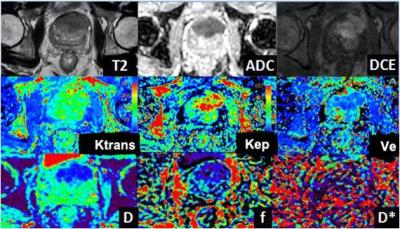
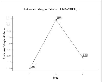
Liver quantification with dynamic contrast-enhanced MRI for evaluation in hepatic function and staging of post-hepatitic liver cirrhosis
Zhang Lan
To evaluate the value of DCE-MR for hepatic reserve function assessment and staging in post-hepatitic liver cirrhosis (PHLC). 10 patients with compensatory PHLC, 10 with decompensatory PHLC and 10 healthy volunteers were performed DCE-MRI scanning. All data were calculated by Extended Tofts model fitting with pharmacokinetic curve and the permeability and perfusion parameters were measured in quantification. The new method validated the feasibility of using quantitative parameters of DCE-MRI with pharmacokinetic model to assess liver cirrhosis. In conclusion, DCE-MRI quantitative parameters can be used for diagnosing and staging liver cirrhosis.


1993.  
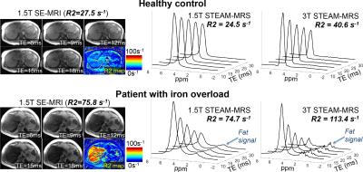
Spectroscopy-Based R2 Relaxometry for Liver Iron Quantification at 1.5T and 3.0T
Diego Hernando, Changqing Wang, Ryan Mattison, Takeshi Yokoo, Scott Reeder
R2-based techniques for liver iron quantification using Spin-Echo (SE) imaging require long acquisitions. In contrast, single-voxel Stimulated-Echo Acquisition Mode (STEAM)-MR spectroscopy enables liver R2 measurements in a single breath-hold. However, the accuracy and field strength dependence of STEAM-MRS R2 quantification are unknown. This study evaluated the accuracy and field strength dependence of STEAM-MRS for R2 quantification in healthy controls and patients with liver iron overload. At 1.5T, STEAM-MRS R2 was in close agreement with SE-MRI-based R2. Further, STEAM-MRS R2 measurements were highly correlated across field strengths. Finally, STEAM-MRS R2 measurements at 1.5T and 3.0T were calibrated to liver iron concentration. 


2001.   
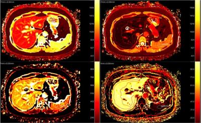
Assessment of the Hepatocyte Fraction for Estimation of Liver Function Based on a Simple Pharmacokinetic Model
Ruo-kun Li, Fu-hua Yan, Jin-wei Qiang, Hui-min Lin, Weibo Chen, Tomoyuki Okuaki, Eunju Kim
There were 16 consecutive patients (12 men, 5 women; mean age, 45.7 years; range, 33–65 years).   Imaging was performed on clinical 3T scanner (Philips Ingenia) using 32ch body/cardiac coil.  A hepatocyte fraction (HF) map and K map were derived. The HF and K values for Gd-EOB-DTPA were correlated with Child-Pugh scores.  Patients with Child-Pugh class B disease showed significantly lower liver FA value and K value. HF value and K value were positively correlated with the Child–Pugh scores  (r=0.752 to 0.855, p<0.05). HF value and K value had the largest AUC of 0.975 and 0.78 for distinguishing the Child–Pugh class A of cirrhosis from class B.  K value had the most area under receiver operating characteristic curve (AUC) of 0.99 for identifying the presence of liver cirrhosis.The study suggested that hepatocyte fraction and hepatic uptake derived from Gd-EOB-DTPA-enhanced MRI can quantify liver function. 


2003.   
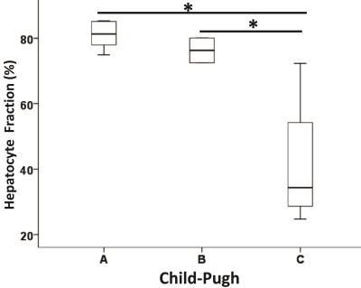
Quantitative Assessment of Hepatic function with Hepatocyte Fraction
Mengqi He, Yingjie Mei, Jing Zhang, Zeyu Zheng, Hongxiang Li, Tomoyuki Okuaki, Eunju Kim, Yikai Xu
Gadoxetic acid has been shown to evaluate liver function as it is known to be actively taken up by hepatocytes via organic anion transporters (OATPs). The measurement of hepatic parenchymal enhancement during the hepatobiliary phase (HBP) may accurately reflect liver function. This study evaluated the liver function of the subject in the Child-Pugh classification method which is still the most commonly applied method for evaluating liver function in clinical. The hepatocyte fractions were significantly different among groups classified according to Child–Pugh scores between A and C, B and C. The hepatocyte fraction could be considered as a promising method for the quantitative assessment of liver function.


2020.  
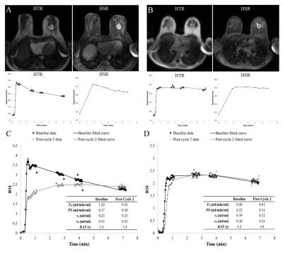
Vascular Input Function Correction with Inflow Relative Enhancement Quantification in Liver DCE-MRI
Jia Ning, Tilman Schubert, Huijun Chen, Chun Yuan, Scott Reeder
Dynamic contrast enhanced magnetic resonance imaging (DCE-MRI) with pharmacokinetic modeling can help to quantify the perfusion and function of liver. The accurate pharmacokinetic modeling relies on the accurately and reliably captured vascular input function (VIF)s. However, due to the fast blood velocity, the blood in the large vessels including abdominal aorta and the main branch of the portal vein experiences only a limited number of excitations and hasn’t reached a steady state. This introduces bias in the VIFs, and consecutively bias in pharmacokinetic parameters. In this study, we sought to correct the inflow effect on VIF acquisition in liver DCE-MRI.


2004.   
Assessment of interrater agreement and reliability for characterization of respiratory motion related artifacts at gadoxetate disodium-enhanced liver MRI
Kristina Ringe, Julian Luetkens, Rolf Fimmers, Renate Hammerstingl, Guenter Layer, Martin Maurer, Claas Naehle, Sabine Michalik, Peter Reimer, Christina Schraml, Andreas Schreyer, Patrick Stumpp, Thomas Vogl, Frank Wacker, Winfried Willinek, Guido Kukuk
In this prospective multicenter study, interrater agreement and reliability for characterization and grading of respiratory motion artifacts related to the injection of gadoxetate disodium were evaluated. Interrater agreement and reliability for scoring of motion artifacts in the arterial phase was excellent among experienced abdominal radiologists from different European tertiary referral centers with an intraclass correlation coefficient of 0.983 and 0.985, respectively. Characterization and grading of respiratory motion artifacts can thus be performed with a high level of confidence, which is a prerequisite for assessing the incidence of this phenomenon in larger multicenter studies. 


2005.   
The predictive value of diffusion-weighted imaging on radiation-induced liver injury of hepatic malignancies
Xiaohong Ma, Shuang Wang, Yongjian Zhu, Han Ouyang, Chunwu Zhou, Xinming Zhao
Radiation-induced liver injury (RILI) is one of the most dreaded complications of radiation therapy (RT),which prevents radiation dose escalation and limits the effectiveness of the treatment . Therefore, it was important to predict and evaluate the RILI on hepatic malignancy using the RT in the early term. In this study, the ADCmid-RT value was significantly highest, and had negative correlation with the radiation dose. As the ADCmid-RT value was less than 1.29 x10⁻³mm2/s, the RILI could possibly occur. Therefore, the ADCmid-RT value may serve as a biomarker for predicting RILI in patients with hepatic malignancies treated with RT.


2018.  
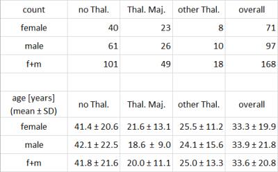
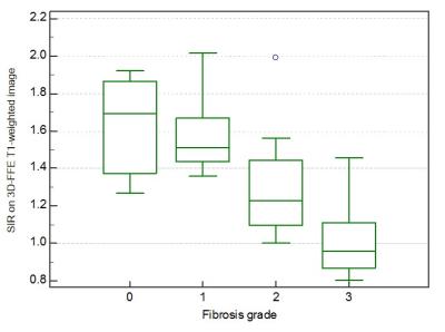
Liver Iron Concentration determined with Gradient Echo MRI by Signal Intensity Ratio: Effects of Patient Characteristics
Arthur Wunderlich, Holger Cario, Isabelle Tomczak, Meinrad Beer, Stefan Schmidt
To investigate the relation between signal intensity ratios gained from gradient echo (GRE) MRI and liver iron concentration (LIC), we studied the influence of patient characteristics. 168 patients (71 f, 97 m; 49 with Thalassemia major, 101 without Thalassemia) suspected for liver iron overload were scanned according to Ferriscan® with spin echo MRI to obtain reference LIC values, and GRE protocols suitable for LIC determination. GRE analysis by manually drawn liver and muscle ROIs yielded liver-to-muscle signal intensity ratios (SIR). Correlation analysis of ln (SIR) to reference LIC revealed differences between patient subgroups concerning disease, gender and age.


2007.   
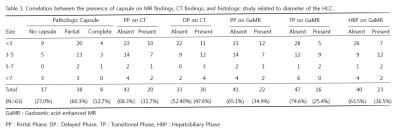
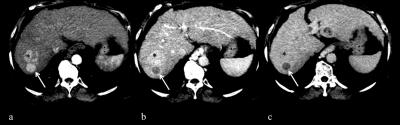
Hepatocellular Carcinoma with Capsule Appearance in Gadoxetic Acid-enhanced MR Images : Correlation with Dynamic CT and Pathologic Fibrous Capsule
Jei Hee Lee, Bohyun Kim, Young Bae Kim
For visualization of capsule appearance on HCC, GaMR is comparable with dynamic CT. And pathologic fibrous capsule can be seen as hypointense rim in HBP. Lower visualization of capsule appearance in TP on GaMR seem to be parenchymal enhancement during the transitional phases. Hyperintensity rim in T2 weighted images with hopointense rim on HBP shows disruption of fibrous capsule with extension of tumor cells across the fibrous capsule in pathology. Hypointense rim on the HBP in the 2014 version of LI-RADS should not be considered as capsule appearance, and further study is also needed.


2010.   
HCC detection in pre-transplant patients: A comparative retrospective study between multiphasic CT, gadoxetic acid-enhanced MRI and extracellular gadolinium based contrast-enhanced MRI
Sahar Semaan, Christopher Song, Sara Lewis, Manjil Chatterji, M. Isabel Fiel, Cecilia Besa, Bachir Taouli
In this study, we present preliminary data comparing the diagnostic performance of multiphasic contrast-enhanced CT with contrast enhanced MRI using a liver specific gadolinium based contrast agent (GBCA), gadoxetic acid (Gd-EOB-DTPA) and extracellular (EC) GBCAs, using explant pathologic data as the reference. We also assessed the added value of delayed hepatobiliary phase (HBP) imaging and DWI in HCC detection. Our data suggests that CT and MRI have similar overall sensitivities for HCC detection and that the addition of DWI and HBP improved HCC detection when using Gd-EOB-DTPA. 


2011.  
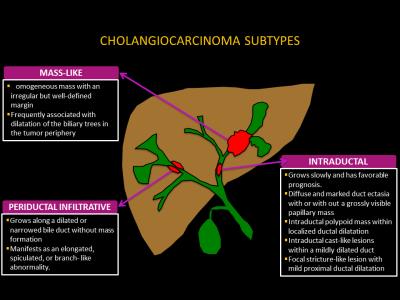
The value of magnetic resonance elastography in differential diagnosis of hepatocellular carcinoma and intrahepatic cholangiocarcinoma
kan liu, qi zhang, hong mei zhang, han ouyang, xinming zhao
To determine the value of MRE in the differential diagnosis of hepatocellular carcinoma and intrahepatic cholangiocarcinoma with 3.0-T MR scanner . The MRE were performed in 36 patients(26 hepatocellular carcinomas and 11 intrahepatic cholangiocarcinomas) by using 60-Hz mechanical waves and Spin echo echo planar sequence.Intrahepatic cholangiocarcinomas had significantly greater mean shear stiffness than hepatocellular carcinoma (9.08 ±2.13kPa vs 6.54 ±1.84kPa). They have statistically significant differences(p<0.01).MRE can help the differentiation of hepatocellular carcinoma and intrahepatic cholangiocarcinoma.


2012.   
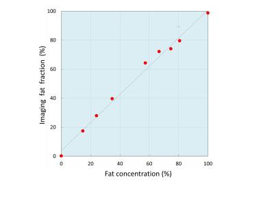
Hepatic fat quantification using automated six-point Dixon methods: comparison with conventional chemical shift based gradient-echo sequences and computed tomography.
Tomohiro Namimoto, Masataka Nakagawa, Kie Shimizu, Takeshi Nakaura, Kosuke Morita, Yasuyuki Yamashita
To compare automated six-point-Dixon(6-p-Dixon) with dual-echo GRE chemical shift imaging(CSI) for quantification of hepatic fat fraction(FF) with CT. In a phantom study, various FF vials were performed to validate the accuracy. In clinical study, fifty-nine patients were examined both 3.0T MRI and CT. Quantitative measurements were calculated SI-index of CSI and imaging-FF of 3D-6-p-Dixon. In phantom study, linear regression between FF and imaging-FF/SI-index showed good agreement(imaging-FF R2=0.992:0-100%FF;SI-index R2=0.978:0-34.7%FF). In clinical study, linear regression between imaging-FF and SI-index showed good agreement(R2=0.890). CT attenuation value was strongly correlated with imaging-FF(R2=0.852) and SI-index(R2=0.812). Imaging-FF of 6-p-Dixon has potential for automated hepatic fat quantification.


2013.  
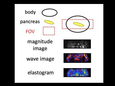
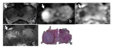
Value of Noncontrast MR Imaging with Diffusion-weighted Imaging for Detection of Primary Small (= 20mm) Solid Pancreatic Tumors and Prediction of Pancreas Ductal Adenocarcinoma
Hyun Jeong Park, Kyung Mi Jang
With recent advances of MRI in abdominal imaging, improved performances on T2-weighted image and diffusion- weighted imaging (DWI) are achieved for pancreatic tumors. We hypothesized that diagnostic performance for detection of primary solid tumors of pancreas on noncontrast MRI with DWI could be sufficiently high and noncontrast MRI with DWI would be useful for pancreas screening. We conducted this study to determine the diagnostic performance of noncontrast MRI with DWI for detection of primary small (≤20mm) pancreatic solid tumors and prediction of ductal adenocarcinoma in comparison with pancreas CT and pancreas MRI with MR cholangiopancreatography.


2006.  
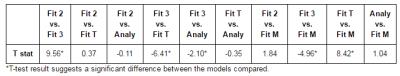
Inter- and intra-reviewer agreement of region-of-interest-based quantification of liver R2* in patients with iron overload
Camilo Campo, Diego Hernando, Tilman Schubert, Andrew Van Pay, Scott Reeder
This study evaluated the inter- and intra-reviewer agreement of different region-of-interest (ROI) sampling methods for the quantification of liver R2* (1/T2*) in patients with iron overload. 37 MRI datasets from patients suspected of having liver iron overload were retrospectively analyzed using ROI sampling methods that have been previously reported. Our results demonstrate that the inter- and intra-reviewer agreement of liver R2* quantification improve when using ROIs that are large in size and number. We conclude that researchers and clinicians should strive to sample as much area of the liver by using multiple large ROIs.


2014.   
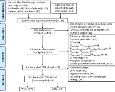
Gadoxetic acid disodium-enhanced magnetic resonance imaging for staging of liver fibrosis: a meta-analysis
Yuelang Zhang, Xiang Li, Haitian Liu, Chenxia Li, Jian Yang
To collect and summarize parameters used by gadoxetic acid-enhanced MR imaging (GD-EOB-DTPA-enhanced MRI) for staging of liver fibrosis (LF) and evaluate diagnostic performance. A systematic literature search was performed in PubMed, Web of science, Embase and Medline database. Studies used frequently-used parameter were included. Pooled weighted mean difference (WMD) was applied to determine the clinical significance. Pooled sensitivity, specificity, and summary receiver operating characteristics (SROC) curve were calculated to evaluate diagnostic performance. Finally, 5 studies were included, and contrast enhancement index (CEI) was the most frequently-used parameter, which was considered to be an efficient biomarker in the staging of LF.


2015.   
Apparent Diffusion Coefficient of Hepatocellular Carcinomas on Diffusion-Weighted Imaging: Correlation with Histopathologic Tumor Grade versus Arterial Vascularity during the Dynamic MRI
In Kyung Park, Jeong-Sik Yu, Eun-Suk Cho, Joo Hee Kim, Jae Joon Chung
Depending on the difference of cellular densities related to the histopathologic grades of HCCs, some investigators have recently suggested apparent diffusion coefficient (ADC) on diffusion-weighted imaging (DWI) as an effective biomarker for the prediction of the patients’ prognosis before the treatment of HCC. In the present study, ADCs of well- or poorly differentiated HCCs were lower than moderately differentiated HCCs; meanwhile the degree of arterial phase enhancement during the dynamic imaging rather well stratified the ADCs of the lesions. We concluded that ADC could not be independently used to estimate the histopathologic grades of HCCs.


2016.   
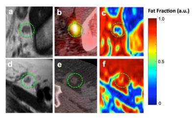
Quantification of liver fat after Gd-EOB-DTPA injection: a IDEAL-IQ feasibility study
Yuan Tian, Pengfei Liu, Lizhi Xie
This article is to explore the feasibility of liver fat quantification via dynamic liver enhancement scanning after injection of Gd-EOB-DTPA. IDEAL-IQ was used to quantify liver fat in 65 patients who were injected with contrast. IDEAL-IQ was performed four times to determine the fat fraction (FF) and R2*. One-way repeated-measures analysis was conducted to evaluate the difference between the four time points of the FF. The assessment of FF at four time points in liver, spleen and spine showed no significant differences. However, after injection of contrast agent, R2* was increased, and the IDEAL-IQ result was relatively stable.


2017.   
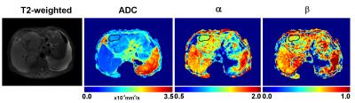
A Preliminary Study of the Clinical Value of FM Model in Malignant Tumor of Liver
huang can
Several models have been proposed to explain the anomalous diffusion in biological tissues. Among them, the fractional motion (FM) model was considered more appropriate. In this study, the FM model was applied to assess its feasibility for diagnosing malignant tumors of liver. It was found that the FM model could improve the diagnostic accuracy in differentiation normal liver tissue and tumor lesion, indicating the potential of the FM model to facilitate future studies of pathological changes in clinical populations.


2019.  
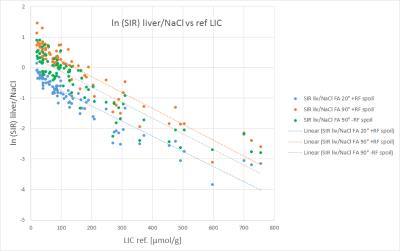
Liver Iron Concentration determined by Gradient Echo MRI using Signal Intensity Ratios: Impact of Acquisition Parameters and Image Quality
Arthur Wunderlich, Holger Cario, Isabelle Tomczak, Meinrad Beer, Stefan Schmidt
Tissue signal intensity ratio (SIR) has been used for a long time to determine liver iron concentration (LIC) based on gradient echo MRI. We studied the influence of acquisition parameters FA, RF spoiling and saturation regions, as well as image quality score, on the correlation of natural logarithm of SIR values to reference LIC obtained with spin echo. In our cohort of 85 patients, no significant influence on the slope of linear regression line was found, neither of acquisition protocol settings nor image quality, whereas the intercept was dependent on parameters influencing T1 sensitivity, namely FA and RF spoiling.


2021.   
Gadoxetate disodium (Gd-EOB-DTPA) DCE-MRI of the Liver for the Assessment of Parenchymal Alterations in Primary Sclerosing Cholangitis
Sarah Keller, Jan Sedlacik, Fabian Kording, Gerhard Adam, Christoph Schramm, Jin Yamamura
This study evaluates the feasibility of  dynamic contrast-enhanced magnetic resonance imaging (DCE-MRI) with Gd-EOB-DTPA for detection of hepatic inflammation/fibrosis in comparison to Ultrasound elastography in primary sclerosing cholangitis (PSC).


2022.   
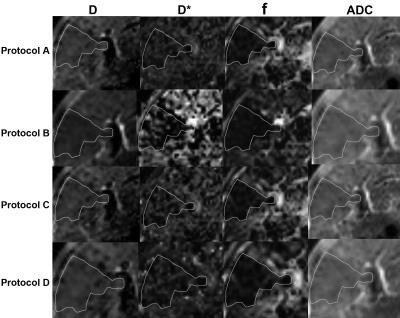
Evaluation of Intravoxel Incoherent Motion with Different b Values in Patients with Hepatocellular Carcinoma
Li Yang, Mengsu Zeng, Xuhao Song, Caixia Fu, Xu Yan
Intravoxel incoherent motion (IVIM) model provides both pure water motion and microcirculation by using multiple b values. IVIM imaging has been shown to be useful for assessment of liver diseases, including liver fibrosis and hepatocellular carcinoma. However, the clinical implementation of IVIM imaging is limited by long acquisition time. We compared IVIM imaging with different b-values to determine the combination of b-values in IVIM imaging that allows the relatively short acquisition time to obtain reproducible values of the IVIM parameters in patients with hepatocellular carcinoma. Our results showed at least 10b-values should be used in IVIM imaging for the assessment of hepatocellular carcinoma, and 5b-values IVIM imaging might increase errors in the perfusion-related f and D* values.


2008.   
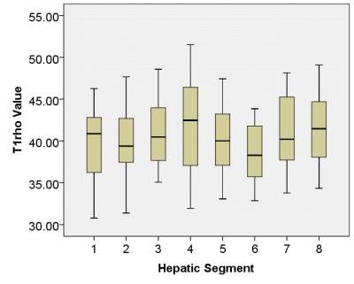
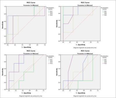
Liver T1rho Distribution across Eight Functionally Independent Segments
Weibo Chen, Xin Chen, Li Yang, Shanshan Wang, Queenie Chan, Guangbin Wang
ROI-based analysis may suffer from sampling errors due to the user-defined placement of the ROI and the choice of slice location. The aim of our study was to find an approach that allow the detection and measurement of the T1rho values of the hepatic segments.


2023.  
Pancreatic MRI associated with pancreatic fibrosis and postoperative fistula: comparison between pancreatic cancer and non-pancreatic cancer tissues
Yoshifumi Noda, Satoshi Goshima, Natsuko Suzui, Tatsuhiko Miyazaki, Kimihiro Kajita, Hiroshi Kawada, Nobuyuki Kawai, Hiromi Koyasu, Masayuki Matsuo
Pancreatic cancer (PC) occurs a histopathologically stronger pancreatic fibrosis compared to other non-PC. The risk factors of postoperative pancreatic fistula (POPF) were reported as soft or normal pancreatic parenchyma, and ampullary or duodenal disease. In this study, in patients with non-PC, frequency of POPF was greater due to lower grade of pancreatic fibrosis, and our results suggest that T1 signal and ADC value of the pancreas may link to the POPF. The T1 signal and ADC value of the pancreas may be a potentially useful imaging biomarker for the assessment of pancreatic fibrosis and POPF.


2025.   
 
Pancreatic changes in patients with liver cirrhosis and diabetes: a 3-T MRI evaluation
Tomohiro Sato, Katsuyoshi Ito, Tsutomu Tamada , Akira Yamamoto, Akihiko Kanki
The liver plays a pivotal role in glucose metabolism, so 60-80% of patients with cirrhosis experience impaired glucose tolerance and hyperinsulinemia, and 10-50% develop diabetes. This study was intended to clarify the extent to which pancreatic MRI findings are affected by the presence of diabetes mellitus in patients with liver cirrhosis. On 3-T MRI, size of the pancreas was significantly increased, the grade of pancreatic lobulation was significantly reduced, and pancreatic SIRs on T2WI with fat suppression were significantly increased in cirrhotic patients complicated with diabetes as compared to cirrhotic patients without diabetes.


2026.  
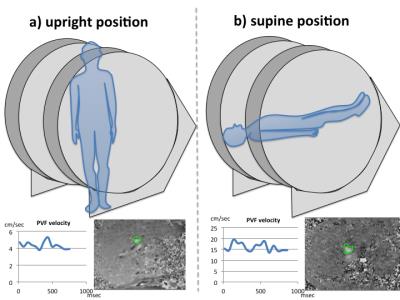
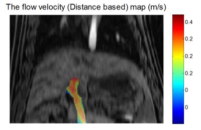
Evaluation of gravity effect on portal venous flow using multi-posture MRI
Yoshisuke Kadoya, Tosiaki Miyati, Naoki Ohno, Satoshi Kobayashi, Toshifumi Gabata
Portal venous flow (PVF) seems to be affected by gravity, ie., it depends on the body posture. We validated the effect of gravity on PVF in supine and upright positions using an original multi-posture MRI. We compared maximum PVF, PVF velocity, PVF volume, and cross-sectional area of portal vein between supine and upright positions. The mean PVF velocity, PVF volume, maximum PVF, and cross-sectional area in the upright position were significantly lower than those in the supine position. Gravity reduces PVF velocity and volume, and these differences between postures potentially provide new diagnostic information.


2024.   
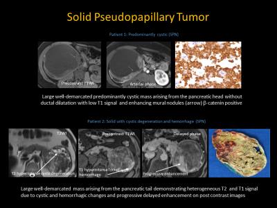
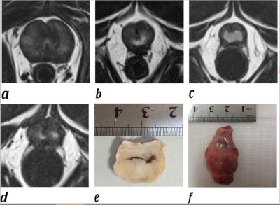
Magnetic Resonance Imaging: Solid Pseudopapillary Neoplasm of the Pancreas, Mimics and Histopathological Correlation
Nikhar Kinger, Peter Harri, Lauren Alexander, Courtney Moreno, Pardeep Mittal
SPN is a rare epithelial neoplasm of low grade malignant potential for local and metastatic spread, occurring predominantly in young females. Differential diagnosis of SPN includes a wide spectrum of cystic and solid entities in the pancreas including pancreatic neuroendocrine, serous or mucinous cystadenoma or carcinoma, intrapancreatic splenules etc. Contrast enhanced MRI plays a key role in characterization of SPN and its mimics , helps to reach a specific diagnosis and narrows the differential, which is complimentary to EUS and biopsy when findings are equivocal to reach an accurate diagnosis which is of utmost importance for management and treatment planning


2027.   
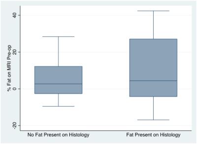
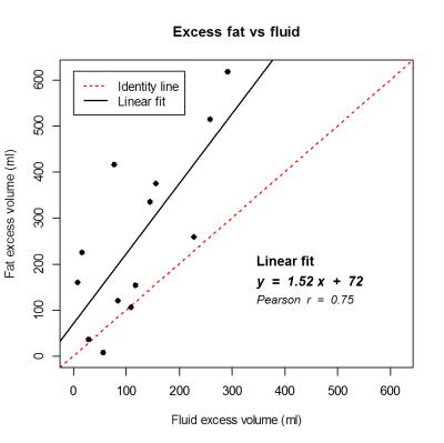
Pre-operative MRI quantification of hepatic fat and its correlation with histology, BMI and length of peri-operative stay following resection of liver metastases
Davinia Ryan, Alessandra Borgheresi, Simone Krebs, Sarah Eskreis-Winkler, Lorenzo Mannelli
We correlate pre-operative quantification of hepatic fat on MRI in patients with colorectal cancer metastatic to the liver with the presence of fat on histology of the resected specimen, patient BMI and hospital length of stay. 


2002.   
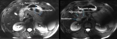
Quantitative T1 and T2 measurements of pancreas at 7 Tesla using a multi-transmit system
Mariska Damen, Quincy van Houtum, Maarten van Leeuwen, Peter Luijten, Andrew Webb, Dennis Klomp, Catalina Arteaga de Castro
Inversion recovery and echo time series were obtained at 7T with a multi-transmit system to determine the T1 and T2 relaxation times of the healthy pancreas. These parameters are crucial when optimizing MR protocols. The T1 and T2 values found were in average 921+/-98 ms and 57+/-12 ms respectively. Excellent T2 contrast is obtained for the pancreas at TE/TR=80ms/17s.


2028.  
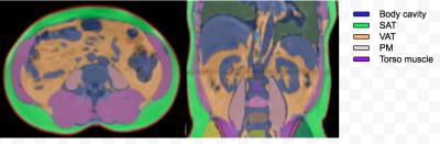
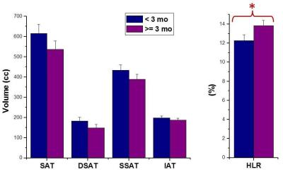
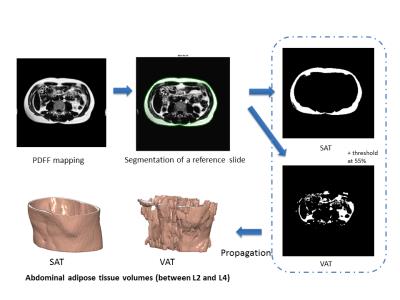
The effects of a 2 week hyperenergetic high carbohydrate or high fat diet on subcutaneous, visceral fat and metabolism
Mehri kaviani , Carolyn Chee, Caroline Hoad, Stephen Bawden, Peter Mansell, Sally Cordon, Aithal Guruprasad, Ian Macdonald, Penny Gowland
With the rise in obesity globally, there is great interest in quantifying body composition and in particular metabolically active visceral adipose tissue (VAT).  Increased accumulation of VAT has been linked with an increase in risk factors for cerebrovascular disease  and Type 2 Diabetes (T2DM) .  We investigated the degree to which weight, subcutaneous fat visceral fat,  liver and lipid markers are affected by 2 weeks of overfeeding at 25% excess energy given as either carbohydrate or fat. 2 weeks of 25% excess energy overfeeding of either carbohydrate of fat does not alter subcutaneous or visceral abdominal fat in line with no changes in weight. MRI measurements of SAT and VAT can be used in longitudinal studies of diet to ascertain any changes in abdominal body fat deposition.


2029.   
Liver T1rho detects liver fibrosis without impact of fatty liver in patients with chronic hepatitis B: a prospective study
shuangshuang xie, qing li, zhizheng zhuo, yu zhang, yue cheng, wen shen
This study investigated the merit of liver T1rho values for detecting fibrosis in patients with chronic hepatitis B and the potential impact of fatty liver on T1rho measurements. Eighteen healthy control subjects, eighteen patients with clinically diagnosed simple fatty and eighteen with liver fibrosis were underwent T1rho MRI and mDIXON-Quant. Mean T1rho values and fat fraction (FF) were compared among the three groups. Receiver operating characteristic (ROC) curve analysis was performed to evaluate the merit of T1rho values for detecting liver fibrosis. T1rho values were correlated with FF and clinical data. Our results showed significant differences in T1rho values among the three groups and T1rho had moderate diagnostic efficacy to detect fibrosis. T1rho values were not correlated with FF, subject age, or body mass index. We conclude liver T1rho values can be used as a MR biomarker for liver fibrosis and are not influenced by fatty liver severity.


2030.   
Diffusion-Weighted Imaging and Variable Flip Angle T1 Mapping in Progression Assessment of Liver Fibrosis Caused by Hepatitis B Virus Infection: A longitudinal study
Peng Hu, Jihong Sun, Fangfang Lv, Borui Pi, Fangping Xu, Guocan Han, Xi Hu, Yue Wang, Ning Huang, Xia Wu, Yong Zhang, Jun Yang, Peipei Pang, Xiaoming Yang
Liver fibrosis caused by Hepatitis B virus (HBV) is a worldwide health problem. However, liver biopsy as the gold standard of diagnosing and evaluating therapy of liver fibrosis is an invasive examination with possible errors from sampling and intra- and inter-observer interpretation (1, 2). The non-invasive MRI may be a promising modality to evaluate the liver fibrosis. We used diffusion-weighted imaging (DWI) and variable flip angle (VFA) T1 mapping to explore the progression of liver fibrosis caused by HBV infection in a longitudinal study.
 
Body Imaging Novel Techniques & Indications
Traditional Poster
Body: Breast, Chest, Abdomen, Pelvis

 
Wednesday, 26 April 2017
Exhibition Hall  08:15 - 10:15


2031.  
Effect of fitting model on the accuracy of T2 and T2* magnetic resonance based liver iron concentration
Tiffany Lin, Shirong Zhang, Michael Liu, Sachin Jambawalikar
Literature has suggested mixed results regarding agreement between T2 relaxivity based liver iron concentration (LIC) and T2* relaxivity based LIC The purpose of the following study is to determine whether or not choice of relaxivity fitting models will impact the agreement of the results.


2032.  
Comparison of 2D and 3D Magnetic Resonance Elastography (MRE) Wave-Image Quality Across a Range of Body Mass Indices
Yesenia Covarrubias, Jonathan Hooker, Ethan Sy, Saya Igarashi, Jennifer Cui, Cheng Hong, Nikolaus Szeverenyi, Jeffrey Schwimmer, Rohit Loomba, Scott Reeder, Kevin Glaser, Meng Ying, Richard Ehman, Claude Sirlin
This analysis of 129 adults compared the wave-image quality of two-dimensional (2D) and three-dimensional (3D) magnetic resonance elastography (MRE) across a range of body mass indices (BMIs). Wave-image quality of each scan was measured quantitatively by region of interest (ROI) areas. We found that 3D MRE provides higher wave-image quality compared to 2D MRE and that wave-image quality was not statistically associated with BMI (p=0.3422). Further research with larger BMI cohorts is needed to confirm our findings. 


2033.   
Gadolinium-enhanced high-flip-angle R2* mapping enables estimation of a greater range of R2* values
Jonathan Hooker, Yesenia Covarrubias, Cheng Hong, Soudabeh Dehkordy, Adrija Mamidipalli, Ethan Sy, Gavin Hamilton, Claude Sirlin
The range of R2* values estimated from MR sequences using low flip angles (FA) is limited by the background signal decay. The administration of contrast allows the use of higher FAs and increases the signal to noise ratio (SNR), allowing the estimation of a greater range of R2* values. We compared pre- and post-contrast R2* values in 158 patients and observed large discrepancies at R2* values greater than 300s-1. Our findings suggest that in patients with severe iron overload, post-contrast high-FA R2* mapping may be the preferred sequence for iron quantification.


2038.  
Abdominal Fast Advanced Spin Echo Diffusion-Weighted Imaging
Takeshi Yoshikawa, Katsusuke Kyotani, Yoshimori Kassai, Kouya Nishiyama, Shinichiro Seki, Yuji Kishida, Yoshiharu Ohno
To reduce distortion on abdominal EPI-DWI, we developed Fast Advanced Spin Echo (FASE)-DWI for abdominal 3T imaging. FASE-DWI improved distortion and showed equivalent diagnostic performance and be used as an alternative to EPI-DWI.


2034.  
Comprehensive dynamic contrast-enhanced 3D MR imaging of the breast with fat/water separation and high spatiotemporal resolution using Dixon Radial Volumetric Encoding (Dixon-RAVE)
Thomas Benkert, Kai Block, Samantha Heller, Melanie Moccaldi, Daniel Sodickson, Sungheon Kim, Linda Moy
Conventional clinical breast MRI consists of several separate T1-weighted scans, including both pre-contrast fat-suppressed and non-fat-suppressed acquisitions as well as the acquisition of dynamic contrast-enhanced phases.

Here, we show how this entire exam can be replaced by a single comprehensive scan, therefore reducing overall scan time and simplifying the clinical workflow. This is achieved by using radial stack-of-stars sampling in combination with a model-based fat/water separation technique, which takes into account the off-resonant blurring of fat and integrates both compressed sensing and parallel imaging. The approach is validated in 24 patients and results were evaluated by two radiologists.


2035.  
Free-Breathing Hepatobiliary Phase Imaging: Comparison of Five Free-Breathing Scans with Conventional Breath Hold Scan
Kimihiro Kajita, Satoshi Goshima, Yoshifumi Noda, Hiroshi Kawada, Tomoyuki Okuaki, Masatoshi Honda, Nobuyuki Kawai, Hiromi Koyasu, Masayuki Matsuo
We applied five free-breathing scan sequences to gadoxetic acid-enhanced hepatobiliary phase imaging. Breath hold eTHRIVE demonstrated the highest SNR on the liver, although it is also revealed the disadvantage for the patients with unstable breath hold. On the other hand, free-breathing 3D VANE with gate and track demonstrated the highest image quality with equivalent SNR to breath hold eTHRIVE. It is notable that free-breathing 3D VANE produced steady and effective image quality for all patients regardless of breath hold ability.


2036.   
BOLD MRI of Activated Human Pancreas
Bozhu Chen, Jian He, Zhengyang Zhou
Glucose ingestion activates the pancreatic functions and increases the oxygen consumption, we hypothesized that BOLD MRI could detect the alterations during the glucose challenge. BOLD MRI was performed in 12 volunteers before and after glucose ingestion. A transient but significant decrease in pancreatic T2* values was observed after glucose ingestion. BOLD MRI may serve as a non-invasive tool to detect the activation of human pancreas.


2037.   
Intravoxel incoherent motion (IVIM) and diffusion kurtosis imaging (DKI) for differential diagnosing hepatocellular carcinoma (HCC) from hepatic hemangioma (HHA).
Ye Ju, Ailian Liu, Qingwei Song, Meiyu Sun, Lihua Chen, Lizhi Xie
Intravoxel incoherent motion (IVIM) imaging is an extension of diffusion weighted imaging (DWI) that can be used to investigate both diffusion and perfusion changes in tissues. DKI adapts a kurtosis based model to depict the non-Gaussian diffusion process, which could be caused by the presence of different barriers in cellular complex structures (e.g. cell membranes and organelle compartments).Initial application of DKI focused on neuroimaging, Recently, it has also been reported that DKI may help to assess response to treatment in HCC. Comparing the IVIM and DKI parameters between carcinoma (HCC) and hepatic hemangioma(HHA) we found that IVIM and DKI can supply many meritorious parameters, combining with the IVIM and DKI may help in increasing the sensitivity and specificity of antidiastole.


2039.  
Computed Diffusion-Weighted Image for Abdominal MRI
Takeshi Yoshikawa, Katsusuke Kyotani, Yoshimori Kassai, Kouya Nishiyama, Shinichiro Seki, Yuji Kishida, Yoshiharu Ohno
The purpose of this study was to assess capability of computed DWI in evaluation of various abdominal diseases. We found cDWI can improve image quality and malignant lesion contrast, conspicuity, and detection. cDWI is a useful post-processing tool for abdominal MRI.


2040.   
Optimisation for Pulmonary R2* Quantification and Repeatability Evaluation
Alex Weller, Matthew Orton, David Collins, James D'Arcy, Nandita de-Souza
In 5 patients with lung cancer, UTE-MRI was used to derive pulmonary R2* in lung and establish its repeatability. Plausible R2* values were obtained only when using TEs of 0.08 and 0.2 ms: higher TEs produced implausible mean negative R2* within individual patients and a cohort R2* not significantly different from zero, due to lack of signal decay beyond TE=0.2ms. Pulmonary R2* values derived using TEs of 0.08 and 0.2ms were higher than prior reports where longer echo-times were employed. Test-retest limits-of-agreement were +90.5%to -47.5% indicating that a 90% increase in R2*is required post-radiotherapy to reliably demonstrate radiation-induced change.


2041.   
Initial Experience of MR Elastography Using Spatially Selective Excitation for the Pancreas.
Yohei Itoh, Yasuo Takehara, Naoki Ooishi, Masataka Sugiyama, Ikumi Igarashi, Maho Hayashi, Satoshi Usami, Takasuke Ushio, Yuki Hirai, Nobuko Yoshizawa, Shuhei Yamashita, Hatsuko Nasu, Tetsuya Wakayama, Atsushi Nozaki, Hiroyuki Kabasawa, Harumi Sakahara
We tried reduced-FOV MR elastography using a local excitation technique called FOCUS for the pancreas. Comparing conventional-MRE with FOCUS-MRE, there was no significant differences of the measurable are of pancreas.  In both methods, the higher the body mass index was, the smaller the measurable area of the pancreas.


2042.   
Test-Retest Repeatability of MR Elastography (MRE) Stiffness Measurements of Liver Phantoms
Jun Chen, Phillip Rossman, Kevin Glaser, Richard Ehman
All the previous test-retest human liver MRE studies could involve physiological stiffness change . In this study, our goal was to evaluate MRE test-retest repeatability with liver phantoms to avoid the possible influence of physiological changes in humans. We hypothesized that the variations of test-retest MRE stiffness measurements of phantoms were smaller than that of human livers.  


2043.   
The assessment of gallstones using three dimensional ultra-short echo time in vivo
Mamoru Takahashi, Yasuo Takehara, Norihiro Tooyama, Katsutoshi Ichijo, Tomoyasu Amano, Takuya Matsumoto, Tomoyuki Okuaki, Yukiko Fukuma, Harumi Sakahara
First clinical study using 3D dual echo UTE sequence for suspected gall stone patients was performed. All gallstones were able to be detected as positive signal in-vivo. Our study may indicate that UTE has an added value of depicting impacted stones or hepatolithiasis as positive signal.  


2044.  
Magnetic resonance elastography for uterine fibroids
Yoshie Omiya, Shintaro Ichikawa, Utaroh Motosugi, Hiroshi Onishi
 We evaluated the feasibility of magnetic resonance elastography (MRE) for uterine fibroids and the effect of imaging sequences (i.e., spin-echo echo-planar imaging [SE-EPI] and gradient-echo [GRE]) on stiffness measurements. All MRE were performed in two cross-sectional planes (i.e., axial and sagittal). SE-EPI-MRE showed lower fibroid stiffness values than GRE-MRE. Each MRE method is a valid and reliable technique for measuring the stiffness of uterine fibroids.


2045.   
MRI to evaluate the response of the locally advanced cervical cancer to CCRT: MRS(magnetic resonance spectroscopy), DWI(diffusion weighted image), and T2WI
Byung Chul Kang, Hye Ran Hyun
MRS(MR Spectroscopy) as well as T2Wi, and  diffusion weighted images may be used to evaluate the response of LACC to CCRT.


2046.  
 
Comparative analysis of image in changing breath hold method of B1 calibration at 3.0T abdomen MRI
Cho Ja Ryong, Park Jong Bin, Cho Seoung Bong
Evaluation of image analysis according to changing breath hold methods of B1 calibration .


2047.   
Using 2D-PACE to trigger scan and supervise motion on Breath-Hold imaging
Qiong Zhang, Yuanyuan Kang

A simple 2D pace module was implemented in a Breath-Hold imaging work flow to trigger the normal scan and supervise the  motion.  Our preliminary experiments show that within the newly added module, images are more immune to respiratory motion artifacts and the detail  of small structures are clearer.


2048.  
Quiet Motion-Robust Technique for 4D T1-Weighted Fat-Suppressed Abdominal Imaging
Ty Cashen, Naoyuki Takei, Kang Wang, Tao Zhang, Lloyd Estkowski, Ersin Bayram
Acoustic noise generated by MR systems represents one of the major barriers to patient comfort, particularly for the pediatric population. Reducing the slew rate of the gradient field is one way to effectively decrease acoustic noise; however, this comes at the expense of scan time. For breath-hold abdominal imaging, longer scan times mean a more stressful breath-hold for the patient, reduce temporal resolution for multi-phase imaging, and may allow for more motion artifact due to suboptimal breath-holding. This work describes an accelerated 3D multi-phase technique compatible with fat saturation for both quiet and motion-robust abdominal imaging.


2049.  
High-resolution 3D lung 1H-MRI in rodents at 9.4T with an optimized multi-echo gradient echo sequence
Dhaval Shah, Nicola Bertolino, Marilena Preda, Robert Zivadinov, Ferdinand Schweser
Preclinical MR lung imaging has been challenging due to lower tissue density compared to other organs. Current practice has been 2D spin-echo or gradient echo with relatively lower resolution. In this work we present an optimized high resolution 3D Multi Gradient Echo and demonstrate the results by comparing it with state of the art 2D Gradient echo.


2050.  
Receiver operating characteristic analysis of fat-fraction is effective in differentiating identifying an optimal threshold to differentiate between brown and white adipose tissue ex vivo and in situ in rats using 3-point IDEAL MRI
Terence Jones, Narendra Reddy, Sarah Wayte, Thomas Barber, Charles Hutchinson
Brown adipose tissue (BAT) has lower fat content than white adipose tissue (WAT), which has been exploited using Dixon-based MRI to identify BAT. We sought to identify the optimal threshold to differentiate between BAT and WAT in rats on the basis of fat fraction.

Fat fraction within BAT was significantly lower than WAT in rodents. Receiver operating characteristic analysis showed that differentiating BAT and WAT on the basis of fat fraction had excellent accuracy in both ex vivo and in situ. The optimal cut-off to separate BAT and WAT was significantly lower in rats exposed to cold, likely secondary to lipolysis.


2051.   
Cyclic Changes of the Boundary Sharpness of Uterine Zonal Structures Visualized by High-resolution T2-weighted Images in Young and Middle-aged Females during the Menstrual Cycle
Yong-Lan He, Ning Ding, Ya-Fei Qi, Tianyi Qian, Yuan Li, Huadan Xue, Zhengyu Jin
This study aimed to demonstrate the cyclic changes of the boundary sharpness of uterine three zonal structures of young and middle-aged women on 3T using a 3D T2-weighted SPACE sequence during the menstrual cycle. Peri-ovulatory phase exhibited the clearest boundary sharpness of corpus zonal structures following by FP, LP and MP, while that of the cervix were almost well-defined during the menstrual cycle.


2052.  
Resistance and susceptibility to diabetes - characterising the ‘Thin on the Outside Fat on the Inside’ (TOFI) profile using magnetic resonance imaging: a pilot study in Asian and Caucasian women
Ivana Sequeira, Wilson Yip, Louise Lu, Reza Nemati, Dech Dokpuang, Jun Lu, Sally Poppitt
Prediction of risk for dysglycaemia and adverse metabolic health is difficult, with some outwardly lean individuals having greater susceptibility to type 2 diabetes (T2D) than obese resilient individuals. Using 3T MR Imaging and spectroscopy we showed, in 10 Asian Chinese and 6 European Caucasian healthy or prediabetic women (18 – 70 years and BMI 25 – 50 kg/m2), that greater storage of fat within riskier ectopic sites, i.e. pancreas and liver, may in part explain the reported increased risk of T2D in Asian Chinese populations compared to their Caucasian counterparts at the same BMI and younger age. 


2053.  
Parameter Optimization for Non-contrast-enhanced Renal MR Angiography and Its Age-dependent Preliminary Study
Wansha Wu, Ke Ren, Jiannan Shang, Wenge Sun, Yi Liu, Songbai Li, Ke Xu
Our purpose was to investigate the feasibility of NCE-MRA using Time-SLIP technique on healthy volunteers by determining the optimized TI value and its preliminary relationship with age as well. 61 healthy volunteers were recruited and divided into two age groups. The acquired data using six differernt TIs sequences were measured and analyzed to get three parameters including VKR, grade of renal artery branches and grade of imaging quality. In conclusion, the Time-SLIP technique is able to obtain renal MR angiography with optimized TI value 1500ms. Moreover, the age of individual subjects can affect the optimized TI value. 


2054.  
A new technique of SPIO-enhanced MRI: delayed recovery of T2*-weighted signal intensity as a novel diagnostic marker for visualization of irradiated liver parenchyma.
Toshihiro Furuta, Masayuki Yamaguchi, Manabu Minami, Osamu Abe, Hirofumi Fujii
Visualization of irradiated liver parenchyma may assist safety margin assessment in radiotherapy. We demonstrate that an MR imaging technique has the ability to visualize irradiated liver parenchyma after 30-Gy irradiation in a tumor-bearing rat model. In this technique, superparamagnetic iron oxide (SPIO) is administered to label Kupffer cells (KCs) before, rather than after irradiation. A dose of 30-Gy is a lower, more clinically relevant dose than that used in the previous studies. Our results suggest that 30-Gy irradiation delays the recovery of hepatic T2*-weighted signal after SPIO administration. Presumably, irradiation delays degradation process of SPIO in the KCs.


2055.  
Changes in fat distribution and composition during ketogenic diet investigated by MRI and MRS.
Martin Buechert, Thomas Lange, Peter Deibert, Paul Urbain
Changes in fat distribution and composition during ketonic diet were investigated in a sub group of twelve volunteers of a larger study. Liver fat concentrations in the examined cohort turned out to be surprisingly low compared to patient cohorts with similar BMI measured in other studies. The hypotheses that intra-hepatic fat may be reduced during a ketogenic diet could not be confirmed. However the composition of the sub group may have biased the outcome. While the MRI protocol and analysis worked well, MRS analysis of data acquired with the given protocol in subjects with low hepatic fat reaches its limitations.


2056.  
Ethnic variation in body composition of men with type 2 diabetes from automated analysis of Dixon MRI
Haris Shuaib, Brandon Whitcher, Kevin Keraudren, David Greer, Geoff Charles-Edwards, Louise Goff
This work explores the role of body composition and its relationship with the development of type 2 diabetes in White European and Black African men. An investigation of differences in body composition between these two groups was performed via automated image segmentation and analysis of Dixon MR images. Our initial results suggest visceral adipose tissue may not be a principal determinant in the development of type 2 diabetes in Black African men as it is with White European men.


2057.   
Quantitative MRI parameters for adipose tissue characterization in obese patients
Nikita Garnov, Stefanie Lehmann, Ulf Retschlag, Nicolas Linder, Alexander Schaudinn, Arne Dietrich, Andreas Oberbach, Thomas Kahn, Harald Busse
In obesity research and clinical practice, disease progress or response to therapy is typically evaluated by the reduction of body weight, adipose tissue (AT) volume or comorbidity remission rates. However, little is currently known about therapy-induced changes in subcutaneous and visceral AT composition and how to quantify them. Lean control subjects had recently been shown to have significantly higher T1 relaxation times in both SAT (301 ms) and VAT (360 ms) than severely obese patients (275 and 294 ms, respectively). Aims of this study were to characterize AT by quantitative MRI and evaluate the intra-individual changes at two time points.


2058.   
The investigation of the relationship between obstetrical risk factors and pelvic floor injuries: a MRI-based study
Limei Guo, Yujiao Zhao, Zhizheng Zhuo, Wen Shen
Vaginal childbirth is an important cause to pelvic floor injuries. In this study, we aim to identify various forms of the injuries and the association with obstetrical risk factors in primiparous women. The results showed that there were significant differences about the proportions of various patterns of the pelvic floor injuries and the severity of LAM injuries between the groups with and without obstetrical risk factors.


2059.  
Phosphorus MRSI reveals subtle changes in metabolic profile of hepatic tissue in insulin resistant population.
Lorenz Pfleger, Peter Wolf, Martin Gajdošík, Sabina Smajiš, Marek Chmelík, Anton Luger, Siegfried Trattnig, Michael Krebs, Martin Krššák
This study employed ultra-high field  (7T)  1H and 31P MRS for the characterization of subtle hepatic tissue changes in insulin sensitive and insulin resistant group of healthy volunteers. Hepatic lipids, the forward rate of ATP synthesis and profile of metabolites containing 31P were assessed. Decreased inorganic phosphate was found in the subgroup of volunteers with increased hepatic fat accumulation and increased phosphocholine was found in the insulin sensitive subgroup. Concentration of 31P containing metabolites found here are well within the range of previous publications and subtle differences point towards their role in pathophysiology of metabolic syndrome and/or hepatic steatosis.


2060.  
Renal perfusion is decreased in kidneys with multiple renal arteries as demonstrated by renal DCE MRI
Anneloes de Boer, Margreet Sanders, Nico van den Berg, Peter Blankestijn, Tim Leiner
The presence of multiple renal arteries per kidney is associated with hypertension. The smaller vessel diameter is thought to lead to decreased renal perfusion, which activates the renin-angiotensin-aldosterone system (RAAS), resulting in increased systemic blood pressure. We measured renal blood flow (RBF) using dynamic contrast enhanced (DCE) MRI to investigate the relation between number of renal arteries, RAAS activity and RBF. The number of renal arteries was associated with reduced RBF and increased RAAS activity. In all patients, we observed that reduced RBF was associated with increased RAAS activity.


2061.  
Development of a lesion-wise metric for evaluation of predictive models of prostate cancer on multiparametric MRI
Ethan Leng, Jin Jin, Lin Zhang, Joseph Koopmeiners, Gregory Metzger
A novel lesion-wise metric was developed to evaluate the quality of predictive models of prostate cancer that use quantitative multiparametric MR data to perform prediction on a voxel-wise basis. The metric is based on the Jaccard similarity coefficient and emphasizes overlap and co-localization of ground truth and predicted lesions. Experiments to characterize the metric demonstrated that it qualitatively reflected the goodness of predictions and was more accurate and informative than voxel-wise measures of sensitivity and specificity. We propose that the metric may be customized to select the best predictive models for specific clinical applications such as performing targeted prostate biopsies.


2062.  
Why is the Peripheral Zone of the Normal Human Prostate High in ADC Value and T2-Weighted Signal Intensity?
Edward Johnston, Colleen Bailey, Elisenda Bonet-Carne, Hayley Pye, Susan Heavey, Dominic Patel, Ashwin Sridhar, Bernard Siow, Thomy Mertzanidou, William Devine, Jagadish Kalasthry, Joey Clemente, David Hawkes, Hayley Whitaker, Manuel Rodriguez-Justo, Greg Shaw, Daniel Alexander, Alexander Freeman, Roger Bourne, Eleftheria Panagiotaki, Shonit Punwani
The biophysical basis of MRI signal in the normal human prostate is uncertain, whereby the normal peripheral zone has high ADC values and returns high signal on T2-weighted imaging. In this study, we use MRI in combination with quantitative digital histopathology to offer an explanation. Paired scans were performed at 3T on a human prostate, prior to and following prostatectomy and changes in zonal morphology and MRI characteristics were measured. The peripheral zone collapsed and reduced in T2 signal intensity and ADC value ex-vivo. Digital histopathological analysis suggested the peripheral zone stores more ejaculatory fluid than the transition zone.


2063.   
Coronal View Renal Perfusion FAIR-ASL Measurements in Mice
Fabian Gutjahr, Thomas Kampf, Stephan Günster, Patrick Winter, Volker Herold, Wolfgang Bauer, Peter Jakob
Renal perfusion measurements in coronal view using FAIR-ASL can be problematic in small animals as the selective inversion slice can label a large fraction of the inflowing blood. In this work, an alternative orientation for the selective inversion slice is shown to increase the sensitivity of the measurement.
 
Body: Cancer
Traditional Poster
Body: Breast, Chest, Abdomen, Pelvis

 
Wednesday, 26 April 2017
Exhibition Hall  08:15 - 10:15


2064.   
Diagnostic accuracy of 3-T magnetic resonance imaging with Star VIBE: versus computer tomography in pulmonary nodules
Nan Yu, Chuangbo Yang, Qi Yang, Shaoyu Wang, Yong Yu, Taiping He
Star VIBE (MRI) sequence in obtaining scan under free breathing can provide high-resolution imaging. Therefore, we assessed the accuracy of magnetic resonance imaging (MRI) for detecting pulmonary nodules by comparing the detection rate of high-resolution Star vibe sequence with the MSCT results.    We concluded that pulmonary nodules with the maximum diameter more than 3mm can be detected by star-vibe (MRI) sequence with a satisfactory accuracy. Although pulmonary nodules with the max diameter less than 3mm had relatively low sensitivity, the accurate treatment decisions may also be made.


2065.   
Histogram analysis of pharmacokinetic parameters of DCE-MRI: differentiating malignant from benign solitary pulmonary nodules
Feng Feng, Ganlin Xia, Peng Cao
A histogram analysis approach has been shown to be a premising tool in discriminating malignant and benign SPNs in terms of their heterogeneity. The purpose of our study was thus to primarily assess the diagnostic performance of DCE-MRI for stratifying the malignant and benign using histogram analysis. The results showed that the mean value of Ktrans and Kep, Kurtosis and skewness assessments from Veof DCE-MRI histograms may be useful for differentiating malignant from benign SPNs.


2066.  
A case-based approach to MR imaging patterns of cholangiocarcinoma and post-intervention features
Peter Harri, Juan Camacho, Lauren Alexander, Pardeep Mittal
Cholangiocarcinoma may demonstrate typical imaging manifestations and common patterns of organ involvement, guiding diagnosis, and facilitating imaging follow up after therapy. Adequate knowledge of tumoral biology in cholangiocarcinoma and current image-guided therapeutic approaches, along with imaging appearance of cholangiocarcinoma before and after image-guided interventions is crucial for adequate diagnosis and surveillance .MR imaging plays a key role for patient management, assessing therapy response and patient surveillance.


2067.   
Treatment Response of Target Tumors and Its Impact on Local Control in Patients with Hepatocellular Carcinoma after Stereotactic Ablative Radiotherapy: Serial Changes of MRI Measurements
Li Ya-Hui, Chen Ran-Chou, Huang Wen-Yen, Chang Wei-Chou, Tang Zun-Cheng
Functional imaging techniques have a potential role in evaluation of treatment response in patients with HCC after SABR. After SABR, the increase of ADC value > 10% had marginally improved local control. A further large-scale study to identify the predictive value of parameters in functional MRI in validated.


2068.   
Diagnostic Value of Calculated High B Value DWI for Prostate Cancer Detection
Mathilde Wagner, Idoia Corcuera, Sara Lewis, Martin Kang, Stefanie Hectors, Ardeshir Rastinehad, Yasmina Chaibi, Bachir Taouli
We compared image quality, lesion conspicuity and diagnostic performance of calculated and acquired b1600 DW images for prostate cancer (PCa) detection and assessed the added value of calculated b2000 images for PCa detection. We showed that calculated b1600 had equivalent image quality and diagnostic performance for PCa detection compared to acquired b1600 images with improved tumor-to-PZ contrast ratio, which suggests that calculated b1600 could be an alternative to acquired b1600 to decrease acquisition time. We also showed that calculated b2000 had better tumor conspicuity than b1600. 


2069.   
Impact of an additional endorectal imaging coil on MR image quality and cancer detection in the prostate
Josephin Gawlitza, Martin Reiss-Zimmermann, Gregor Thörmer, Alexander Schaudinn, Nikita Garnov, Lars-Christian Horn, Minh Do, Roman Ganzer, Jens-Uwe Stolzenburg, Thomas Kahn, Michael Moche, Harald Busse
Multiparametric MRI has been shown to improve detection, localization and characterization of patients with suspected prostate cancer (PCa). The current PIRADS guideline (v2, 2015) states that the additional use of an endorectal imaging coil (ERC) increases SNR at any magnetic field strength which may be particularly useful for staging and inherently lower SNR sequences (DWI or DCE). On the other hand, phased array coils with 16 or more elements may also provide adequate SNR. This work assesses the potential benefit of an ERC for the detection of prostate cancer– within the same patients.


2070.   
An mpMRI derived Logistic Regression Model for Gleason 4 Pattern Prediction in Peripheral Zone Prostate Cancer
Michela Antonelli, Edward Johnston, Manuel Cardoso, Sebastien Ourselin*, Shonit Punwani*
Gleason grade is the most important determinant of prognosis and survival in prostate cancer, and is determined using prostate biopsy. Here we investigate whether multi-parametric MRI can be used to classify Gleason grade non-invasively with logistic regression (LR) models, classifying tumours into 3+3 and those containing a 4 component. A selection of clinical and quantitative MRI metrics were used. The LR model was trained in ninety-nine patients and tested following a Leave-One-Out (LOO) analysis on a temporal separated cohort of nineteen patients. LR models were shown to predict the presence of Gleason 4 component in cancer lesions both before and after LOO analysis.


2071.   
Evaluation of T1/T2 ratios in a pilot study as a potential biomarker of biopsy - proven benign and malignant breast lesions in correlation with histopathological disease stage.
Marina Malikova, Jaroslaw Tkacz, Priscilla Slanetz, Adam Aakil, Chao-Yu Guo, Hernan Jara
The T1/T2 ratios obtained by qMRI  provide measures that strongly correlate with histopathological findings. This quantitative information of tissue properties can provide basis for improving the specificity of diagnostic breast imaging and serve as a tool to assess response to treatment and contralateral breast involvement.


2072.  
 
Using preoperative MRI to prevent unnecessary lymphadenectomies in patients with grade 1 endometrial cancer decreases operating room times and post-operative complications.
Elizabeth Maddox, Ashley Cahoon, Jessica Robbins, Krupa Patel-Lippmann, David Kushner, Ahmed Al-Niaimi, Elizabeth Sadowski
MRI can exclude myometrial invasion (MI) and cervical invasion (CI) with high accuracy in endometrial carcinoma. Women with small tumors, no MI, no CI and no evidence of extra-uterine spread have a very low risk of lymph node metastasis, and lymphadenectomy can be avoided.  The goal of our study was to demonstrate how using preoperative MRI to prevent unnecessary lymphadenectomy can affect operating room time (OR) and post-operative complications.  We performed an IRB approved retrospective study evaluating MRI results, lymphadenectomy status, operative times, and post-operative complications.  This demonstrated a statistically significant decrease in OR times and post-operative complications.


2073.   
Assessment of intravoxel incoherent motion diffusion-weighted MR imaging in solitary pulmonary nodules : comparison and correlation with dynamic contrast-enhanced MR imaging.
Shuchang Zhou, Liming Xia
The study aim to compare the intravoxel incoherent motion (IVIM) and DCE-MRI for distinguishing benign pulmonary nodules and lung cancer and  evaluate the diagnostic performance of two methods. We found ADCtotal,D,D*from IVIM and Tmax,SLE from DCE-MRI valuable for differential diagnosis ,with D and Tmax have better sensitivity and accuracy.But parameters between the two methods show poor correlation.Combination of IVIM and DCE-MRI can get excellent diagnostic performance.


2074.  
Variable Refocusing Flip Angle Single-Shot Fast Spin Echo Imaging of Liver Lesions: Improved Lesion Contrast and Speed
Robert Hicks, Andreas Loening, Shreyas Vasanawala, Michael Ohliger, Thomas Hope
Variable refocusing flip angle single-shot fast spin echo (vrfSSFSE) decreases T2-decay related blurring, allows increased K-space coverage while maintaining clinically relevant echo times, and reduces specific absorption rate.  We evaluated clinical image quality and acquisition time of vrfSSFSE for imaging liver lesions in comparison with conventional SSFSE in patients undergoing 68Ga-DOTA-TOC PET on a 3T time-of-flight PET/MRI.  Quantitative analysis of 53 lesions in 27 patients demonstrated higher mean CNR with vrfSSFSE (9.9 vs. 6.7, p<0.001) and 1.7-fold increase in speed compared to SSFSE. vrfSSFSE improves liver lesion contrast with shorter scan times acquired using a single 20 second breath hold.


2075.  
Reduction of the variation in parameter estimation from atypical signal intensity decay or its variation near tumor in low b factors using a ROI-based analysis method in IVIM model for prostate diffusion imaging
In Chan Song, Sang Youn Kim, Jeong Yeon Cho, Seung Hyup Kim
In an IVIM technique, atypical signal intensity decay in low b factors or its variation at neighboring pixels at tumor lesion may be caused by tumor heterogeneity or spatial mismatch due to image distortion in EPI sequence, which can make estimated IVIM parameters be unreliable in conventional pixel-by-pixel method. Thus, to obtain more reliable IVIM outputs for prostate IVIM MR imaging, we suggest a new and simple ROI-based analysis method using all data of surrounding pixels in estimation and our study demonstrates a ROI-based analysis method decreased variation in IVIM parameters and can provide more reliable IVIM map images.


2076.   
Prediction of low-risk breast cancer using quantitative DCE-MRI and its pathological basis
Tingting Xu, Guangyu Tang

Purpose: This study aimed to evaluate the difference in dynamic contrast enhanced magnetic resonance imaging characteristics between low-risk and non-low-risk breast cancers and to explore the possible pathological basis.

Materials and methods: Approval from the institutional review board and informed consent were acquired for this study. The MR images of 104 lesions with pathologically proven breast cancer were retrospectively analyzed.

Result: Low-risk tumors showed significantly lower Ktrans and Kep values and higher ADC value than non-low-risk breast cancers.

Conclusion: The prediction parameter using Ktrans, Kep, and ADC obtained on DCE-MRI and diffusion-weighted imaging could facilitate the identification of low-risk breast cancers.


2090.   
The predictive value of preoperative examination in differenciating high and low Gleason score of radical prostatectomy specimens
Wang Huihui, Wang Xiaoying
To evaluate the value of different preoperative examniations in differenciating high/low Gleason score, prostate volumev(PV), PSA, PSA density (PSAD), DRE, multiparametric MRI (mpMRI) and transrectal ultrasound guided biopsy were analyzed. The area under curve (AUC) of PSAD was 0.911 with the cut-off level of 0.25, which was even better than biopsy(AUC=0.887). MpMRI performed good diagnostic accuracy according to PI-RADS v2 by Reader 1(AUC=0.831) and 2(AUC=0.933).


2077.  
Using ngram-based features to explore the correlation of prostate MR findings and PI-RADS classification
Shuai Ma, Yi Liu, Ge Gao, Rui Wang, Yahui Shi, Zuofeng Li, Juan Wei, Xiaoying Wang
The decision tree trained on MR descriptions by natural language processing (NLP) method represents a desirable performance in identifying low-risk PI-RADS 2-3 classes with high precision and high-risk PI-RADS 5 class with high recall. From the decision path, several specific features are adopted to make decision and the identification of key indicator contributes to distinguish PI-RADS 2 class from PI-RADS 3 class.


2078.  
"Push-button" PET/MRI using a continuous scan 3D quantitative T2 MRI sequence
Isabel Dregely, Claudia Prieto, Radhouene Neji, Camila Munoz, Rene Botnar, Andrew Mallia, Gary Cook, Vicky Goh
To avoid overtreatment and repeated use of invasive biopsies, there is a need for improved diagnostic tools for prostate cancer risk stratification. Simultaneous Positron Emission Tomography / Magnetic Resonance Imaging (PET/MRI) systems are promising, however, fundamental differences in PET and MR imaging methodology currently limit full integration and thus the true potential of simultaneous PET/MR: While PET is a rather straightforward, single 3D scan, the MRI exam is complex and current clinical practice consists of several 2D sequences with different image contrast weightings and spatial coverage, performed in a serial fashion. Here, we propose a single, 3D, quantitative T2-MRI sequence that parallels the continuous scan workflow of PET data acquisition. 


2079.   
Prostate Cancer: Differentiation of Transition Zone Cancer from Benign Prostatic Hyperplasia by Using Diffusion Kurtosis imaging, Intravoxel Incoherent motion and ultra-high b-value apparent diffusion coef?cient techniques
Yan Zhong, Lu Ma, Yanguang Shen, Yingwei Wang, Jingjing Pan, Haiyi Wang, Huiyi Ye
To evaluate the value of the (kurtosis, perfusion and diffusion) parameters in the differentiation of TZ cancer from benign GH.30 patients underwent preoperative 3-T magnetic resonance (MR) imaging. Each parameter in TZ carcinomas and GH were compared using Student's t test, receiver operating characteristics (ROC) analysis. All parameters except for D* in TZ carcinoma and GH were significantly different, and showed the same sensitivity for differentiating TZ carcinoma from GH (92.3%), and K and ADCs with equal specificity (96.7%).The monoexponential ultra-high b value ADC calculation is more practical to discriminate TZ carcinoma from GH.


2080.   
Differential Diagnosis of Ovarian Tumor and Degenerated Subserous Leiomyoma Using Diffusion-tensor Imaging
Xijia Deng, Ailian Liu, Jinghong Liu, Meiyu Sun, Bing Wu, Lihua Chen, Anliang Chen, Jiaojiao Zhu
Ovarian cancer is one of the most common malignant tumor of female reproductive organs, which is the first cause of death in gynecological malignancies. A mixed cystic and solid appearance of an ovarian mass is usually difficult to differentiate from degenerated subserous leiomyoma. In this study, DTI MR measurements were performed to investigate the difference of the ADC and FA values in ROIs of the soild component region between ovarian tumors and uterine fibroids.


2091.   
Evaluation of Prostate Imaging Reporting and Data System version 2 (PI-RADS v2) in predicting clinically significant prostate cancer: a whole-mount step-section analysis
Wang Huihui, Wang Xiaoying
The diagnostic accuracy of PI-RADS v2 for clinically significant cancer was evaluated by using whole-mount step-section slides as standard of reference. All significant cancers could be identified and the accuracy of PI-RADS 3 to 5 for predicting significant cancer was 91.7%, with high sensitivity (100%) and negative predictive value (100%). 


2081.   
Improved lymph node staging using MRI mDixon fat fraction measurements in patients with intermediate and high-risk prostate cancer
James O'Callaghan, Edward Johnston, Arash Latifoltojar, Harbir Sidhu, Magdalena Sokolska, Jamshed Bomanji, Alan Bainbridge, Shonit Punwani
The staging of lymph nodes in prostate cancer is important for the planning and monitoring of treatments.  However, there is currently a paucity of techniques that can accurately identify the presence of metastases in small nodes.

In this study, we investigate the usefulness of signal fat fraction from MRI mDixon acquisitions in discriminating between benign and metastatic nodes using 18F-Choline PET as a reference standard.

We present data suggesting that in comparison to commonly used diameter measurements, mDixon fat fraction may be better at discriminating benign from involved lymph nodes that are <10mm in short axis diameter. 


2082.   
The Histogram analysis of quantitative  Dynamic enhanced and Diffusion-weighted Intravoxel Incoherent Motion MRI for  pathologic Gleason grading of prostate cancer
Ru wen, Junkang Shen, Wenlu Zhao, Peng Cao, Jiangfen Wu
There are different treatments for low-risk and high-risk prostate cancer patients in clinical, NCCN guidelines recommend proactive monitoring management as the preferred treatment for patients with  low risk PCa(GS ≤6 ),and active measures for intermediate/high-risk patients. Our research found that both DCE and IVIM quantitative parameters Histogram analysis results can successfully distinguish  LG from HG PCa ,and their diagnostic performance was not statistically significant. Considering the method we used to acquire quantitative parameters of  DCE-MRI , IVIM may provide  us a new way for those who cannot bear the invasive intravenous injection of contrast agent .


2083.  
Association of breastfeeding duration with abdominal fat distribution and adipose tissue hydration at 4.5 years
Suresh Anand Sadananthan, Wei Wei Pang, Navin Michael, Mya Thway Tint, Kuan Jin Lee, Lynette Shek, Yap Fabian, Keith Godfrey, Melvin Leow, Yung Seng Lee, Peter Gluckman, Christiani Jeyakumar Henry, Marielle Fortier, Yap Seng Chong, S. Sendhil Velan
The association of the duration of breastfeeding with childhood obesity is equivocal. The first year of life is a period of hypertrophic expansion of adipocytes with little increase in adipocyte number. Hence, over-nutrition during this period could potentially result in persistent changes in adipocyte size that may last until adulthood. In this study, we have investigated the association of breastfeeding duration with abdominal fat distribution and adipose tissue cellularity at 4.5 years. Our results indicate a weak protective effect of longer breastfeeding duration on abdominal subcutaneous adipose tissue volumes and subcutaneous adipose tissue morphology.


2084.   
Noninvasive Quantification of Prostate Cancer Using IVIM:evaluation of IVIM perfusion-related parameters by comparing to quantitative DCE-MRI
Yu Guo, Penghui Wang, Chao Chai, Zhizheng Zhuo, Yu Zhang, Wen Shen
The purpose of the study was to evaluate the clinical usefulness of IVIM technique for the PCa and furtherly investigate the diffusion and perfusion characteristics among PCa, normal peripheral zone (PZ) and central gland (CG) comparing to pharmacokinetic parameters based on quantitative DCE-MRI. The IVIM was performed at 11 b values of 0, 10, 20, 30, 50, 75, 100, 250, 500, 750 and 1000s/mm2. The D value in prostate cancer were significantly lower than those in the PZ and CG. The perfusion fractions in PCa were significantly higher than those in the PZ. There were no significant differences in the PCa, PZ and CG for the D*, which had large SDs. D showed significant negative correlations with Ktrans and Kep respectively, while f showed a significantly positive correlations with Ktrans and Kep. IVIM can provide more detailed information on perfusion and diffusion of prostate cancer noninvasively without intravenous contrast agent administration. 


2085.  
Quantification of abdominal subcutaneous and visceral fat by magnetic resonance imaging of the proton at 3T: application to an overfeeding protocol.
Angeline Nemeth, Hélène Ratiney, Benjamin Leporq, Kevin Seyssel, Bérénice Segrestin, Pierre-Jean Valette, Martine Laville, Olivier Beuf
Overweight and obesity are a major worldwide health problem increasing the risk to develop pathologies such as diabetes and cardiovascular disease. The metabolic risk is modulated by the adiposity distribution and the fatty acid composition. Quantitative MRI to assess the fat volumes and composition is still little used. This study shows, using a single 3D multiple gradient echo sequence, that overfeeding has a significant effect on fat storage in the body with an increase of visceral and subcutaneous abdominal adipose tissues volumes, fat storage in the liver and seems to have an effect on stored fat composition.


2086.  
Magnetic Resonance Imaging Studies of a Testosterone and Estrogen-supplemented Experimental Mouse Model of Lower Urinary Tract Symptoms
Devkumar Mustafi, Erin McAuley, Rebecca Valek, Erica Markiewicz, Marta Zamora, Aytekin Oto, Donald VanderGriend, Gregory Karczmar
Lower urinary tract symptoms (LUTS) affect many elderly men and cause urinary obstruction and prostatic enlargement. The aim of this study was to use functional/anatomic MRI for serial imaging of LUTS development in a testosterone/estrogen-supplemented experimental mouse model.  These mice exhibited a greater increase in prostate volume and decrease in urethra volume than control mice, and changes in urethral and prostatic volume were strongly inversely correlated. Results suggest that serial MRI could improve understanding of initiation and development of LUTS as well as the origin of clinical symptoms of benign prostatic hyperplasia, and evaluate effects of therapy in mouse models.


2087.   
Region-adaptive Deformable Registration for MRI/CT Pelvic Images via Bi-directional Image Synthesis
Xiaohuan Cao, Jianhua Yang, Yaozong Gao, Guorong Wu, Dinggang Shen
Registering pelvic CT and MRI can help propagate accurate delineation of pelvic organs (prostate, bladder and rectum) from MRI to CT, since it is difficult to directly obtain accurate organ labels from CT due to its low soft-tissue contrast. We propose to use image synthesis to first eliminate the appearance gap between modalities by performing image synthesis in bi-directions in order to provide more anatomical information for guiding the registration. Then, a hierarchical region-adaptive registration framework is proposed to utilize the significant anatomical information from each modality to guide accurate MRI/CT deformable registration.


2088.   
On the influence of susceptibility-related field inhomogeneities caused by intestinal gases on the distortion in prostate and cervix
Julian Emmerich, Rebecca Schilling, Sina Straub, Asja Pfaffenberger, Frederik Laun
Due to field inhomogeneities, MR-images suffer from image distortions in frequency-encoding direction. Using MRI as a tool for dose planning in MR-guided radiation therapy, image distortions play an important role in defining safety margins and the planning target volume (PTV). To investigate the influence of susceptibility-related field inhomogeneities on the distortion in prostate and cervix, distortions maps based on phase images were calculated for data acquired in a MR-guidance study. It was found that maximum distortions in prostate and rectum can be as large as 0.6 mm or 1.0 mm respectively, which should be considered when defining PTVs.


2089.   
The Value of Apparent Diffusion Coefficient Map based on PI-RADS v2 in Predicting Clinically Significant Prostate cancer
Wang Huihui, Wang Xiaoying
To determine the value of ADC map in identifying clinically significant prostate cancer, the ADC PI-RADS scores and mean ADC values of index lesion based on PI-RADS v2 were measured. ADC PI-RADS score may be good as overall PI-RADS v2 score in predicting clinically significant PCa. The clinically significant cancers had higher ADC scores and lower ADC values. An ADC value of 619-889×10-6 mm2/s may be useful in the identification of clinically significant cancers.  


2092.  
3D T2-weighted fast spin echo (FSE) with outer volume suppression versus 2D FSE in the prostate: Comparison of lesion detection in the transition zone and image quality
Paul Stoddard, Valentina Taviani, Suchandrima Banerjee, Bruce Daniel, Anshul Haldipur, Brian Hargreaves, Shreyas Vasanawala, Andreas Loening
3D T2-weighted imaging (T2WI) of the prostate for prostate cancer (PCa) is often hampered by motion artifacts and signal starvation. However, incorporating reduced phase field of view capability using outer volume suppression (OVS) into 3D fast spin echo (FSE) can overcome these obstacles. In the prostatic transition zone (TZ), superior contrast and diminished blur were found over standard 2D FSE, with preserved SNR and anatomic detail. Diagnostic confidence was greater with 3D, whether used alone or in combination with 2D. Furthermore, less equivocal diagnoses resulted with the combination of 2D and 3D.


2093.  
Test-Retest Reliability of in bore MRI Guided Prostate Biopsy: a pilot study to optimize the current repeated biopsy paradigm in patients on Active Surveillance?
Kareem Elfatairy, Christopher Filson, Adeboye Osunkoya, Rachel Geller, Sherif Nour
Consistent results throughout repeated biopsy sessions is an essential requirement for any tool used for active surveillance. TRUS biopsy showed inconsistent results in repeated biopsy sessions. Regarding MRI guided biopsy, the reliability of repeated biopsies needs to be established. 5 patients with 17 lesions which were repeatedly biopsied under direct MRI guidance were included. Kappa statistics showed moderate agreement. Negative predictive value for 2nd biopsy was 93% and for 3rd biopsy was 90%. Consistent biopsy results may obviate the need for the current paradigm of obtaining annual prostate biopsies in patients undergoing active surveillance


2094.   
Computer-Aided Diagnosis of Prostate Cancer on Multiparametric MRI: the Application for Cancer Localization
Ge Gao, Chengyan Wang, Xiaoying Wang, Jue Zhang, Yajing Zhang, Yajing Zhang
Multiparametric MRI (mpMRI), including T2WI, DWI/ADC and DCE, is becoming a promising noninvasive tool for prostate cancer (PCa) detection, localization and stage. Although PI-RADS has provides recommendations for image reading and reporting, the interpretation of mpMRI is still challenging for clinical work, for poor interobserver agreement and strong experience dependence. We therefore developed a machine learning model that combines features derived from mpMRI for PCa detection and localization. The model predicted the transition zone (TZ) and peripheral zones (PZ) separately and compared with whole-mount step-section slide. The computer-aided diagnosis (CAD) achieved excellent performance both in PZ and TZ.


2095.   
Can MR quantitative fat fraction technique evaluates bone marrow toxicity during radiotherapy and chemotherapy?
Jingling Li, Xiaocheng Wei, Shun Qi, Hong Yin, Haitao Zhao
Bone marrow toxicity is very common side effect during radio-chemotherapy treatment of pelvic tumors. In this study, six patients with cervical cancer were included. The Bone marrow fat faction of the subjects were evaluated using quantitative fat fraction MR technique before each week’s treatment and at the end of whole five weeks’ therapy. The results indicated that MRI was sensitive to marrow composition changes and can evaluate the real time bone marrow toxicity during radio-chemotherapy. This could potentially benefit patient with a more optimized treatment plan.


2096.   
Whole-Tumor Quantitative Apparent Diffusion Coefficient Histogram Analysis in Differentiating intrahepatic mass-forming cholangiocarcinoma from poorly differentiated hepatocellular carcinoma
Xianlun Zou, Yaqi Shen, Yao Hu, Zhen Li, Daoyu Hu
Differentiating intrahepatic mass-forming cholangiocarcinoma (IMCC) and poorly differentiated hepatocellular carcinoma(pHCC) is often difficult for radiologists, but it is important for providing appropriate treatments. In our study, we use noncontrast MRI with diffusion-weighted imaging in combination with whole-tumor quantitative apparent diffusion coefficient (ADC) histogram analysis to differentiate IMCC from pHCC. The result reveals that ADC histogram analysis based on the whole-tumor can be considered a useful and noninvasive method to help differentiate IMCC from pHCC. Especially ADC25% of histogram analysis allows differentiation of IMCC from pHCC with higher accuracy.


2097.   
 
Preoperative MRI of Uterine Malignant Mixed Mullerian Tumors versus Endometrial Carcinomas with Emphasis on Dynamic Enhancement Characteristics
Sandra Garza, Tara Sagebiel, Wei Wei, Jingfei Ma, Priya Bhosale
Distinguishing uterine malignant mixed mullerian tumors (MMMTs) from endometrial carcinomas preoperatively by pelvic MRI may help with surgical and treatment planning of this highly aggressive tumor. Our study found that prolapse of tumor through the external cervical os, delayed iso- or hyper-enhancement, high mean tumor: myometrium positive enhancement integral (PEI) ratio and low tumor signal enhancement ratio (SER) are more commonly seen in patients with MMMT and may alert the radiologist to the possibility of this diagnosis. 


2098.   
Differentiation of Fat-Poor Renal Angiomyolipoma from Other Renal Tumors with Low Signal Intensities on T2-weighted MR Image
Deuk Jae Sung, Ki Choon Sim, Na Yeon Han, Beom Jin Park, Min Ju Kim, Sung Bum Cho
A low T2 signal intensity is a well-known feature of fat-poor angiomyolipoma and papillary renal cell carcinoma. However, many other renal tumors showing low T2 signal intensity are encountered in daily practice. So, a low T2 signal intensity is not a pathognomonic finding for fat-poor angiomyolipoma and papillary RCC. Even though renal mass biopsy might be considered to establish a diagnosis for tumors without typical imaging features of renal cell carcinoma, the decision to biopsy or not sometimes seems to be difficult in small renal tumors. Accurate MR imaging characterization of renal masses is essential for ensuring appropriate management and avoiding unnecessary surgical procedure.


2099.   
Usefulness of Subtracted Images from 4 Minutes to 1 Minute in Dynamic Contrast-enhanced (DCE) Magnetic Resonance (MR) Imaging for Prostate Cancer; Pilot Study
Sung Kyoung Moon, Hyug-Gi Kim, Kyung Mi Lee, Joo Won Lim
The purpose of our study is to assess the usefulness of subtracted images from 4-1 min in DCE MR imaging for the prostate cancer diagnosis. Subtracted images from 4-1 min were assessed for the visual washout scores of prostate cancer and normal PZ, and compared with the time-contrast media curve patterns in each patient. A subtraction image set of DCE MRI 4-1 min mirrored time - concentration curve patterns of DCE MR imaging in prostate cancer and normal PZ. This subtraction image set can be a more simple method to display the DCE characteristics without any additional post-processing.


2100.   
Dynamic contrast-enhanced MRI for uterine cervical cancer: correlations with clinical staging and pathologic types
Xiaoduo Yu, Meng Lin, Lizhi Xie, Han Ouyang, Chunwu Zhou
Tumor blood supply is closely related to the tumor occurrence, development, metastasis and prognosis. MRI is the optimal imaging method for investigating uterine cervical carcinoma, which provides excellent morphological information using conventional series. Additionally, the tumor perfusion information can be quantitatively assessed by dynamic contrast-enhanced MRI (DCE-MRI). DCE derived parameters include MaxSlop, CER, IAUGC, Ktrans, Kep and Ve.  Our results showed mild negative correlation to clinical FIGO stage based on mean and maximum of Ktrans and Kep, increased minimum MaxSlop, and increased max- and mini-mum Ktrans in squamous cell carcinoma than those of adenocarcinoma. DCE-MRI is a significant supplement to provide valuable morphological information that contributes to clinical decision-making and prognosis prediction.


2101.   
Value of DWI and dynamic contrast-enhanced MRI in differentially diagnosing stage-?a endometrial carcinomas and endometrial polyps
Yuan Chen, Jingliang Cheng
Yuan Chen,female,graduated from Zhengzhou University,Master's degree in reading at the First Affiliated Hospital of Zhengzhou University.


2102.   
Conductivity imaging for assessing the treatment outcome of MR-HIFU ablation of uterine fibroids
Sin-Yuin Yeo, Ulrich Katscher, Young-Sun Kim, Holger Gruell
MR-HIFU is a non-invasive thermal therapy used to treat symptomatic uterine fibroids. During therapies, clinicians utilize information provided by MRI for treatment planning and to ensure ablation of fibroids using non-invasive temperature monitoring via PRFS thermometry. Thereafter, the therapeutic outcome is determined by measuring the non-perfused volume (NPV) following contrast agent administration. We present a case study using Electric Properties Tomography (EPT) for assessment of treatment outcome by correlating the change in conductivity to the NPV. An increase in conductivity of up to 20% was observed. Thus, EPT is a promising approach for assessment of treatment outcome.


2103.   
Benign prostatic hyperplasia after prostatic arterial embolization in a canine model: a 3T multi-parametric MR imaging and whole-mount step-section pathology correlated longitudinal study
Basen Li, Liang Wang
Prostatic arterial embolization (PAE) is a minimally invasive procedure developed in the recent years. Recent studies reported clinical applications of PAE in the treatment of BPH, and its safety and efficacy was confirmed. For the basic theory research, some scholars have made preliminary animal experiments in beagle dogs and pigs.To the best of our knowledge, there is no longitudinal study regarding BPH morphological and functional characteristics of BPH in different periods after PAE with multi-parametric magnetic resonance imaging (mp-MRI).
 
Breast Cancer
Traditional Poster
Body: Breast, Chest, Abdomen, Pelvis

 
Wednesday, 26 April 2017
Exhibition Hall  08:15 - 10:15


2104.   
Estimating breast tumor blood flow during neoadjuvant chemotherapy using interleaved high temporal and high spatial resolution MRI
Leonidas Georgiou, Nisha Sharma, David Broadbent, Daniel Wilson, Barbara Dall, Anmol Gangi, David Buckley
An ideal breast MRI protocol might include high spatial and high temporal resolution images acquired following a single contrast agent injection. Here we present an acquisition strategy to acquire both for clinical reporting and tracer kinetic analysis. The approach was evaluated using simulations and tested through application in patients undergoing neoadjuvant chemotherapy. Radiologists could adapt similar protocol strategies to examine the physiological characteristics of tumors and their associated changes during treatment without significantly compromising the data used for clinical reporting.


2105.  
Reducing computation time for registration in Breast DCE-MRI: Effects of percent sampling on kinetic analysis model parameters, uncertainties, and goodness of Fit
Matthew Mouawad, Heather Biernaski, Muriel Brackstone, Michael Lock, Anat Kornecki, Olga Shmuilovich, Ilanit Nachum, Frank Prato, R. Terry Thompson, Stewart Gaede, Neil Gelman
Patient movement during dynamic contrast enhanced breast MRI acquisition can degrade signal enhancement curves. Non-rigid image registration can improve enhancement curves, but computation time can be long, especially if all voxels are sampled for cost function estimation. This work investigates the influence of the percentage of voxels sampled (PS) on goodness-of-fit, kinetic model parameter values and uncertainties. The spatial distribution of parameter values was more strongly influenced by registration and PS compared to global tumor measures. Registration with very low PS values increased parameter uncertainties compared to unregistered. 5 PS provided similar performance to 100 PS with reduced computation time.


2106.   
Detection and Morphology of Breast Lesions with Very Early Phase of Ultrafast Dynamic Contrast Enhanced MRI using Compressed Sensing Reconstruction
MASAKO KATAOKA, Natsuko Onishi, Shotaro Kanao, Hajime Sagawa, Mami Iima, Makiko Kawai, Akane Ohashi, Rena Sakaguchi, Ayami Kishimoto, Marcel Nickel, Masakazu Toi, Kaori Togashi
Using ultrafast DCE MRI (UF-DCE)with compressed sensing reconstruction, detection rate and morphology of breast lesions on very early phase images within 1 minutes post contrast injection were compared to those on full diagnostic protocol (FDP). Almost all (95%) of the lesions reported on FDP were identified on UF-DCE. Size of the lesions were slightly smaller on UF-DCE but within-2mm difference in 96% of masses. Considering that wash-in kinetic information can be obtained by UF-DCE, the current data of equivalent lesion detection, size and morphology evaluation on UF-DCE support its application to abbreviated breast MRI protocol.


2107.  
10 Second Temporal Resolution of Early Enhancement Visualization: Framework for Fast Breast MRI Screening
Jorge Jimenez, Roberta Strigel, Kevin Johnson, Leah Henze Bancroft, Scott Reeder, Walter Block
In this work, we present a new methodology for an abbreviated dynamic contrast enhanced (DCE) MRI breast screening protocol. The methodology relays on a novel dynamic reconstruction scheme, local low-rank, applied to breast MR imaging for the first time. Our work provides the framework for a high resolution (0.83 mm isotropic), ultra-fast (10 second volumetric frame rate) imaging technology to deliver detailed information of the early enhancement phase of breast lesions, preserving diagnostic accuracy while shortening exam times. We demonstrate the feasibility of the proposed approach.


2108.   
3D Volumetric Noncompressive Breast MR Elastography
Jun Chen, Roger Grimm, Anshuman Panda, Bhavika Patel, Judy James, Kevin Glaser, Jennifer Kugel, Yuan Le, Alvin Silva, Richard Ehman
Sternum drivers for breast MRE can produce shear waves in breasts without breast compression effect, which can result in increased breast stiffness due to tissue nonlinearity. A recent 5-slice 2D GREMRE sequence study found sternum driver MRE was a very reproducible method. In our current study, we were able to use 40-slice 3D GREMRE volumetric MRE sequence to compute volumetric breast elastograms in patients.


2109.   
T2* Mapping for Breast Tumor categorizing
Xiaoqi Wang, Lan Liu
Utilizing the state of art water-fat separation method with multi-echo acquisition in mDIXON quant to explore the relationship between magnetic resonance transverse relaxation time (T2*) and the pathological type of breast tumors.


2110.   
MRI can assess breast cancer related lymphoedema tissue composition and guide management strategy
Marco Borri, Kristiana Gordon, Julie Hughes, Erica Scurr, Dow-Mu Koh, Martin Leach, Peter Mortimer, Maria Schmidt
Here we present an MRI-based approach to assess breast cancer related lymphoedema tissue composition (fluid, fat, and muscle volumes) and we evaluate a cohort of 13 patients. Our measurements indicate that fat, which cannot be eliminated by first line treatment, was the predominant component of the swelling. Furthermore, changes in tissue composition were not uniform along the arm. Quantification of the volume and spatial distribution of different tissue components could greatly improve treatment delivery and patient selection for optimal treatment. With a 6 minute MRI scan and automated post-processing, this methodology shows potential for implementation in clinical practice.


2111.  
Accuracy of Multi-Expert algorithm for segmenting the breast
Artem Mikheev, Laura Heacock, Jean Logan, Henry Rusinek
Breast density, defined as fraction of fibroglandular tissue (FGT), and post-contrast FGT enhancement (background parenchymal enhancement) are considered cancer risk factors. These MRI measures are recommended for radiologic reports and are promising cancer biomarkers. There is a general agreement that isolating the breasts from the chest wall (CW) is the most difficult to automate step in the FGT segmentation pipeline. Various methods for this task have been reported, but all show significant limitations. We have previously developed a semi-automated FGT segmentation tool that required approximately 7 min per case. We are reporting a new algorithm based on six overlapping Experts that significantly improves segmentation speed and accuracy.


2112.  
Quantifying Fibroglandular Tissue Volume using Chemical-Shift Encoded MRI: Validation in a Phantom
Leah Henze Bancroft, Diego Hernando, Xiaoke Wang, Scott Reeder, Roberta Strigel
Increased breast density is a known risk factor for the development of breast cancer.  Quantitative MRI methods have the potential to provide accurate, volumetric measures of breast tissue volume and density.  A novel, confounder-corrected chemical shift encoded (CSE) MRI technique designed to provide accurate fibroglandular tissue volume and density quantification is validated using a fat and water phantom.  The CSE MRI technique provided accurate quantification of water and volumes  and was robust to changes in spatial resolution and complexity of fat/water interfaces, indicating this method is expected to produce accurate and robust quantification of fibroglandular and adipose tissue in-vivo.


2113.   
T2 Relaxation Times of Breast Fibroglandular Tissue Measured From High Resolution, Non-Fat Saturated MR Imaging
Meredith Sadinski, Dana Haddad, Michelle Zhang, Elizabeth Morris, Elizabeth Sutton
Reported T2 values in breast tissue are sparse and often disagree. In this study we estimate the T2 relaxation time of fibroglandular breast tissue including fatty compartments intermixed within the fibroglandular matrix. T2 mapping was performed at high spatial resolution for 15 patients at 3T with 4 TEs from 12 to 160 ms and intra- and inter-patient T2 heterogeneity were evaluated. T2 within the finroglandular environment is on the order of 100 ms with a mean standard deviation per patient of 29.6 ms. We hypothesize that this heterogeneity may reflect differences in the fibroglandular microenvironment related to breast cancer risk.


2114.   
Application of Whole-lesion Histogram Analysis of Pharmacokinetic Parameters in Dynamic Contrast-Enhanced MR Imaging of Breast lesions with CAIPIRINHA-Dixon-TWIST-VIBE Technique
Yiqi Hu, Tao Ai, Xu Yan, Dominik Nickel, Liming Xia
As the use of neoadjuvant chemotherapy is gradually increased in the treatment of breast cancer, evaluating its therapeutic effect is gaining importance. However, tumors are markedly heterogeneous in cells, micro-environmental factors and vasculature structures, which manifests as radiologic heterogeneity. A few studies have been trying to extract heterogeneity metrics from regions of interest (ROIs) such as by means of histogram analysis. Thus, the aim of our study is to investigate the feasibility of whole-lesion histogram analysis of pharmacokinetic parameters in breast T1-weighted dynamic contrast-enhanced MR imaging (DCE-MRI) for differentiating the malignant from benign breast lesions.


2115.  
Comparison of methods for high spatial-resolution breast diffusion imaging
Jessica McKay, Sudhir Ramanna, Steen Moeller, Edward Auerbach, Gregory Metzger, Michael Nelson, Kamil Ugurbil, Essa Yacoub, Patrick Bolan
Diffusion weighted imaging (DWI) has applications in the screening, diagnosis, and treatment monitoring of breast cancer, but its clinical value in is limited by the low resolution and artifacts of standard methods. In this work we compared a standard method with two high-resolution techniques: read-out segmented EPI (RS-EPI) and single-shot simultaneous multi-slice EPI with in-plane slice encoding (SMS-IPSE). Both the SMS-IPSE and RS-EPI methods can produce high-resolution, accurate diffusion-weighted images at the cost of decreased SNR within a clinically practical 5-minute time window, enabling the detection of smaller lesions.


2116.   
Advanced Ultrafast Dynamic Contrast Enhanced Breast MRI with Compressed Sensing VIBE
Suzan Vreemann, Alejandro Rodriguez-Ruiz, Dominik Nickel, Marnix Maas, Nico Karssemeijer, Elisabeth Weiland, Berthold Kiefer, Ritse Mann
Previous work showed that ultrafast breast DCE-MRI enables assessment of the contrast inflow curve while providing images at diagnostic spatial resolution. However, the slice thickness (~2.5mm) prevented multiplanar reconstructions and therefore did not yield the same morphological information as obtained with conventional T1-weighted series. We evaluate a compressed sensing VIBE sequence (CS-VIBE) for ultrafast breast MRI that enables high spatio-temporal resolution for both dynamic inflow analysis and morphological evaluation. Two reader-studies were conducted to evaluate image quality and lesion morphology assessment. Our results show that CS-VIBE combines the advantages of both high-spatial and high-temporal resolution of clinically available sequences. 


2117.   
Dynamic contrast-enhanced magnetic resonance imaging in discriminating invasive ductal carcinoma and fibroadenoma: 2D maximum diameter versus 3D whole-tumor
Ting Liang, Hongwen Du, Gang Niu, Peng Cao, Chenxia Li, Heng Liu, Miaomiao Wang, Jian Yang
DCE quantitative measurement plays an important role in the identification of breast tumors. However, different ROI placement can directly affect the inspection results. However, there is no clear standard protocol for clinical routine use. This study aims to evaluate the effect of 2-dimensional maximum diameter (2DMD) and 3-dimensional whole-tumor (3DWT) on quantitative DCE-MRI in differentiating invasive ductal carcinoma (IDC) and breast fibroadenoma which are confirmed by surgical pathology, and determined the more efficient approach for ROI measurement. Our results suggested that 2DMD mean value has more diagnostic performance than 3DWT assessment in distinguishing IDC from fibroadenoma.


2118.   
Comparison of whole tumor and single slice ROIs for measuring ADC in breast cancer
Jessica Gibbs, Ella Jones, Lisa Wilmes, David Newitt, John Kornak, Melanie Regan, Nola Hylton
Diffusion weighted imaging is a promising technique to monitor treatment response in patients undergoing pre-surgical chemotherapy, but manually drawn whole tumor measurements are time-consuming and subject to inter-user variability. In this study of 60 patients with sequential DWI exams during treatment, we measured apparent diffusion coefficient (ADC) from whole tumor ROIs and from a single central slice of the tumor. We found strong agreement in ADC between the two methods, suggesting that a slice from the center of the tumor may accurately represent the ADC of the entire tumor.


2119.  
Abbreviated Breast Magnetic Resonance Imaging (MRI) for Extent of Disease Evaluation in Newly Diagnosed Breast Cancer
Stephanie Lee-Felker, Lindsey Storer, Bo Li, Anne Hoyt, Melissa Joines
            In this retrospective study, an abbreviated protocol consisting of pre-contrast T1 and first post-contrast T1 sequences with fat saturation had near perfect detection of index and secondary cancers, as well as suspicious axillary and internal mammary lymph nodes, in women with newly diagnosed breast cancer.  In conjunction with clinical histories and prior imaging examinations, an abbreviated breast MRI protocol is adequate for ipsilateral extent of disease and contralateral breast screening in newly diagnosed breast cancer. 


2120.   
Dynamic Contrast-Enhanced Breast MRI using A Chemically Fat-Suppressed View-Sharing Technique
Kang Wang, Naoyuki Takei, Courtney Morrison, Leah Henze Bancroft, Ping Ni Wang, James Holmes, Ersin Bayram, Roberta Strigel, Frederick Kelcz , Frank Korosec
In clinical breast MRI, the dynamic contrast-enhanced (DCE) T1-weighted fat-suppressed scan plays an essential role for lesion detection and characterization. In order to improve temporal resolution of the dynamic scan, view-sharing techniques are typically used along with Dixon-based water-fat separation methods. However, there are several limitations and drawbacks of using Dixon-based techniques. In this work, we proposed to use chemical fat suppression with view-sharing to improve the temporal resolution of DCE breast MRI.


2121.  
Image quality of silicone-specific STIR Cube-FLEX MRI for breast implant imaging
Kanae Miyake, Debra Ikeda, Jafi Lipson, Jeong Seon Park, Lloyd Estkowski, Bruce Daniel
3D Fast-Spin-Echo MRI with Short Tau inversion recovery fat suppression and 2-point Dixon decomposition of water and silicone signal (STIR Cube-FLEX or “Si-Cube”) is a recently developed silicone-specific sequence providing isotropic, high-resolution 3D datasets that are easily reformatted into any plane. We performed a reader study to evaluate the image quality of Si-Cube in 39 females, and found Si-Cube is robust to artifacts and has equivalent image quality to conventional 2D silicone-specific sequences (C-2D). The ability to reformat Si-Cube images in any plane potentially obviates the need to obtain C-2D sequences in other planes, streamlining the overall implant imaging protocol.


2122.   
Comparing the diagnosis efficiency of different parameters of diffusion kurtosis imaging model in benign and malignant breast lesions
Ying-ying Wang, Yan Zhang, Jingliang Cheng , Baohong Wen, Dandan Zheng
In order to comparethe diagnosis efficiency of different parameters derived from diffusion kurtosis imaging model in benign and malignant breast lesions, eighty patients were analyzed in this study . DKI data with six b-values and 15 directions were acquired using single-shot SE-EPI sequence. The values of mean kurtosis (MK), axial kurtosis (AK) and radial kurtosis(RK) in malignant lesions group were significantly higher than those in benign lesions groups. The diagnosis efficiency of these parameters were also analyzed. The results showed that DKI-derived parameters can be used to distinguish the malignant lesions and benign lesions, especially AK and MK


2123.   
Ductal carcinoma in situ of the breast: Investigation of quantitative MRI features of lesion and normal tissue to predict recurrence after treatment
Averi Kitsch, Brian Johnston, Savannah Partridge, Habib Rahbar
Ductal carcinoma in situ (DCIS) is a pre-invasive breast cancer that may be overtreated due to lack of reliable clinical and pathological prognostic features. Kinetics parameters on dynamic contrast-enhanced MRI have shown promise as breast cancer risk biomarkers. We compared imaging parameters of lesions and normal breast tissue between patients with DCIS recurrence and matched controls and found that patients with larger lesions with higher signal enhancement ratio (SER) and higher background parenchymal enhancement (BPE) on preoperative MRI were more likely to recur. These MRI biomarkers show promise for decreasing DCIS overtreatment and warrant further study in larger cohorts.


2124.  
Breast Background Parenchymal Enhancement as a Marker of Breast Cancer Recurrence Risk
Dania Daye, Dorothy Sippo, Elkan Halpern, Vishala Mishra, Constance Lehman, Aditya Bardia
Emerging studies suggest that imaging features could complement standard pathologic variables in cancer prognostic assessment. Studies on the association of background parenchymal enhancement (BPE) with breast cancer prognosis remain limited. The goal of this study was to investigate the complementary value of breast BPE as a prognostic marker for breast cancer recurrence risk assessment. DCE-MRI images were retrospectively analyzed from 100 women with breast cancer. Oncotype DX score was used as a surrogate for cancer recurrence. Our results suggest that BPE could complement histopathologic factors in predicting breast cancer recurrence risk and could potentially improve breast cancer prognostication.


2125.   
Perfusion Parameters at Dynamic Contrast-enhanced Breast MR Imaging are Associated with Disease-Specific Survival in Patients with Triple-Negative Breast Cancer
Vivian Park, Eun-Kyung Kim, Min Jung Kim, Jung Hyun Yoon, Hee Jung Moon
We investigated the association between perfusion parameters in pretreatment MR imaging and survival outcome. This retrospective study included 61 consecutive patients (median age, 50 years; range, 27-77 years) diagnosed with TNBC who underwent pretreatment DCE breast MR imaging and definitive surgery. The median follow-up time was 46.1 months. Among pretreatment variables, a higher ve value and higher peak enhancement at pretreatment MR imaging were significantly associated with worse disease-specific survival in patients with TNBC. With further validation, these perfusion parameters have the potential to aid in the pretreatment risk stratification of patients with TNBC and in evidence-based clinical decision support.


2126.   
Quantitative Breast MRI Background Parenchymal Enhancement for Predicting Response to Chemotherapy
Vignesh Arasu, Paul Kim, Roy Harnish, Cody McHargue, Wen Li, David Newitt, Ella Jones, Laura Esserman, Bonnie Joe, Nola Hylton
The purpose of this study was to investigate how background parenchymal enhancement (BPE) may additively improve an MR tumor model for prediction of non-pathologic complete response (non-PCR) patients in the neoadjuvant setting. BPE identified 24-36% of non-PCR patients independent of tumor factors while maintaining a low misclassification of PCR patients.  In conjunction with a tumor model using tumor and treatment factors, addition of BPE may improve residual cancer prediction of up to 60% of patients, but results were not statistically significant. 


2127.   
The Significance of Joint Clinical Application of Digital Mammary Gland 3D Tomosynthesis with Magnetic Resonance Imaging for Diagnosis of Breast Cancer
Wenwen Fan, Han Ouyang, Chunwu Zhou, Xinming Zhao, Lizhi Xie
Synopsis In recent years, the incidence rate of breast cancer in China has been rising rapidly, which highlights the importance of early diagnosis. Therefore, how to improve the detection rate and reduce the recall rate become significant for the breast cancer prevention and treatment. Considering the tissue overlapping effect from conventional digital mammary gland photography technology may lead to false positive and negative results. 


2128.  
Multicenter study of intravoxel incoherent motion (IVIM) metrics in breast cancer with software comparison.
Gene Cho, Elizabeth Sutton, Linda Moy, Lucas Gennaro, Artem Mikheev, Henry Rusinek, James Babb, Daniel Sodickson, Elizabeth Morris, Sunitha Thakur, Eric Sigmund
This study compares data collected from different MR vendor systems using different software packages to better understand the robustness and reproducibility of IVIM metrics. Patient data from 2 sites (Site 1 & 2) from 1.5/3T systems (GE/Siemens) were analyzed with 2 software packages to derive IVIM biomarkers and their intersite/software variability. Results show that metrics of IVIM average and histogram analysis are robust quantitative imaging biomarkers for breast cancer.


2129.   
Using Natural Language Processing to Explore the Correlation of Breast MR Findings and BI-RADS classification
Yuan Jiang, Yi Liu, Yahui Shi, Zuofeng Li, Juan Wei, Xiaoying Wang
The decision tree trained on MR descriptions by natural language processing (NLP) method shows desirable capability in identifying the high-risk BI-RADS 5-6 class.From the decision path, we identify the key indicators to distinguish BI-RADS 5-6 from the relatively low-risk classes. And the inner heterogeneity of BI-RADS 4 cases makes it difficult to build a general model for this class.


2130.   
Quantitative analysis of background parenchymal enhancement in whole breast on MRI: influence of menstrual cycle and comparison with qualitative analysis
Tae Hee Kim, Doo Kyoung Kang, Sun Young Park, Joo Sun
We quantitatively analyzed the background parenchymal enhancement of whole breast on MRI using in-house software with MATLAB.

The mean values of BPE were well correlated with qualitative grades of BPE. 

The mean and ninety percentile values of BPE were lowest in 2nd week of menstrual cycle and highest in 4th week with statistical significance (p=0.005 for mean values and p=0.003 for nienty percentile, respectively). 


2131.  
Gradient tracing for segmentation of low resolution, low T1-weighted breast MR images
Jacob Johnson, Leah Henze Bancroft, Ryan Zea, Diego Hernando, Scott Reeder, Roberta Strigel
Segmentation of breast MR images remains a challenge and a necessity for a variety of quantitative applications. We present a semi-automatic methodology for segmentation of breast tissue for the special case of low resolution, low flip angle chemical shift encoded MRI (CSE-MRI) with water-fat separation. User interaction is required to set the bounds of the segmentation, while the chest wall and skin are segmented automatically. The results differed with corrections by an experienced radiologist by 4.2% average error per case. The method exhibits comparable accuracy to published methods and high agreement between non-expert reviewers.


2132.  
Sub-millimeter bSSFP isotropic T2 weighted breast imaging - results of a prospective clinical study to determine if specificity of breast MRI can be improved.
Frederick Kelcz, Leah Bancroft, Jorge Jimenez, Walter Block
Although DCE-MRI is the mainstay of breast MRI diagnosis, specificity is limited due to multiple enhancing benign lesions.  Specificity can by improved by viewing T2 information, but conventional T2 imaging is limited by spatial resolution.  We performed a prospective clinical study of a novel T2-like imaging sequence with sub-mm spatial resolution to determine if a breast radiologist could improve his assessment of benignity. Results show that excellent spatial resolution was achieved, but at the cost of increased noise and loss of T2 contrast.  Ultimately, the radiologist felt less confident in more cases than he felt more confident regarding benign lesions.


2133.   
 
Association of Preoperative MR Imaging Features with Positive Resection Margins in Breast Conservation Surgery
Min Sun Bae, Luca A. Carbonaro, Elizabeth J. Sutton, Elizabeth A. Morris
Breast conservation surgery (BCS) is a standard treatment for early-stage breast cancer, and includes a complete removal of the tumor with a margin of tumor-free breast tissue. If margins of the resected tissue are involved at final surgical pathology, patients undergo additional surgery. Although MR imaging is the most sensitive modality for breast cancer, the effect of preoperative MR imaging on the improvement of surgical outcomes is controversial. There are only a few studies evaluating association between preoperative MR imaging features and positive resection margins (RMs) in patients with invasive breast cancer.  


2134.   
Quantitative assessement of MRI Background Parenchymal Enhancement in comparison with qualitative assessment – Can it predict breast cancer?
Barbara Bennani-Baiti, Pascal Baltzer
While breast density is a recognized risk factor for breast cancer, the role of background parenchymal enhancement (BPE) is still controversially discussed. Since BPE reflects hormonally active breast tissue, it may serve as a biomarker for malignancy. Current assessment of BPE, however, is hampered by the subjective nature of its assessment. We therefore tested an automated approach that quantified the percentage of enhancing breast tissue of the entire contralateral breast. This pilot study finds the amount of quantitatively assessed enhanced breast parenchyma as a percentage of the entire breast to inversely correlate with breast cancer risk, while visually estimated BPE did not correlate with breast cancer. 


2135.   
Is ADC heterogeneity helpful in characterizing ductal carcinoma in situ (DCIS) at 3.0T breast MRI
Oi Lei Wong, Gladys Goh Lo, Jing Yuan, Helen Hei Lun Chan, Ting Ting Wong, Polly Suk Yee Cheung
In this study, we intended to investigate the relationship between ADC heterogeneity of DCIS lesions and DCIS lesioxn morphology, histological grade and BIRADS classification using 3T DW breast MRI. Increasing heterogeneity was observed with increasing DCIS histological grade and increasing BIRADS, but not reaching significance level, was observed based on our results. This study was mainly limited in the small numbers of DCIS lesions, so statistical power has to be further strengthened in future studies with larger sample size.
 
Lung
Traditional Poster
Body: Breast, Chest, Abdomen, Pelvis

 
Wednesday, 26 April 2017
Exhibition Hall  08:15 - 10:15


2136.  
Utility of MRI for the Evaluation of Acute Pulmonary Embolism
Anthony Jedd, James Costello, Shannon Urbina, Bobby Kalb, Diego Martin
CT-PA is the gold standard for diagnosis of pulmonary embolism, but requires use of radiation and iodinated contrast.  There is an overall low incidence of positive studies (5%) in the patient population 18-45.  Our study evaluates the negative predictive value of MRI as the primary imaging modality for exclusion of suspected PE in patients <40 y/o and/or relative contraindication to iodinated contrast.  We found a negative predictive value of 99% in our patient population.  These results suggest that MRI is an effective first-line imaging modality to exclude PE, while reducing exposure to ionizing radiation and iodinated contrast.


2137.   
Evaluation of ventilation changes in lung transplant recipients with hyperpolarized helium-3 MR imaging: Comparison with pulmonary function tests
Lucia Flors, Talissa Altes, John Mugler III, G Miller, Jaime Mata, Sarah Kilbourne, Hannah Mannem, Max Weder, Yun Shim
Purpose: To determine if the changes in lung ventilation using HP 3He-MRI can provide in vivo pulmonary physiology highly relevant in defining CLAD phenotypes among lung transplant patients, phenotypes which otherwise are undetectable by the usual PFT parameters such as FEV1. Methods: Thirteen lung transplant recipients underwent ventilation HP 3He MR lung imaging and spirometry; the latter was compared to baseline spirometry. Time from transplant was 2.5 ±2.5 yrs. Results/ Conclusion: Declined lung function after lung transplant correlated well with decreased ventilated lung volume in the transplanted lung found with HP 3He MRI. 


2138.  
3D Mapping of Whole Lung Morphometry with 129Xe Diffusion-Weighted MRI and Compressed Sensing: Comparison with 3He
Ho-Fung Chan, Neil Stewart, Juan Parra-Robles, Guilhem Collier, Jim Wild
3D whole lung morphometry maps were acquired with 129Xe DW-MRI and compressed sensing. Prospective three-fold undersampled 3D 129Xe lung morphometry (LmD) maps were derived using the stretched exponential model (SEM) and compared with equivalent 3He datasets. Five healthy volunteers were imaged using a range of 129Xe diffusion times and the most agreeable 129Xe and 3He LmDresults were obtained with a 129Xe diffusion time of 8.5 ms. These results indicate that the LmD values derived from the SEM are dependent on diffusion time and that 129Xe could present a clinically-viable alternative to 3He for whole lung morphometry mapping. 


2139.  
Rapid acquisition of co-registered 3D xenon-129 and proton images of the human lung in a single breath-hold using compressed sensing
Guilhem Collier, Paul Hughes, Felix Horn, Ho-Fung Chan, Graham Norquay, Neil Stewart, Jim Wild
The feasibility and suitability of using compressed sensing to accelerate the acquisition of 3D lung ventilation images with hyperpolarized 129Xe and to enable same-breath anatomical 1H imaging was investigated. Fully sampled and prospective data were acquired from one healthy smoker. Retrospective simulations showed a good agreement between fully sampled and reconstructed images using different error metrics. The method was further validated by comparing quantitative imaging metrics; percentage ventilated volume, distribution of signal intensity and maps of coefficient of variation in prospectively acquired data. The results indicate that the method can be implemented for clinical evaluation in patients with lung diseases.


2140.  
Large-scale production of highly-polarized 129Xe
Graham Norquay, Guilhem Collier, Madhwesha Rao, Adam Maunder, Oliver Rodgers, Neil Stewart, Jim Wild
Rapid production of large volumes of highly polarized 129Xe with continuous-flow spin-exchange optical pumping (SEOP) 129Xe polarizers is vital for high-throughput hyperpolarized (HP) 129Xe lung imaging and emergent clinical applications with dissolved 129Xe, e.g. brain perfusion. However, the production rate is limited by cell volume, previously between 300-1500 cm3. Here we present a custom-built 129Xe polarizer designed with a SEOP cell volume of 3500 cm3 which can produce 129Xe polarized to 35% at a Xe production rate of 1200 mL/hour, enabling high-SNR 129Xe lung imaging of naturally abundant Xe and high-SNR 129Xe brain imaging with isotopically-enriched Xe.   


2141.   
Double tracer gas single breath washout (SBW) lung imaging with hyperpolarized Xe-129 and He-3
Felix Horn , Guilhem Collier, Ho-Fung Chan, Neil Stewart, Laurie Smith, Jim Wild
Single breath washout (SBW) is an emerging pulmonary function test due to its relative simplicity and speed. Most significant outcome parameter, phase III ‘alveolar’ slope (tracer gas decay during mid-exhalation) is sensitive to ventilation heterogeneity. In this work, a method is presented for SBW-imaging with two tracer gases with inherently different physical properties: hyperpolarized 129Xe and 3He. Findings in our study are in agreement with trends seen in SBW from pulmonary function lab: the heavier, less diffusive gas (129Xe) has a steeper phase III slope. This indicates increased regional ventilation heterogeneity due to a lesser degree of diffusional mixing.


2142.   
Assessment of Repeatability of Disease Burden and ADC estimates in Malignant Pleural Mesothelioma using Diffusion Weighted Imaging
Lin Cheng, Matthew Blackledge, David Collins, Nina Tunariu, Matthew Orton, Martin Leach, Dow-Mu Koh
We demonstrate the repeatability of tumour volume and apparent diffusion coefficient (ADC) estimates; obtained by combining 3D semi-automatic segmentation with a global ADC threshold using DW-MRI in malignant pleural mesothelioma. The results of our classification of solid tumour show excellent repeatability of mean and median ADC estimates and tumour volume. Our methodology provides a clinical tool for radiologists to evaluate tumour burden of MPM in a fast and highly repeatable way.


2143.   
Dissolved phase Hyperpolarized Xenon-129 pulmonary imaging in the presence of gaseous Xenon signal
Jeff Kammerman, Andrew Hahn, Scott Robertson, Bastiaan Driehuys, Sean Fain
Dissolved-phase hyperpolarized Xenon-129 imaging shows promise as a means to evaluate gas transfer from the airspaces of the lungs to parenchymal tissue and the blood stream.  This typically requires selective excitation of dissolved-phase 129Xe, but its short T2* requires the use of short RF pulses. This reduces the achievable spectral selectivity and often leads to unwanted excitation of gas-phase Xenon. In this work, we present a method to selectively remove gas-phase contamination from dissolved-phase images.  Our method is developed and validated with guidance from simulated data using a digital phantom and shown to be feasible in human subject scans.


2144.   
Optimizing data efficiency in SENCEFUL-based lung perfusion studies
Andreas Weng, Tobias Wech, Lenon Mendes Pereira, Simon Veldhoen, Andreas Kunz, Thorsten Bley, Herbert Köstler
SElf-gated Non-Contrast-Enhanced FUnctional Lung imaging (SENCEFUL) allows assessment of lung ventilation and perfusion without the use of contrast agent or ionizing radiation. The original implementation, however, is rather inefficient in terms of data usage when reconstructing perfusion weighted datasets, as it analyzes data from a single breathing state only. In this study we present an approach that uses data from all breathing states, aiming at an improved quality of the resulting perfusion maps. A registration algorithm was applied for this purpose.


2145.  
Assessment of lung inflation state on the repeatability of hyperpolarized gas ventilation MRI
Paul Hughes, Laurie Smith, Felix Horn, Alberto Biancardi, Neil Stewart, Graham Norquay, Guilhem Collier, Jim Wild
Repeatability of inflation levels, and the imaging metrics derived from them, is important in hyperpolarized gas MRI, particularly when attempting to measure a response from interventions. This work presents same-session repeatability of 5 different inflation levels and their accuracy in comparison to plethysmography measures. Further the effect of inflation level on percent ventilated lung volume and coefficient of variation was investigated. The most repeatable lung volumes were total lung capacity, functional residual capacity plus 1 liter and residual volume. Percent ventilated lung volume was repeatable to within a maximum of 2% error.


2146.   
Optimization of free breathing radial DCE-MRI protocol for quantitative clinical evaluation of pleural malignancies
Thomas Ng, Ravi Seethamraju, Ritu Gill
Clinical diagnosis of pleural malignancies and evaluation of their treatment response to novel anti-angiogenic agents would benefit from quantitative clinical DCE-MRI. Implementation of robust DCE-MRI of the thorax is challenging given the presence of significant respiratory motion. We optimized a clinical DCE-MRI protocol based upon the Radial Stack of Stars acquisition scheme to obtain 3-dimensional motion insensitive DCE-MRI of pleural malignancies. When compared to other commonly used DCE-MRI protocols with cartisian acquisitions, our free breathing protocol demonstrated good SNR, minimal motion down  to a spatial resolution of 2mm3.


2148.  
Reproducibility and Methodological Considerations for Dissolved-Phase 129Xe Spectroscopy in Patients with Idiopathic Pulmonary Fibrosis.
Neil Stewart, Nicholas Weatherley, Ho-Fung Chan, Laura Saunders, Madhwesha Rao, Guilhem Collier, Laurie Smith, Matthew Austin, Graham Norquay, Stephen Renshaw, Stephen Bianchi, Jim Wild
The reproducibility of quantitative parameters of pulmonary gas-exchange function derived from 129Xe chemical shift saturation recovery (CSSR) and high-resolution spectroscopy (HRS) was evaluated in patients with idiopathic pulmonary fibrosis. Of the CSSR-derived parameters, surface-area-to-volume ratio was found to be most reproducible (intraclass correlation κ=0.756). Furthermore, the ratio of 129Xe signal in pulmonary red blood cells (RBC) to tissue/plasma (TP) from HRS exhibited good reproducibility (κ=0.760). The clinical interpretation of these results is discussed along with methodological considerations and their bearing on the future clinical potential of these techniques.


2147.  
3D UTE Cones for high resolution MR lung imaging and lung density visualisation
Konstantinos Zeimpekis, Klaas Prüssmann, Florian Wiesinger, Patrick Veit-Haibach, Gaspar Delso
The primary objective of this study is to test 3D UTE Cones clinically for MR lung imaging. The purpose of the investigation is twofold. Primary goal is to test whether or not Cones can be used for high resolution lung anatomical imaging that might be useful for imaging vessels or pulmonary nodules.  We test also the possibility for detecting and visualising the lung density that can also be used for imaging parenchyma diseases and more importantly can lead to more accurate extraction of lung attenuation maps for PET/MR attenuation correction.


2149.  
Implications of B0 and B1 inhomogeneity for bSSFP imaging of hyperpolarized media
Neil Stewart, Jim Wild
The effect of B0 and B1 transmit inhomogeneity on 3D bSSFP lung imaging with hyperpolarized 129Xe was simulated using flip angle and off-resonance frequency maps in combination with the matrix product operator approach to predict 129Xe magnetization dynamics and associated bSSFP signal distributions. B1-related signal drop-off was predicted in posterior and some anterior regions, whilst central regions were generally robust to flip angle variations. Regions of high off-resonance frequency near the diaphragm resulted in low simulated bSSFP signal, corresponding spatially to banding artifact locations. When combined, the two factors led to mean bSSFP image intensity variations ~15-20%.


2150.  
Comparison of quantitative algorithms for calculating VDP from hyperpolarized 129Xe MRI – testing reproducibility of a biomarker of airway obstruction
Wei Zha, Mu He, Bastiaan Driehuys, Sean Fain
There is a need to establish robust quantification pipelines to analyze 129Xe ventilation MRI for multi-center studies. Moreover, there is increasing interest in quantifying not only ventilation defect percent, but also regions of low and high ventilation. To this end, we sought to determine inter-method agreement between two different semi-automated quantitative mapping approaches — linear binning and adaptive K-means. The results suggest that once bias field corrections are applied consistently, both ventilation analysis methods agree well when classifying ventilation into 4 bins. Thus, with key steps outlined here, either method can be readily deployed in multi-center studies.


2151.   
Density-adapted UTE for SF6 visualisation in small animal lung imaging
Marta Tibiletti, Armin Nagel, Volker Rasche
Fluorinated gases as hexafluorane (SF6) may be used to visualize ventilations in lungs clinical and preclinical acquisitions. SF6 is characterized by T1 ~ T2* ~ 1 ms, therefore is best visualized with sequences allowing for short TR and TE, such as 3D UTE. SNR is generally limited by low spin density and low thermal polarization. In this work, we applied a density-adapted sampling scheme, previously developed for 23Na imaging, to the visualization of SF6 in phantom and in-vivo lung mice acquisitions. We verified that it increases image SNR and sharpness with respect to regular sampling without significant drawbacks.


2152.  
Quantifying Changes in Time-Resolved Hyperpolarized 129Xe Spectroscopy among Healthy and IPF Subjects
Elianna Bier, Scott Robertson, Rohan Virgincar, Mu He, Ziyi Wang, Geoff Schrank, Rose Marie Smigla, Craig Rackley, H. Page McAdams, Bastiaan Driehuys
The spectral parameters of 129Xe in airspaces, interstitium and red blood cells (RBCs) are sensitive to disease. We sought to test how these parameters change during inhalation, breath-hold, and exhalation, and identify dynamic signatures that distinguish healthy subjects from patients with idiopathic pulmonary fibrosis (IPF). We find in all subjects that the RBC amplitude oscillates at the cardiac pulsation frequency. However, in IPF patients, this oscillation is also prominent in the chemical shift and phase of the RBC resonance. These dynamic metrics are potentially useful biomarkers for disease progression, as well as discriminating between different pathologies that impact gas exchange.


2153.  
Continuous Cryogen-Free Up-Concentration of Hyperpolarized $$$^{129}$$$Xe Gas
Wolfgang Kilian, Lorenz Mitschang, Sergey Korchak, Jan Wind
A semipermeable membrane was utilized to separate the process gas helium from a continuous gas stream containing hyperpolarized $$$^{129}$$$Xe. By this the xenon partial pressure was increased by a factor of ten and the gross amount of helium was removed from the gas stream  which was fed from the polarizer to a liquid probe within an NMR spectrometer. Using a sample of dissolved cryptophane-A cage allowed to separate the effects of up-concentration and polarization loss. The increase in xenon partial pressure was still a twofold higher than the polarization loss we have seen and improvements are obvious from the analysis.


2154.  
Diaphragm displacement during ABC controlled breath holding: is there an optimal inspiratory threshold?
Evangelia Kaza, David Collins, Matthew Orton, Martin Leach
Diaphragm motion range during controlled breath-holding at three different inspiratory levels was assessed during dynamic MRI employing an Active Breathing Coordinator (ABC). Colour intensity projections displayed diaphragm position with time. Diaphragm displacements were calculated from difference images between the end and beginning of breath-holds. Overall, diaphragm volume displacements were smaller for the 75% deep inhalational volume (DIV) threshold typically applied in lung and breast radiotherapy, than for the shallower inspiratory levels of 50% and 25% DIV. The clinical standard 75% DIV threshold not only expands the lungs more but also decreases diaphragm motion range.


2155.  
Evaluation of the impact of blood inflow on free-breathing 2D dynamic oxygen-enhanced MRI
Jose Ulloa, Alexandra Morgan, Tony Lacey, Geoff Parker
Technical validation of dynamic oxygen–enhanced MRI (OE-MRI) techniques is required for them to become accepted and useful imaging biomarkers. In this work we quantify the impact of using different scanner platforms and protocols on the parameterisation of dynamic single-slice OE-MRI of the lung. Results show that blood in-flow effects consistently provided lower estimates of baseline T1 and higher estimates of maximum change in partial pressure of oxygen, but it does not influence the wash in time estimation. This suggests that sensitivity to variation in ventilation is approximately equivalent using both protocols.


2156.  
Point-resolved spectroscopy with self-navigation for precise reversible transverse relaxation quantification in pulmonary tissue robust to breathing state variation at 1.5 and 3 Tesla
Jascha Zapp, Sebastian Domsch, Sebastian Weingärtner, Lothar Schad
The reversible transverse relaxation time is currently under investigation as a promising biomarker for diagnosis of lung diseases. We propose an enhanced point-resolved spectroscopy sequence for precise relaxation quantification with self-navigation robust to breathing state variation. The obtained relaxation times show evidence for sensitivity to tissue structure alteration during normal breathing. This approach potentially enables a precise assessment of tissue structure in pulmonary diseases such as fibrosis and COPD.


2157.   
Fast Dynamic Lung Ventilation MRI of Hyperpolarized 129-Xenon using Spiral k-space Sampling
Ozkan Doganay, Tahreema Matin, Brian Burns, Rolf Schulte, Fergus Gleeson, Daniel Bulte
We implemented a spiral k-space sampling approach for Dynamic hyperpolarized 129Xe Ventilation Imaging (DXeVI) of human lungs. The gas-inflow effect, susceptibility artifacts, spatial and temporal resolutions for capturing Gas Flow Patterns (GFPs) were quantified in the gas-flow phantom and compared to corresponding simulated GFPs. DXeVI of GFPs are shown to be sensitive to small gas flow changes between the anterior and posterior lung regions in healthy three subject. This technique can potentially be used to detect and quantify ventilation defects associated with early stage COPD or asthma to assess disease severity, response to treatments and to identify disease progression.


2158.  
k-space based Restoration of Hyperpolarized Xenon-129 MRI
He Deng, Junshuai Xie, Huiting Zhang, Xianping Sun, Xin Zhou
The magnetization of hyperpolarized media (such as Xenon-129, Helium-3 and Carbon-13) is nonrenewable, which makes it difficult to achieve both high signal-to-noise ratio and good spatial resolution in reconstructed MR images. Consequently, a k-space based restoration method is proposed to improve the quality of hyperpolarized MR images in this study, aiming to improve the visual quality of such images. Moreover, a new descriptor is proposed to measure the visual quality of hyperpolarized MR images. Experimental results demonstrated the proposed method is beneficial in visualizing detailed structures in pulmonary images, such as ventilation fine defects. 


2159.  
Human Lung Morphometry using Hyperpolarized 129Xe Multi-b Diffusion MRI with Compressed Sensing
Huiting Zhang, Junshuai Xie, Sa Xiao, Xian Chen, Xiuchao Zhao, Ke Wang, Guangyao Wu, Chaohui Ye, Xin Zhou
This work is to investigate the feasibility of compressed sensing (CS) on the morphologic measurement of lung microstructure using hyperpolarized 129Xe diffusion magnetic resonance imaging (MRI). The fully sampled (FS) and prospective CS multi-b diffusion MRI data were obtained from five healthy subjects and one COPD patient, respectively. The maps and global average values of mean linear intercept length (Lm) were calculated using cylinder model. The results were compared between FS and CS method for all subjects. The difference between FS and CS ranged from -2.3% to 7.2%. A highly Person’s correlation (R=0.988) between FS and CS was presented.


2160.   
MRI Characterization of Lung Lesions from Pulmonary Tuberculosis
Liya Wang, Zhou Liu, Tianran Li, Sulan Wei, Xien Bai, Yuzhong Zhang, Hui Mao
This work demonstrated that lung MRI can be applied to imaging and characterize the abnormalities and lesions in patients with history of pulmonary tuberculosis (TB) with the validation of clinical CT.  Results and examples show that MRI not only offers a non-radiation imaging alternative to CT for the lung examination, but also can provide additional information on lung soft tissue properties.


2161.  
Determination of RF and acquisition properties for optimal scan performance in 19F-MRI of inhaled perfluoropropane
Mary Neal, Prosenjit Dutta, John Simpson, Andrew Blamire, Pete Thelwall
19F-MRI of inhaled perfluoropropane gas allows assessment of lung ventilation properties with a thermally polarised tracer gas. Due to the scarcity of signal from the non-hyperpolarised gas-phase imaging agent, optimal scan protocol design plays a critical role in image quality. Acquisition variables and coil power performance were modelled to determine the optimal image acquisition parameters for a spoiled gradient echo pulse sequence for human 19F birdcage and chest surface coils. Application of optimised scan protocols to human studies shows acceptable image quality for assessment of lung function.


2162.   
1H MAGNETIC RESONANCE IMAGING OF THE LUNGS USING PROPANE AS AN INHALATION AGENT
Ashlyn Kopanski, Francis Hane, Tao Li, Mitchell Albert
We used 80% propane/20% oxygen mixture as an imaging agent and conventional 1H MRI to image the lungs of a living rat. We believe that this is the first time propane gas has been used in vivo for imaging of the lungs. We obtained an SNR approaching 50 from the propane in the lungs. Our results demonstrate that propane has a possibility of being used as a lung imaging modality for detection of various pulmonary diseases.


2163.   
A Portable Constant-Volume Ventilator for Rodent Hyperpolarized Gas MRI
Rohan Virgincar, Jerry Dahlke, Scott Robertson, Bastiaan Driehuys, John Nouls
Hyperpolarized 129Xe MRI is a powerful probe of lung ventilation, perfusion and gas-exchange. It can be used in both clinical and preclinical settings, where the latter enables rapid discovery of new applications and early testing of therapies in animal models. However, preclinical hyperpolarized gas MRI has been limited to a few expert sites, owing to the challenge of reliably delivering hyperpolarized gas to small animals. Here, we present a constant-volume ventilator that allows for high-resolution hyperpolarized gas imaging and spectroscopy during precisely controlled multi-breath acquisitions. The ventilator is compact, portable, and easy to duplicate and disseminate.


2164.   
2D-Turbo Spin-Echo Sequence with Incremental Trigger Delay Time for Monitoring Vascular Signal Suppression in Peripheral Pulse Gated Black-blood Lung MR Imaging
Ryotaro Kamei, Yuji Watanabe, Sungtak Hong, Koji Sagiyama, Ryo Murayama, Satoshi Kawanami, Hiroshi Honda
Breath-hold black-blood magnetic resonance imaging of the lung provides promising results in focal lesion detection. Using peripheral pulse gating, we intended to monitor the degree of vascular suppression and the changes in tissue contrast more closely than in the previously reported methods. Black-blood fat-saturated T2-weighted images were acquired for healthy volunteers with incremental delay time points throughout the pulse cycle. The relative ratios of the specific tissue to the muscle were quantified. The systolic phase provided superior black-blood effects and was considered optimal for signal acquisition.
 

The International Society for Magnetic Resonance in Medicine is accredited by the Accreditation Council for
Continuing Medical Education to provide continuing medical education for physicians.