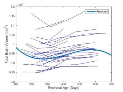
Aging Brain & Dementia
Traditional Poster
Neuro
Wednesday, 26 April 2017
| Exhibition Hall |
13:45 - 15:45 |
|
2338.
 |
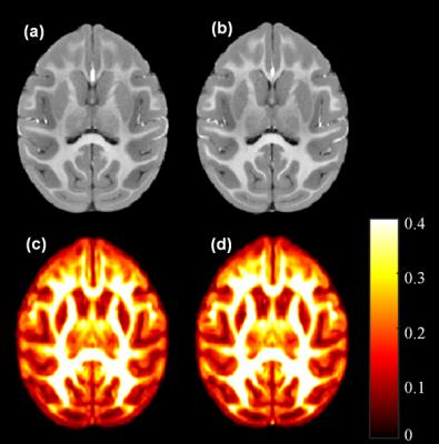
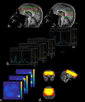
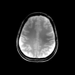
Age-related change of the whole brain T1 relaxation time: voxel-wise study with MP2RAGE
Gosuke Okubo, Tomohisa Okada, Akira Yamamoto, Yasutaka Fushimi, Tsutomu Okada, Kaori Togashi
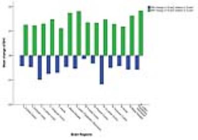
Correlation was investigated between normal aging and T1 relaxation time in the deep gray matter with MP2RAGE sequence using voxel-based analysis (VBA) and regions-of-interest (ROIs) focusing on the deep gray matter (GM). Seventy healthy subjects were included. In VBA, linear correlation was explored, whereas second order regression was added in ROI analysis. The results showed relationship that varied among deep GM structures. Those findings will shed light on further investigation of T1 relaxation time in patient groups.
|
|
2339.
 |
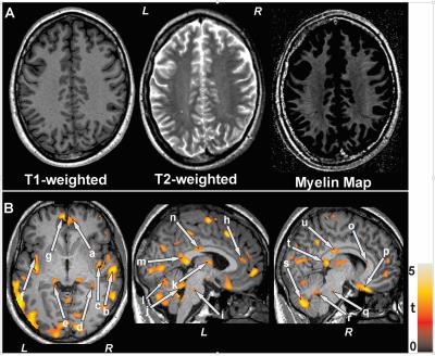
Factors influencing the detection of age-dependent variations of cortical myelin by MP2RAGE at 9.4T.
Gisela Hagberg, Jonas Bause, Thomas Ethofer, Philipp Ehses, Thomas Dresler, Cornelia Herbert, Rolf Pohmann, gunamony Shajan, Andreas Fallgatter, Marina Pavlova, Klaus Scheffler
Detection of subtle variation of the myeloarchitecture within the cerebral cortex may be feasibile with high-field mapping of the longitudinal relaxation time. However, besides myelin other factors like iron or variation of the grey matter volume may impact this MR parameter. In the present work we propose a model that includes and quantifies these factors. We found a regionally dependent, continuous increase from early adulthood into the middle ages that tentatively can be assigned to myelin.
|
|
2340.
 |
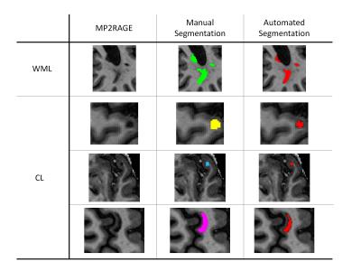
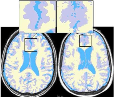
Individual Evaluation System Development Concept Research for Personalized Brain Aging Process Using Machine Learning
Kyung Mi Lee, Hyug-Gi Kim, Sung Kyoung Moon, Eui Jong Kim, Woo Suk Choi
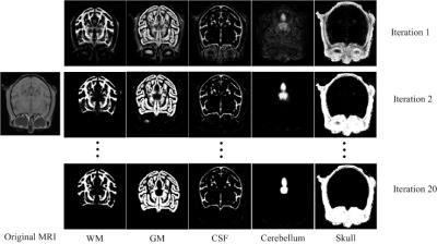
White matter hyperintensities (WMH) is one of the important characteristics of cerebral small vessel disease (cSVD). To diagnosis individual WMH evaluation method and investigate the degree of WMH form using MR image, we proposed that machine learning based on WMH group classification and individual diagnosis system.
|
|
2341.
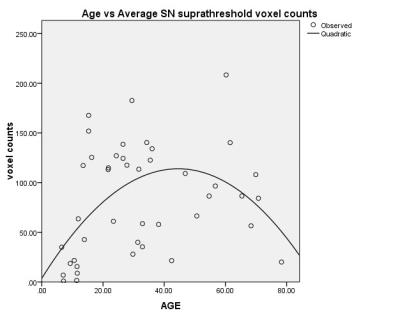 |
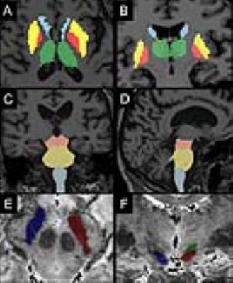
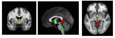
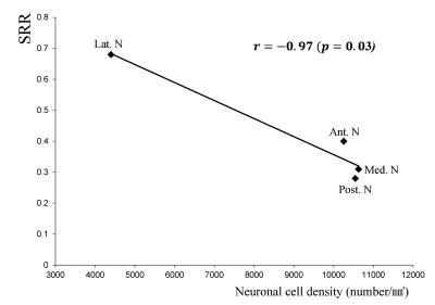
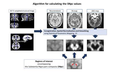
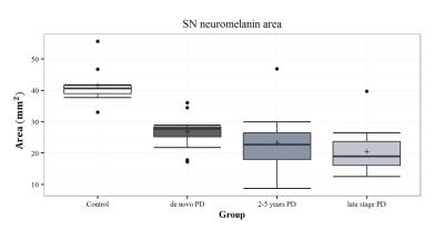
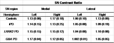
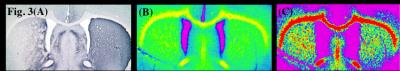
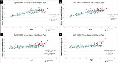
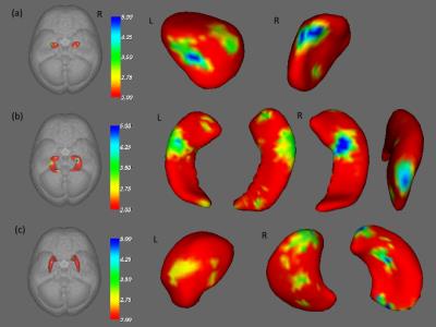
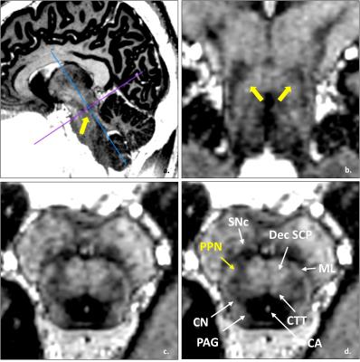
Neuromelanin-weighted MRI in revealing human development and age-related changes in locus coeruleus and substantial nigra
Yue Xing, Abdul Sapuan, Robert Dineen, Andrew Cooper, Dorothee Auer
Rapid neuromelanin-weighted MR imaging sequence was performed to noninvasively inspect the physiological changes of substantial nigra and Locus ceruleus across a wide range of age in healthy subjects using neuromelanin-sensitive MRI for the first time.
|
|
2342.
 |
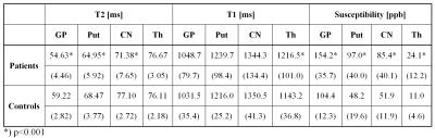
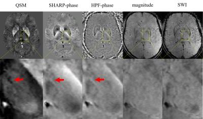
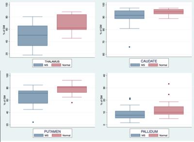
Characterizing brain iron deposition in patients with subcortical vascular mild cognitive impairment using quantitative susceptibility mapping: A potential biomarker
Yawen Sun, Yan Zhou, Yao Wang, Xu Han, Weina Ding, Yong Zhang, Qun Xu, Jianrong Xu
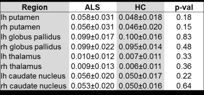
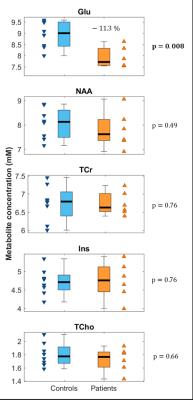
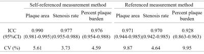
The presence and pattern of iron accumulation in subcortical vascular mild cognitive impairment (svMCI) or their effects on cognition has rarely been investigated. The purposes of the present study were to investigate brain iron deposition in deep gray matter nuclei in svMCI patients using quantitative susceptibility mapping (QSM) and its correlation with the severity of cognitive impairment. Susceptibility values were found to be elevated within bilateral hippocampus and right putamen in svMCI group compared with controls, which were related to cognitive measurements. Our result suggests that brain iron deposition which relation to cognition indicates the clinical relevance of the biomarker.
|
|
2343.
 |
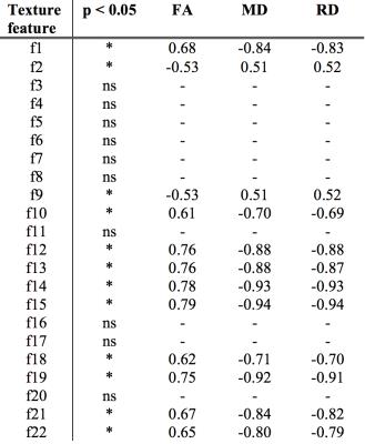
Early detection of Alzheimer's Disease using a combined index of gray matter texture
Subin Lee, Hyunna Lee, Heesoog Kim, Ki Woong Kim
Development of sensitive markers that can detect Alzheimer’s Disease at its pre-dementia stages is of critical importance. We hypothesized that altered cytoarchitecture caused by AD pathology would lead to a subtle altered pattern of voxel intensities on MRI which could be detected by texture analysis. In a training set of 111 AD and 141 normal controls, we extracted a set of texture features from core regions affected by pathology – precuneus, posterior cingulate cortex, hippocampus – that could efficiently discriminate between the two groups(AUC=0.875, p<0.001) as well as predict MCI/AD conversion from the normal stage (AUC=0.716, p=0.031).
|
|
2344.
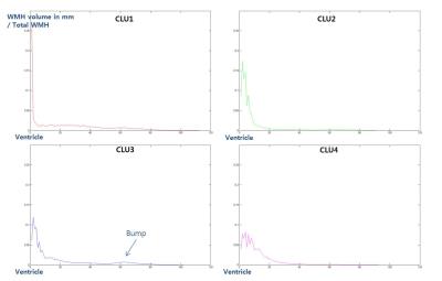 |
The profile pattern of white matter hyperintensities (WMH) has a primary role on discriminating the cognitive function.
Heisoog Kim, Jiwon Han, Hyunna Lee, Ki Kim
The areas in cerebral white matter appearing hyperintense on T2-weighted and fluid-attenuated inverse recovery (FLAIR) MRI are commonly referred to as white matter lesions (WMLs). Diverse scores and rating systems have been used for evaluating WMLs. However, these visual rating scales are still qualitative and subjective, leading to low reliability and reproducibility. The purpose of this study is to investigate the relation between cognitive functions and WMLs clustered by their profiles of distance from the ventricle, establishing the quantitative and objective evaluation of WMLs.
|
|
2345. 
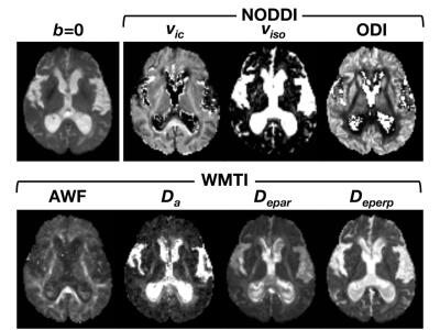 |
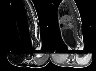
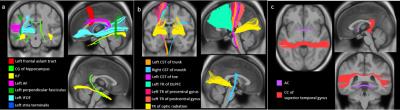
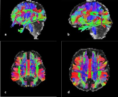
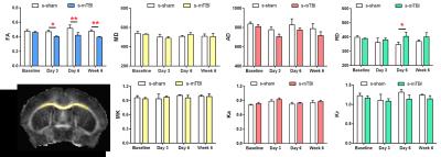
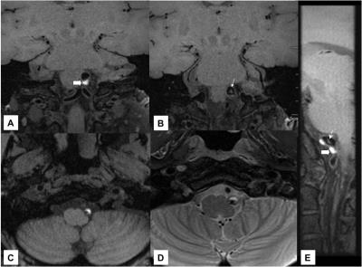
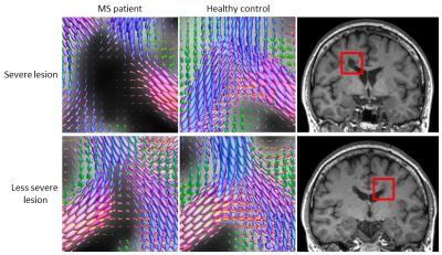
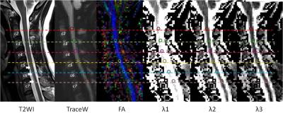
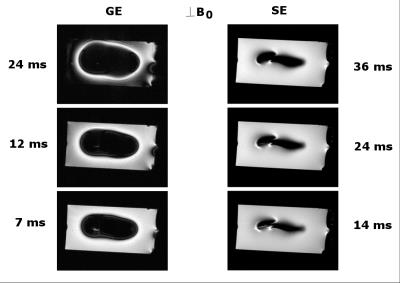
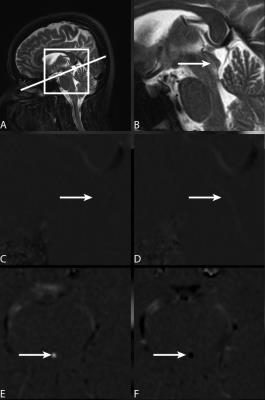
Diffusion microstructural imaging of reversible and irreversible changes within the corticospinal tract in idiopathic normal pressure hydrocephalus
Kouhei Kamiya, Masaaki Hori, Ryusuke Irie, Masakazu Miyajima, Madoka Nakajima, Koji Kamagata, Kouhei Tsuruta, Yuichi Suzuki, Asami Saito, Misaki Nakazawa, Harushi Mori, Akira Kunimatsu, Hajime Arai, Shigeki Aoki, Osamu Abe
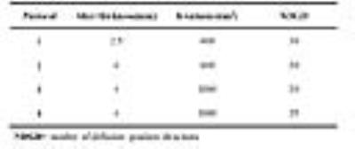
Microstructural changes of the corticospinal tract (CST) in idiopathic normal pressure hydrocephalus (iNPH) before and after CSF shunt surgery were studied using NODDI and WMTI. Pathological increase of orientational coherence and its postoperative normalization were shown, indicating axon stretching and its recovery. To the contrary, decrease of axon density was present in iNPH and remained after the surgery. Simulation using undulating cylinder model demonstrated both NODDI and WMTI can separate the effects from axon density and undulation. These results suggest possibilities of diffusion MRI to distinguish between reversible and irreversible microstructural changes in iNPH, and raise expectation for prediction of treatment outcome.
|
|
2346.
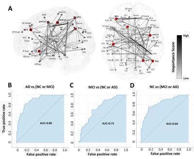 |
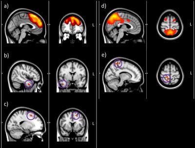
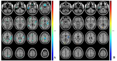
Global Change of Intrinsic Functional Networks as an Imaging biomarker of Alzheimer’s disease
Chia-Feng Lu, Wen-Jin Hsieh, Yu-Chieh Jill Kao, Paul Blakeley, Fei-Ting Hsu, Hua-Shan Liu, Ping-Huei Tsai, Li-Chun Hsieh, Cheng-Yu Chen
The change of whole-brain connectivity during resting state can be a reliable feature in discriminating patients with Alzheimer’s disease (AD) from patients with mild cognitive impairment and cognitively healthy elders. Significant correlations between resting-state functional connectivity and cognitive decline measured by Mini-Mental State Examination (MMSE) were further identified.
|
|
2347.
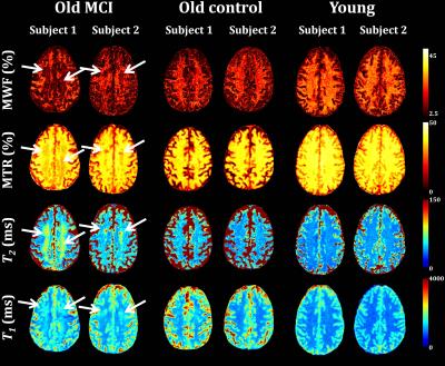 |
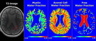
Demyelination in Mild Cognitive Impairment
Mustapha Bouhrara, David Reiter, Christopher Bergeron, Linda Zukley, Susan Resnick, Stephanie Studenski, Josephine Egan, Luigi Ferrucci, Richard Spencer
An emerging hypothesis suggests that the underlying pathophysiology of mild cognitive impairment (MCI) involves alterations in brain myelination. These alterations may represent an important correlate of dementia. Several studies have examined this correlation; however, these earlier analyses were performed using non-myelin-specific methods such as relaxation times, magnetization transfer and diffusion. This greatly complicates the interpretation of such imaging results in terms of myelin content. Our results show direct evidence of MWF alterations and loss in MCI using a direct measure of myelin-bound water.
|
|
2348.
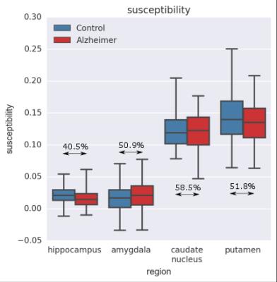 |
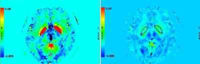
No significant increase of magnetic susceptibility found in subcortical gray matter of patients with Alzheimer’s Disease
Jakob Meineke, Fabian Wenzel, Iain Wilkinson, Ulrich Katscher
Quantitative Susceptibility Mapping (QSM) and volumetry are used to study the deep gray-matter nuclei of patients with Alzheimer’s Disease (AD) and healthy control subjects. QSM is performed using “Joint background-field removal and segmentation-Enhanced Dipole Inversion” (JEDI), which leverages the information from automated model-based segmentation and allows the compact single-step formulation of the ill-posed inversion problem of QSM. For comparison QSM is also performed using L1-MEDI from the MEDI-Toolbox. The tissue magnetic susceptibility shows no significant difference between the Alzheimer group compared to the healthy control group. In contrast, the normalized volume of segmented gray-matter regions is significantly reduced in AD patients.
|
|
2349.
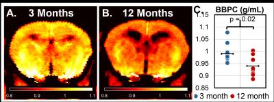 |
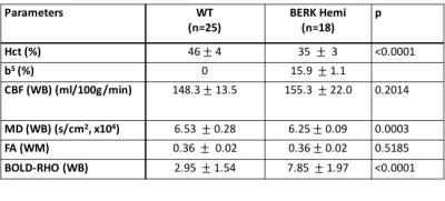
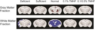
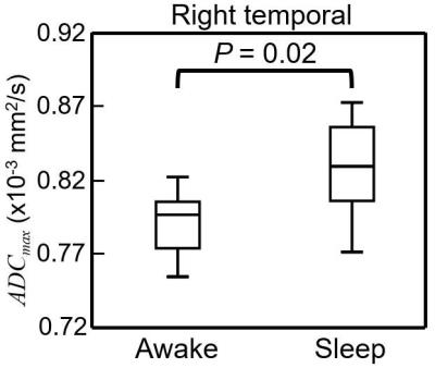
Using Calibrated Proton Density Imaging to Measure Blood-Brain Partition Coefficient in Aging and Alzheimer's Disease Mice
Scott Thalman, David Powell, Andrew Shen, Anika Hartz, Ai-Ling Lin
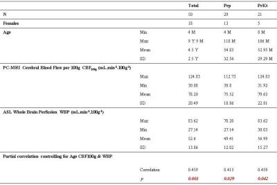
In this study we determine the blood-brain partition coefficient (BBPC) in aging C57Bl6/N mice and the transgenic 129S6/Tg2576 mouse model of Alzheimer’s disease using a calibrated proton density imaging approach. Aging mice demonstrate a 5.5% reduction in BBPC compared to young mice (0.94±0.04 mL/g vs 0.99±0.04 mL/g, p = 0.02), however Tg2576+ mice preliminarily demonstrate an elevated BBPC compared to wild-type controls (01.03±0.04 mL/g vs 1.00±0.05 mL/g). These high quality BBPC maps acquired much faster than previously reported could potentially be used to correct cerebral blood flow measurements derived from arterial spin labeling.
|
|
2350.
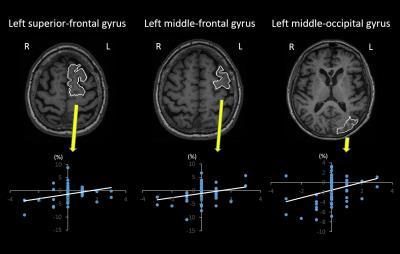 |
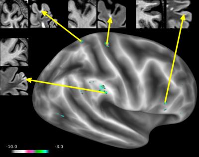
Neuroanatomical substrates that account for worsening performance in the Clock-Drawing Test in mild cognitive impairment
Satoshi Nakajima, Susumu Mori, Kaori Togashi, Kenichi Oishi
The Clock-Drawing Test (CDT) is used to screen and select cognitively impaired individuals for further evaluation. For the clinical interpretation of the CDT, an understanding of the neuroanatomical substrates that account for a decline in the CDT score is essential. We investigated the relationships between regional volume loss and a decline in the CDT score in two years. Atrophy in the left prefrontal and middle-occipital gyri was correlated with a decline in the CDT score. The result validated the use of the CDT, combined with memory tests that evaluate parieto-temporal functions, as part of an overall cognitive screening.
|
|
2351.
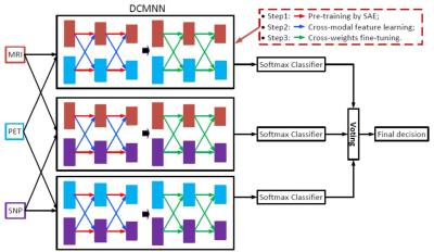 |
Deep Cross-Modal Feature Learning and Fusion for Early Dementia Diagnosis
Tao Zhou, Kim-Han Thung, Dinggang Shen
Studies have shown that neuroimaging data (e.g., MRI, PET) and genetic data (e.g., SNP) are associated with the Alzheimer’s Disease (AD). However, to achieve a more accurate AD diagnosis model using these data is challenging, as these data are heterogeneous and high-dimensional. Thus, we first used region-of-interest based features and deep feature learning to reduce the dimension of the neuroimaging and SNP data, respectively. Then we proposed a deep cross-modal feature learning and fusion framework to fuse the high-level features of these data. Experimental results show that our method using MRI+PET+SNP data outperforms other comparison methods.
|
|
2352.
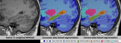 |
Use of population-specific atlases for brain morphometry: benefits in Alzheimer’s Disease diagnosis decision support for Chinese patients
Bénédicte Maréchal, Peipeng Liang, Lin Shi, Tianyi Qian, Defeng Wang, Tobias Kober, Alexis Roche, Kuncheng Li
We investigate the potential of a Chinese-specific brain atlas derived from a large MR database of healthy Chinese subjects to accurately support Alzheimer’s Disease diagnosis using automated brain volumetry in the Chinese population. Our experiments show that disease detection sensitivity is higher than when using either a Westerner-specific atlas and normative ranges or combining a Westerner atlas with Chinese-specific normative ranges. These findings suggest that population-specific models improve the reliability of medical decision support systems based on automated brain volumetry.
|
|
2353.
 |
Prediction of early stage of AD based on functional connectivity network characteristics: an fMRI-based study
Zhizheng Zhuo, Zhuqing Long, Bin Jing, Xiangyu Ma, Han Liu, Jianxin Dong, Xiao Mo, Qi Yan, Haiyun Li
MCI (Mild Cognitive Impairment) is a pre-stage of Alzheimer’s Disease (AD). And the early detection of MCI is important for early treatment of AD patients. In this work, prediction efficiency of early stage of AD based on the functional connectivity network characteristics was evaluated by using a couple of classifiers with AAL_90 and AAL_1024 templates. The results showed that brain functional characteristics were effective in the prediction of MCI with a SVM-based classifier. And a more fine template could improve prediction accuracy.
|
|
2354.
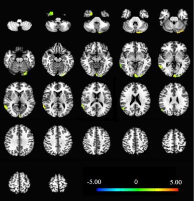 |
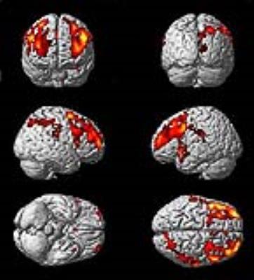
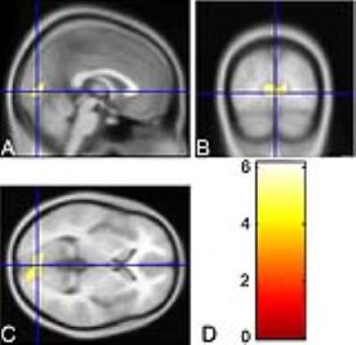
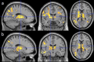
Acupoint-Specific Effect of Acupuncture in Alzheimer’s Disease: A Functional MRI Study
Yi Shan, Yunpeng Bian, Zhiqun Wang, Zhilian Zhao, Mo Zhang, Jie Lu, Kuncheng Li
Acupuncture has been a major therapeutic method in Chinese medicine for treating Alzheimer’s disease (AD) with validation and safety. In this study, we use functional magnetic resonance imaging (fMRI) to investigate the acupoint-specific effect of acupuncture in treating for AD. We found acupuncture at real acupoints activated brain areas primarily in the left uvula, right superior temporal gyrus and right uncus, while acupuncture at sham acupoints only activated areas in the left insula. These results showed that acupoint-specific effect of acupuncture presented by fMRI may help to facilitate its clinical use in AD treatment.
|
|
2355.
 |
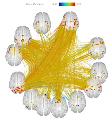
Disrupted brain connectivity networks in Alzheimer's Disease
Xiaoqing Ji, Haiyang Geng, Rui Li, Le He, Chun Yuan
In this study, we used graph theory-based approaches to explore the topological organization of whole brain network in Alzheimer’s Disease. We performed resting state fMRI on 6 AD patients and 19 normal controls. Topological properties such as small-world, efficiency and nodal centrality were calculated and nonparametric permutation tests were further used for group comparisons. The results showed that high-level cognitive regions including parietal cortex, vmPFC, precuneus and emotional regions including caudate and thalamus were changed in the topological metrics, this may be a biomarker for AD classification.
|
|
2356.
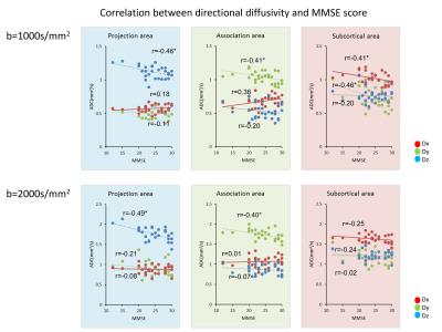 |
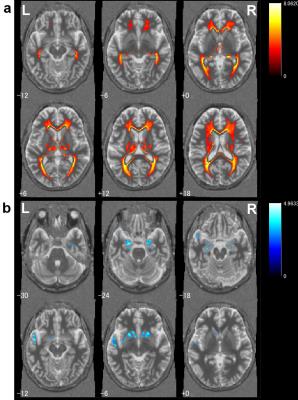
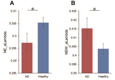
Evaluating glymphatic system by diffusion images: Alzheimer's disease cases analyzed by Diffusion Tensor Image analysis Along Perivascular Space (DTI-ALPS)
Toshiaki Taoka, Yoshitaka Masutani, Hisashi Kawai, Toshiki Nakane, Kiwamu Matsuoka, Fumihiko Yasuno, Shinji Naganawa
We tried to evaluate the activity of human glymphatic system by diffusion images. Our subjects were Alzheimer's disease (AD), in which it is known that the activity of the glymphatic system is impaired in animal experiments. We evaluated the diffusivity along the perivascular spaces as well as projection fibers and association fibers, and correlated them with MMSE score. There were significant positive correlation between diffusivity along perivascular spaces and MMSE score, indicating impaired water diffusivity related to AD severity. Our result may indicate that activity of the glymphatic system can be evaluated by diffusion images.
|
|
2357.
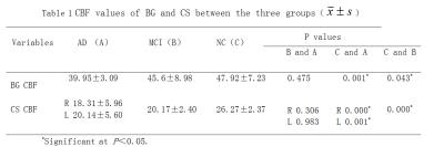 |
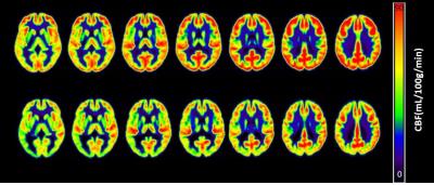
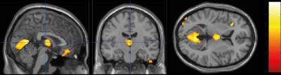
Can EPVS reflect Cerebral Blood Flow and Cognitive State in MCI and AD Patients?
Jin Shang, Liu Yingqiu, Yanwei Miao, weiwei wang
EPVS may reflect underlying cerebral small vessel disease. We assumed the presence of EPVS were associated with cerebral blood flow (CBF)reduction ,and can accurately reflect the cognitive state.The study verifies a good coherence between EPVS and CBF in BG and CS of AD patients , and furthermore reflects of EPVS as the biomarker of cognitive status to a certain extent.
|
|
2358.
 |
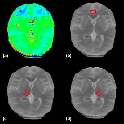
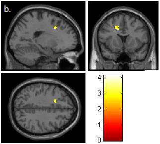
A Potential Biomarker of Alzheimer's Disease: T1sat of the Thalamus
Parshant Sehrawat, H. Michael Gach, Wenna Duan, Andrea Gillman, James T. Becker, Oscar L. Lopez, Weiying Dai
Biomarkers for Alzheimer’s disease (AD) are crucial for early diagnosis and treatment monitoring once therapies become available. In this study, the spin-lattice relaxation time in the rotating frame with (T1sat) and without (T1nosat) off-resonance saturation was measured at 1.5 T in the cardiovascular health study cognition study cohort. Cross-sectional and longitudinal studies of the normalized difference (ΔT1RF) between T1sat and T1nosat revealed regions of statistical significance in the brain that are associated with dementia pathogenesis. ΔT1RF changes in the thalamus were consistent for the cross-sectional and longitudinal studies, indicating that ΔT1RF could be a promising imaging biomarker for AD.
|
|
2359.
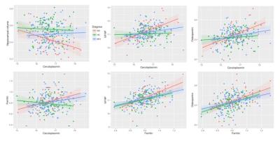 |
Hippocampal atrophy is correlated to cerebrospinal fluid levels of ceruloplasmin, neuroinflammation and total tau in Alzheimer’s disease
Azhaar Ashraf, Dr Po-Wah So
The aim of the study was to characterise the relationship between cerebrospinal fluid (CSF) iron regulatory proteins (ferritin, ceruloplasmin), neuroinflammation and MRI-derived hippocampal volume in healthy controls, mild cognitive impairment and Alzheimer’s disease subjects. Ceruloplasmin positively correlated with neuroinflammation and ferritin in MCI and AD while in the latter group, it was negatively correlated with hippocampal volume. Ferritin positively correlated with neuroinflammation in HC and AD but also with tau levels in MCI. Iron dyshomeostasis, neuroinflammation and tau metabolism may increase hippocampal atrophy and aggravate AD pathogenesis.
|
|
2360.
 |
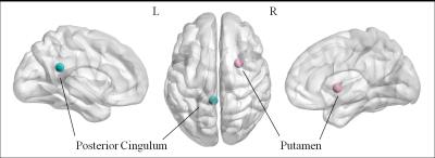
Altered Hippocampal Functional Connectivity with PCC in SCI, MCI and AD
Hui Zhang, Joseph Shiu-Kwong Kwan, Pui-Wai Chiu, Edward S. Hui, Queenie Chan, Henry Ka-Fung Mak
Brain imaging research has elucidated the structural and functional changes at the clinical stages of AD and MCI, however, the characteristics of resting-state functional connectivity of SCI are largely unstudied. Hippocampus is among the first regions targeted by AD pathology. In this study, we employed resting state fMRI to study the connectivity between hippocampus and posterior cingulate cortex in SCI, MCI and AD groups. In the preliminary results, the connectivity between hippocampus and PCC in SCI was found stronger than control and other two neurodegenerative groups which may suggest that compensatory mechanisms providing preserved hippocampal pathway in SCI.
|
|
2361.
 |
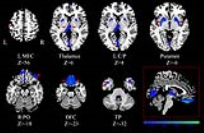
Brain iron load, as measured by Quantitative Susceptibility Mapping, promotes beta-Amyloid associated functional brain change in elderly subjects but not in Super-Agers
Jiri van Bergen, Xu Li, Frances-Catherine Quevenco, Sandra Leh, Anton Gietl, Valerie Treyer, Rafeal Meyer, Alfred Buck, Roger Nitsch, Peter van Zijl, Christoph Hock, Paul Unschuld
To investigate whether brain iron load has an impact on Aβ associated functional brain change, this study investigated a large sample of cognitively healthy adults including 44 Super-Agers (subjects over the age of 85 without cognitive impairments) using simultaneous assessment of Amyloid-PET for Aβ-plaque-density, QSM for estimation of iron load and resting-state-fMRI. Our findings indicate that the combination of Aβ-plaque-density with other neurodegenerative change (iron), has an impact on brain functionality, reflected by significant changes of resting state functional connectivity. Additionally, Aβ-plaque-density had no significant effect on functional connectivity in Super-Agers.
|
|
2362.
 |
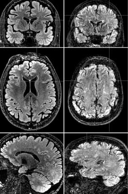
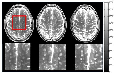
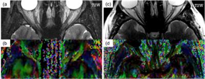
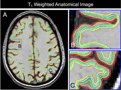
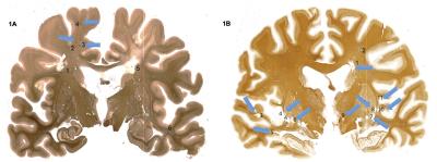
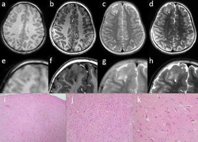
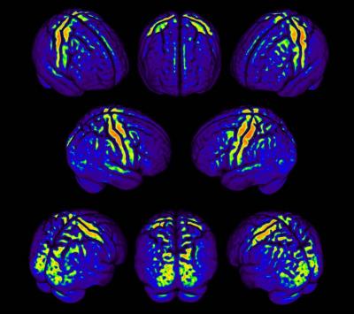
Demonstration of Abnormal Cortical Layers In Alzheimer's Disease Using Subtracted Tissue Attenuated Inversion Recovery (STAIR) Pulse Sequences
Shujuan Fan, Yajun Ma, Xing Lv, Jiang Du, Graeme Bydder, Nikolaus Szeverenyi
The use of subtracted STIR images designed to null white and gray matter respectively is illustrated in formalin fixed brain samples at 11.7T. The images show the normal layers of the cerebral cortex with high contrast. The layers were less well seen, or not seen at all in Alzheimer’s Disease samples. Use of MT pulses with the STIR sequences produces high positive and negative contrast on difference images. The STIR subtraction technique made have general application, and be used with different forms of data acquisition. It may also be useful in other clinical situations for demonstrating changes due to small differences in T1 in the presence of long T1 fluids.
|
|
2363.
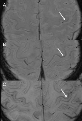 |
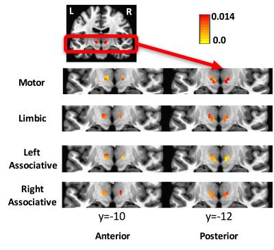
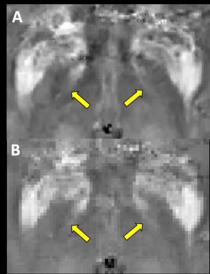
Motor cortex hypointensity on SWI is associated with APOE status in cognitive impaired patients
Mina Park, Yeonsil Moon, Seol-Heui Han, Won-Jin Moon
We evaluated the prevalence and its associated risk factors of motor cortex hypointensity on SWI in cognitive impaired patients. This retrospective study included 116 cognitively impaired patients (28 Alzheimer disease patients and 88 mild cognitive impaired patients). Among them, 83 patients showed positive motor cortex hypointensity on SWI and it was associated with age. Furthermore, the group with positive motor cortex hypointensity on SWI had less APOE4 allele carriers than negative group and this shows (+) APOE4 allele may act as an accelerating factor of cognitive decline even before iron accumulation starts to show changes in the motor cortex.
|
|
2364.
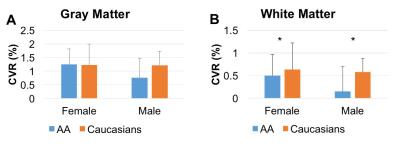 |
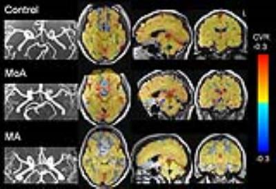
Racial differences in cerebral microcirculatory function and cognitive function
Junjie Wu, Ganesh Chand, Om Sharma, Deqiang Qiu, Ihab Hajjar
We examined racial differences in cerebrovascular reactivity (CVR) to carbon dioxide and their associations with cognitive function among patients with mild cognitive impairment and hypertension. The results provide evidence for association between impaired CVR and executive dysfunction. Hypertensive African Americans have more compromised cerebral microcirculatory function compared to Caucasians.
|
|
2365.
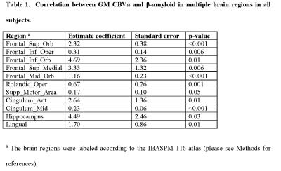 |
Synergistic Effect of ß-Amyloid and Microvascular Abnormality on Longitudinal Cognitive Decline in Elderly Subjects at Risk for Alzheimer’s Disease (AD)
Jun Hua, M Wyss, J. M. G. van Bergen, S. J. Schreiner, S. C. Steininger, A. F. Gietl, A. Buck, R. M. Nitsch, K. P. Pruessmann, H. Lu, P. C. M. van Zijl, M. Albert, C. Hock, P. G. Unschuld
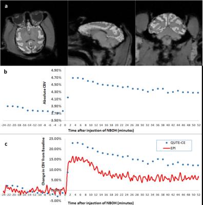
Progressive impairment in multiple cognitive domains is a clinical hallmark of Alzheimer’s Disease (AD), which is the most frequent cause for dementia in the elderly and is neuropathologically characterized by both cerebral β-amyloid (Aβ) accumulation and microvascular abnormalities. Here, we report significant co-localization of regions with microvascular abnormalities measured by arteriolar-cerebral-blood-volume (CBVa) MRI and Aβ accumulation measured by PiB-PET in elderly subjects at-risk for AD. Multiple regression analysis suggested that CBVa and Aβ may have a synergistic effect on longitudinal cognitive decline in these subjects . Both variables may need to be considered for secondary prevention trials in such populations.
|
|
2366.
 |
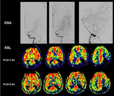
The mediating effects of functional disconnection on the association between structural disconnection and cognitive impairment in symptomatic carotid artery disease
Dewen Meng, Akram Hosseini, Richard Simpson, Robert Dineen, Dorothee Auer
This study investigated the association between functional disconnection of cognitive networks, structural disconnection indexed as microscopic damage of specific white matter tracts, and global cognitive impairment in symptomatic carotid artery disease. The findings regarding the mediating effects of functional disconnection on the association between structural disconnection and global cognitive impairment provided promising implications for future studies that develop therapies for patients with vascular cognitive disorder.
|
|
2367.
 |
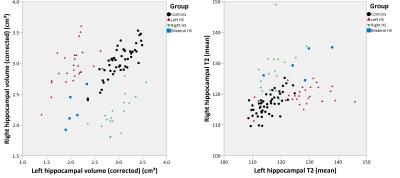
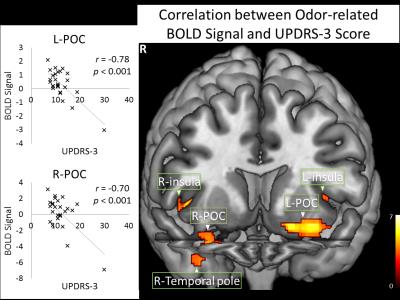
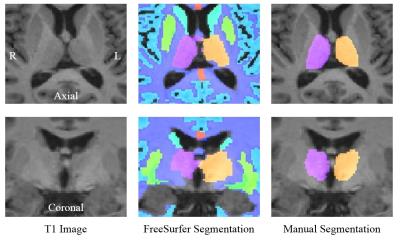
The Correlation of Olfactory Dysfunctions and Hippocampal Atrophy in Patients with Cognitive Impairment: A Potential Clinical Marker for Alzheimer’s Disease
Bing Zhang, Bin Zhu, Yun Xu, Qing.X Yang
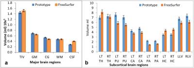
Olfactory deficits have been observed in subjects with Alzheimer’s disease (AD) and its potential as a biomarker has been demonstrated by several recent studies. However, its sensitivity and specificity in detecting early AD have not been validated in the Chinese population where aging population is growing rapidly. In this study, by using a 3.0 T MR scanner, we evaluate the presence of olfactory deficits and hippocampal volume loss measured by FreeSurfer (6.0) based on local population in China, to validate the use of olfactory test battery as a clinical AD marker.
|
|
2368.
 |
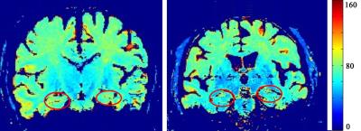
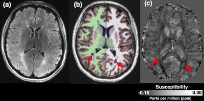
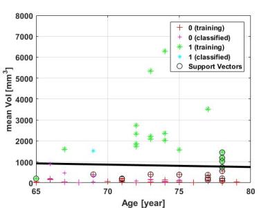
Subcortical Nuclei Iron Deposition of Alzheimer's Patients on MRI-QSM: Maybe a Diagnostic Indicator
Lei Du, Yijiang Zhu, Tianbin Song, Lizhi Xie, Guolin Ma
Based on gradient echo (GRE) magnetic resonance phase data, quantitative susceptibility mapping (QSM) is a novel technique which allows the non-invasive assessment of magnetic tissue susceptibility distribution in mild alzheimer’s disease (AD). In this study ,we investigated the correlation between mini-mental state examination (MMSE) and bulk tissue magnetic susceptibility in subcortical nuclei of 14 mild AD subjects and 14 cognitively healthy controls scanned at 3T . A strong linear correlation between them was found in caudate nucleus and dentate nucleus. Hence, QSM can be used for early AD diagnosis and intervention.
|
|
2369.
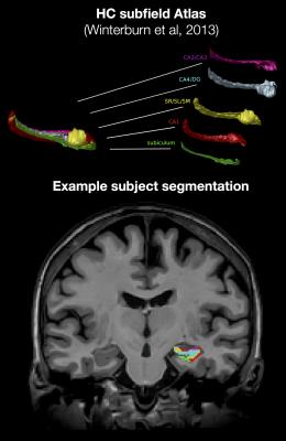 |
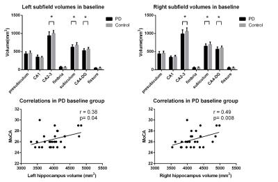
Hippocampal T1-weighted and FLAIR contrast is associated with CSF biomarkers in asymptomatic individuals with parental history of Alzheimer’s disease
Christine Tardif, Robert Amaral, Gabriel Devenyi, Pedro Rosa-Neto, Judes Poirier, John Breitner, M Mallar Chakravarty, The PREVENT-AD Research Group
Cerebrospinal fluid (CSF) β-amyloid and phosphorylated-tau are consistently used as biomarkers related to the pathophysiology and clinical severity of individuals in the earliest phases of Alzheimer’s disease (AD). This study shows that T1-weighted and FLAIR signal intensity in the hippocampal subfields, normalized using the fimbria, are associated with ApoE4 status and CSF biomarkers. The FLAIR results suggest that presence of inflammation in the subiculum of ApoE4 carriers and in the CA1 and molecular layers of the hippocampus in subjects with low CSF β-amyloid burden as tau pathology increases.
|
|
2370.
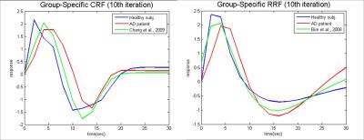 |
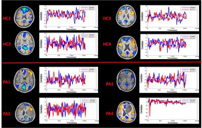
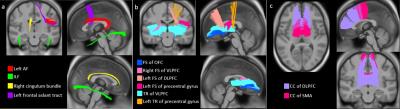
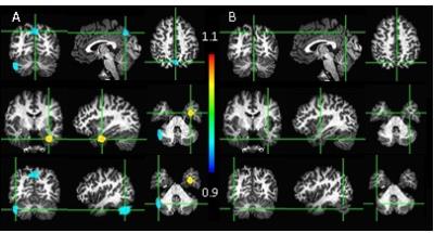
Modulated the Physiological Response Delay to Prevent Overestimating the Disruption of Default Mode Network in Alzheimer’s Disease
Yi-Tien Li, Chun-Yuan Chang, Yi-Cheng Hsu, Jong-Ling Fuh, Fa-Hsuan Lin
This study quantified the impact of physiological noise correction in characterizing the resting-state fMRI in Alzheimer’s disease (AD) patients with age- and gender-matched 17 healthy subjects and 15 AD patients. Using a seed-based correlation method with seeds at posterior cingular cortex and medial prefrontal cortex, we found that the difference in the functional connectivity between AD patients and healthy controls was significantly reduced when physiological noise was suppressed.
|
 |
2371.
 |
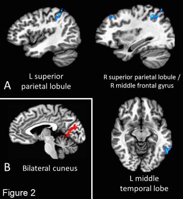
Association between whole brain functional connectivity and cortical thickness throughout healthy aging
Bruno Vieira, Carlos Salmon
The aging process entails morphological and functional alterations in the human brain. Using magnetic resonance imaging data of 130 subjects aged between 18 and 81 years from a publicly available dataset we obtained whole brain cortical thickness estimates and resting state connectivity to study how healthy aging affects these. Additionally, we studied the relationship between cortical thickness and functional connectivity. A heterogeneous thinning profile was observed and also a dominance in increases in connectivity, with few decreases. Connectivity correlates with thickness with temporal and occipital seed ROIs. These results might help to understand how connectivity and thickness relate in neuropathologies.
|
|
2372.
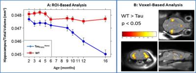 |
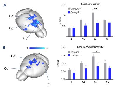
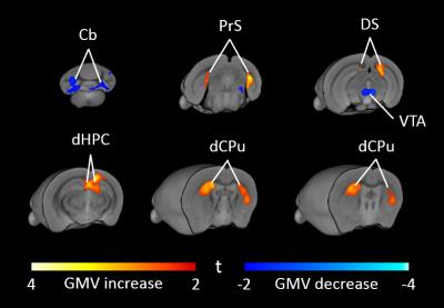
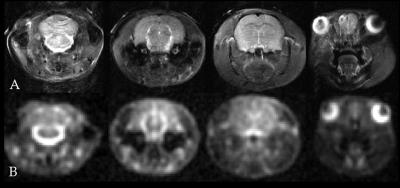
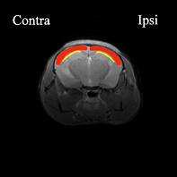
Exaggeration of Tau Pathology by Amyloid-ß production in PS2APPxTauP301L Transgenic Mouse Model of Alzheimer’s Disease
Vineela Gandham, William Meilandt, Kai Barck, Maj Hedehus, Kimberly Malesky, Claire Pichon, Oded Foreman, Gai Ayalon, Kimberly Scearce-Levie, Richard Carano
The PS2APP, PS2APPxTauP301LHet, TauP301LHomo, TauP301LHet transgenic mouse models of AD were characterized using MRI, and various regional brain atrophies were detected using a completely automated analysis by SPM-8. Furthermore, through MRI (both ROI and voxel based morphometry) and histology we showed that the presence of PS2APP mutation in PS2APPxTauP301LHet triple transgenic mouse model exaggerates the Tau pathology. The presented results will enable MRI as a great tool to non-invasively assess the disease progression over time in preclinical therapeutic studies targeting Aβ and Tau pathology.
|
|
2373.
 |
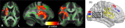
The relationship between brain white matter hyperintensities burden and age-related neuropathologies is location dependent
Chantal Sopacua, Arnold Evia, Aikaterini Kotrotsou, Sue Leurgans, David Bennett, Julie Schneider, Konstantinos Arfanakis
White matter hyperintensities (WMH) are white matter lesions appearing hyperintense in T2-weighted MRI. WMH are common in older adults and have been associated with increased risk of cognitive decline and dementia. Previous efforts have attempted to identify the neuropathologies associated with whole brain WMH burden. However, it is yet to be determined if the relationship between regional WMH burden and age-related neuropathologies is the same, or varies, in different parts of the brain. Therefore, the purpose of this research was to investigate the association between regional WMH burden and neuropathologies in a community cohort of older adults.
|
|
2374.
 |
MRI of longterm changes in vascularization and functional connectivity in a mouse model of vascular cognitive impairment
Philipp Boehm-Sturm, Joseph Kuchling, Susanne Mueller, Marco Foddis, Carsten Finke, Celeste Sassi, Christoph Harms, Stefan Koch, Ulrich Dirnagl, Tracy Farr
Chronic mouse brain hypoperfusion produces white matter damage; a feature of vascular cognitive impairment. Despite growing interest in this model, we have struggled to observe a strong phenotype. The present study aimed to improve the phenotype through extended hypoperfusion (6m). We examined the effect on various MR biomarkers including functional connectivity and vascular remodeling. We found massive structural changes including arterial neovessels, small subcortical strokes, and microbleeds. Animals showed behavioral deficits accompanied by changes in resting state MRI signals of the cingulate cortex, which is functionally connected to regions related to behavior (hippocampus) and emotion (amygdala).
|
|
2375.
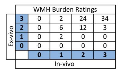 |
White matter hyperintensity burden assessed ante-mortem and post-mortem on the same older adults
Arman Kulkarni, Arnold Evia, Julie Schneider, David Bennett, Konstantinos Arfanakis
White matter hyperintensities (WMH) are commonly observed in brain MR images of older adults. Recently, more and more research studies assess WMH burden using ex-vivo MRI, aiming at directly linking WMH to the underlying neuropathologies detected at autopsy. The purpose of this work was twofold: 1) to investigate the relationship between WMH burden assessed in-vivo and ex-vivo on the same older adults, and 2) to test the hypothesis that WMH burden assessed ex-vivo is higher than that assessed in-vivo for longer ante-mortem intervals (AMI) (i.e. from in-vivo MRI to death).
|
|
2376.
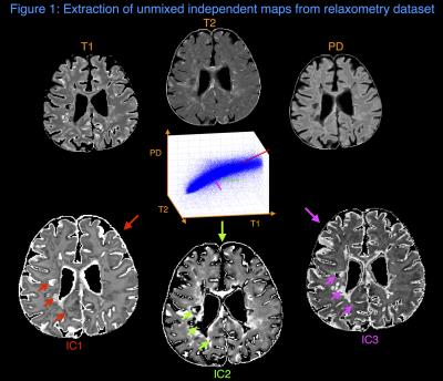 |
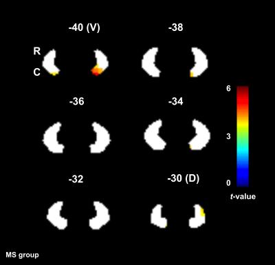
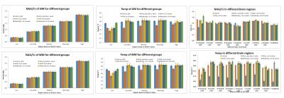
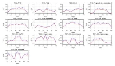
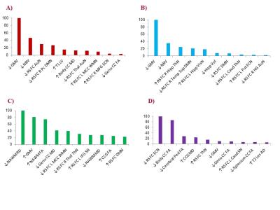
Fast multivariate relaxometry can differentiate neurodegenerative disease processes and phenotypes
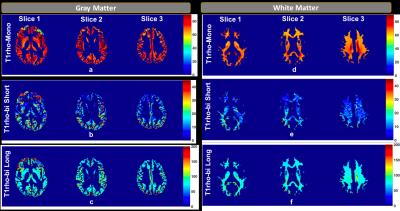
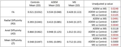
Gabriel Mangeat, Benjamin De Leener, Virginija Danylaité Karrenbauer, Marcel Warntjes, Nikola Stikov, Caterina Mainero, Julien Cohen-Adad, Tobias Granberg
Hereditary diffuse leukoencephalopathy with spheroids (HDLS) and multiple sclerosis (MS) are demyelinating and neurodegenerative disorders that can be hard to distinguish clinically and radiologically. Here, we present a framework to extract independent physiological sources of signal from time-efficient multiple quantitative relaxometry (T1, T2 and PD maps) to characterize varying degrees and mechanisms of tissue disruption. The method can aid in the differentiation of HDLS and MS (p=0.007), as well as identify MS subtypes (p=0.0007), which would be helpful in ensuring a correct diagnosis and treatment of these disorders.
|
|
2377.
 |
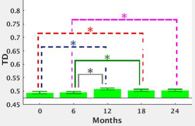
What Happens to the Hippocampus 12-months After Training? A Longitudinal Linear Mixed Effects Model Analysis of Mild Cognitive Impairment in the SMART Trial
Kathryn Broadhouse, Chao Suo, Maria Fiatarone Singh, Nicola Gates, Wei Wen, Perminder Sachdev, Henry Brodaty, Nidhi Saigal, Nalin Singh, Guy Wilson, Jacinda Meiklejohn, Bernhard Baune, Michael Baker, Nasmin Foroughi, Yi Wang, Yorgi Marvos, Michael Valenzuela
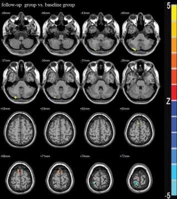
Mild cognitive impairment (MCI) increases future risk of dementia, however, several studies have shown that mental and physical exercise reduce this risk. From the Study of Mental Activity and Resistance Training (SMART) we have previously shown significantly improved global cognitive function immediately after 6 months of progressive resistance training in MCI. In this analysis, we compare longitudinal hippocampal volume change in MCI using linear mixed effects models over an 18-month period comprised of a 6-month training phase and a 12-month post training follow-up. Our results show both isolated cognitive and progressive resistance training significantly diminished the rate of left hippocampal atrophy compared to a double sham intervention across training and an extended follow-up period.
|
|
2378.
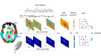 |
Extraction of Both Dynamic Functional and Structural Connectivity from Resting-state fMRI for MCI Classification
Xiaobo Chen, Han Zhang, Lichi Zhang, Dinggang Shen
In this abstract, we show that the diagnosis accuracy of mild cognitive impairment (MCI) can be significantly improved by integrating dynamic information contained in the traditional functional connectivity (FC) from grey matter (GM) regions and the functional correlation tensors (FCT) from white matter (WM) regions, both computed from resting-state fMRI (RS-fMRI). The advantages of our method include: 1) dynamic FC is exploited to reveal rich time-varying information in FC, and 2) the anatomical structure information within WM can be well incorporated in RS-fMRI.
|
|