ISMRM 25th Annual Meeting & Exhibition • 22-27 April 2017 • Honolulu, HI, USA

Traditional Poster Session: Cardiovascular
2719 -2767 Myocardial Tissue Characterization
2768 -2791 Atherosclerosis Imaging
2792 -2823 MR Angiography
2824 -2861 Flow & Velocity
2862 -2887 Function
 
Myocardial Tissue Characterization
Traditional Poster
Cardiovascular

 
Thursday, 27 April 2017
Exhibition Hall  08:15 - 10:15


2719.   
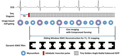
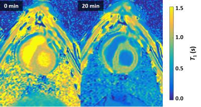
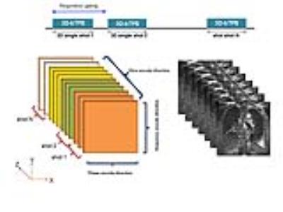
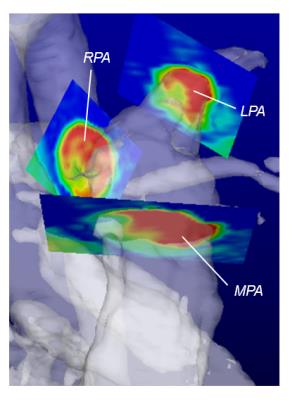
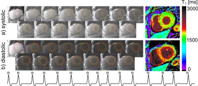
Simultaneous measurements of myocardium T1, T2 map, Cine and synthetic LGE using Inversion Recovery tiny golden angle radial balanced-SSFP within 6 second
Panki Kim, Byoung Wook Choi
In the cardiac MRI study, the quantification of T1, T2 relaxation time has become an important indication as well as the cine and the late gadolinium enhancement. In this work, we presented a novel method for simultaneous acquisition of T1, T2 map and Cine image of the myocardium based on the transient phase of inversion recovery balanced-SSFP imaging, and was proven potential both phantom and in vivo on a healthy volunteer.


2720.  
Accurate Myocardial T1-Mapping in Arrhythmia using Saturation-Recovery during Systole at 3T
Nadja Meßner, Sebastian Weingärtner, Johannes Budjan, Dirk Loßnitzer, Theano Papavassiliu, Lothar Schad, Frank Zöllner
In arrhythmic patients, ECG mis-triggering frequently leads to T1-quantification inaccuracy.

In this study, a heart-rate independent saturation-recovery T1-mapping method was adapted for systolic imaging at 3T by performing magnetization saturation right after the systolic imaging window and prior to R-wave detection. Estimated T1- and ECV- values during systole were (1557±53ms/ 0.21±0.03) compared to (1585±58ms/0.21±0.03) at diastole.

Our results show that SR T1-mapping might be an advantageous alternative to yield accurate T1- and ECV-values in patients with arrhythmia or reduced myocardial wall-thickness.


2727.  
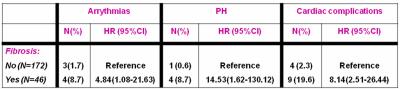
Myocardial fibrosis is a predictor of adverse outcomes in thalassemia intermedia patients
Antonella Meloni, Nicola Giunta, Pietro Giuliano, Stefania Renne, Laura Pistoia, Vincenzo Positano, Calogera Gerardi, Vincenzo Spadola, Petra Keilberg, Alessia Pepe
Myocardial fibrosis detected by LGE has been confirmed as an independent predictor of CV complications in thalassemia intermedia patients. Our finding is consistent with a growing evidence that LGE has a strong prognostic impact in thalassemic patients, warranting a close clinical and instrumental follow-up.


2721.   
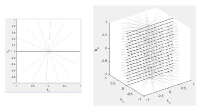
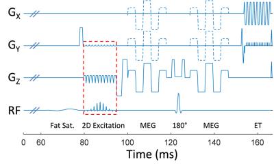
Towards High Success Rate in vivo Cardiac DTI on a Clinical 3T Scanner: Considerations on Heart Rate, Body-to-Mass Index, and Free Breathing
Christopher Nguyen, Sen Ma, Xiaoming Bi, Debiao Li
Clinical translation of cardiac diffusion tensor MRI has been challenging because of the sensitivity to bulk motion and thus, low success rates of scans. Current techniques require either high gradient systems (>40 mT/m) or excess breath holding ( >10 breath holds / slice) to acquire motion free cardiac DT-MRI. We propose a cardiac DT-MRI technique optimized for clinical translation and aimed at achieving high success rates in subjects with high and variable heart rate and high bold-to-mass index under free breathing conditions. Results in subjects with high BMI and variable HR yielded success rates > 90%. 


2722.  
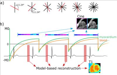
Multi-parametric cardiac MRI for T1 mapping and cine imaging using iterative model-based image reconstruction
Kirsten Becker, Jeanette Schulz-Menger, Tobias Schaeffter, Christoph Kolbitsch
In cardiac MRI, different diagnostic parameters are obtained in separate scans, leading to long examination times. In this work, we present an iterative model-based reconstruction approach for continuously acquired data, which provides native T1 maps and functional cine images within a single breath hold. The continuous acquisition allows for T1 reconstruction for different cardiac phases. Evaluation in a phantom demonstrated accurate T1 values (R˛>0.99) and insensitivity to heart rates, with T1 variations of less than 5% (50 to 90 bpm). In three healthy volunteers T1 maps were assessed for diastole and systole and cine images had a consistent dark-blood contrast.


2723.   
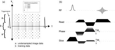
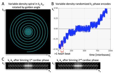
Non-ECG, Free-breathing Joint T1-T2 Mapping in the Myocardium with CMR Multitasking
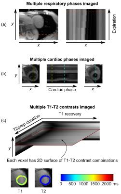
Anthony Christodoulou, Christopher Nguyen, Jaime Shaw, Yibin Xie, Nan Wang, Debiao Li
Myocardial tissue characterization via quantitative T1 and T2 mapping is typically accomplished using ECG-triggering and breath-holding. Here we describe a novel method for non-ECG, free-breathing joint T1-T2 mapping using the cardiovascular low-rank tensor imaging framework for CMR multitasking. This method achieves joint T1-T2 mapping in the myocardium at multiple cardiac and respiratory phases within 1.5 min for one slice. Measurements were within the range reported in the literature, and were repeatable to 3.9% for T1 and 6.1% for T2.


2724.   
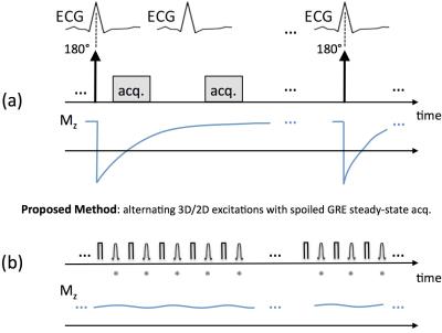
End-systolic myocardial T1 mapping using a spoiled steady-state approach: Towards reducing the confounding effect of intra-myocardial blood on native T1
Zulma Sandoval, Daniel Berman, Noel Bairey Merz, Behzad Sharif
Recent results in myocardial tissue characterization using cardiac MRI have shown that native myocardial T1 values are confounded by intra-myocardial water content, mainly driven by intra-myocardial blood volume. We developed and tested a free-breathing T1 mapping method that, in contrast to MOLLI-based methods, avoids any magnetization preparation and therefore can achieve significantly higher temporal resolution, thereby enabling it to capture purely-systolic or purely-diastolic T1 maps. End-systolic T1 mapping using the proposed steady-state approach has the potential to generate native T1 maps with minimal confounding effect from intra-myocardial blood. 


2725.  
Left Ventricle Remodeling in Bicuspid Aortic Valve Evaluated by ECG, Echocardiography, and Cardiovascular Magnetic Resonance
Kenichiro Suwa, Amir Rahsepar, Ahmadreza Ghasemiesfe, Julia Geiger, Alex Barker, Jeremy Collins, Micahel Markl, James Carr

The relationship between ECG characteristics, left ventricular (LV) diastolic function by echocardiography and Gadolinium extracellular volume fraction (ECV) in bicuspid aortic valve (BAV) are unknown. We aimed to test the hypothesis that BAV has an association with LV hypertrophy, myocardial fibrosis, and diastolic dysfunction. Cardiac magnetic resonance, ECG, and echocardiography were performed in 80 patients with BAV and 34 patients with trileaflet aortic valve (TAV). ECV was significantly higher in BAV than TAV. ECV, Sokolow-Lyon voltage, Cornell product, and e’ demonstrated significant relationships with LV end-diastolic volume index. ECV can be a useful predictor of LV remodeling in BAV.


2726.   
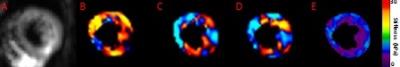
Simultaneous T1 and T2 mapping of the myocardium in normal volunteers using Cardiac MR Fingerprinting
Shivani Pahwa, Jesse Hamilton, Joseph Adedigba, Samuel Frankel, Gregory O'Connor, Ozden Kilinc, Wei-Ching Lo, Joshua Batesole, Seunghee Margevicius, Pingfu Fu, Mark Griswold, Nicole Sieberlich, Vikas Gulani
This study reports normative cardiac T1 and T2 values generated in a single cardiac MRF scan in a cohort of normal volunteers. These values were compared against MOLLI (for T1) and bSSFP (for T2). Our results show that cardiac relaxometry values obtained with MRF at 1.5 T are comparable to values previously reported in the literature and those obtained using MOLLI and bSSFP.


2728.  
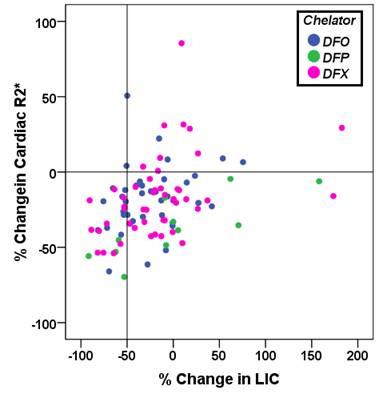
Association between cardiac iron clereance and hepatic siderosis by T2* MRI in thalassemia major patients
Antonella Meloni, Laura Pistoia, Nicolň Schicchi, Gennaro Restaino, Paolo Preziosi, Vincenzo Positano, Monica Benni, Maria Paola Smacchia, Daniele De Marchi, Alessia Pepe
We evaluated in thalassemia major (TM) if the cardiac efficacy of the three iron chelators (Desferrioxamine, Deferiprone, and Deferasirox) was influenced by hepatic iron levels over a follow up of 18 months. In patients treated with Deferasirox and Deferiprone percentage changes in cardiac R2* over 18 months were associated with final liver iron concentration (LIC) and percentage LIC changes.  In no chelation group percentage changes in cardiac R2* were influenced by initial LIC or initial cardiac R2*.


2731.   
Improved Myocardial T1 mapping using a Novel Motion-Insensitive Reconstruction
Céline Smekens, Radhouene Neji, Reza Razavi, René Botnar, Sébastien Roujol
Myocardial T1 mapping sequences are commonly performed under breath-hold conditions. However, motion between T1-weighted images can be observed in ~50% of acquisitions. Image registration algorithms can be used for motion correction but are not available on all scanners, can occasionally fail and remain challenging using low contrast sequences such as saturation-based T1 mapping techniques. In this study we sought to develop and evaluate a novel motion-insensitive T1 mapping reconstruction approach which automatically discards misaligned/artefact T1-weighted images.


2732.   
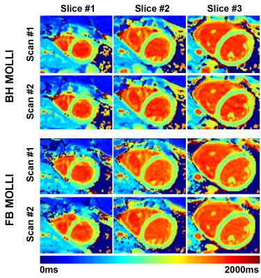
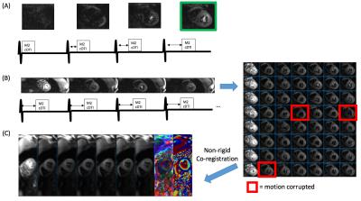
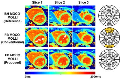
Free Breathing Myocardial MOLLI T1 Mapping using Real-time Slice Tracking and Non-Rigid Image Registration
Céline Smekens, Radhouene Neji, Reza Razavi, René Botnar, Sébastien Roujol
The modified Look Locker (MOLLI) sequence is the most widely used myocardial T1 mapping approach. In this 2D sequence, several T1-weighted images are acquired within a breath hold and used to create a T1 map. However, some patients are unable to sustain stable breath-holds which generates 3D motion between the T1-weighted images. While image registration can potentially be used to correct for in-plane motion, through-plane motion cannot be corrected and may introduce bias in T1 estimates. In this study, we sought to develop and assess a free breathing MOLLI sequence with real-time slice tracking and non-rigid image registration to reduce in-plane and through-plane motion effects.


2733.  
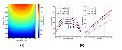
Off-resonance correction in myocardial T1-mapping using Bloch simulations of the MOLLI sequence
Nicola Martini, Andrea Barison, Maria Santarelli, Daniele Della Latta, Francesco Avogliero, Vincenzo Positano, Luigi Landini, Dante Chiappino
Myocardial T1 mapping using MOLLI is influenced by off-resonance effects that lead to underestimation of T1. In this study a method for the correction of the off-resonance influence on T1 mapping, based on the Bloch simulations of the MOLLI sequence, is proposed. In vivo results on a healthy population (N=67) showed that the regional variation of T1 is highly correlated with the distribution of the off-resonance. The proposed method effectively reduced the artifactual T1 underestimation due to off-resonance, especially in myocardial segments that exhibited elevated B0 inhomogeneities.


2734.   
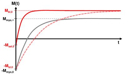
Quasi black blood T1-mapping using slice-selective TRASSI for improved visualization of the myocardium
Daniel Gensler, Tim Salinger, Georg Ertl, Peter Jakob, Peter Nordbeck
Currently available T1 mapping techniques have only restricted capabilities for the visualization of the right myocardium and they have the limitation that after contrast agent application the T1-contrast between blood pool and fibrotic myocardium is partially very low.

The quasi black blood TRASSI sequence shows improved abilities for the visualization and T1-quantification of myocardial structures. So it might be suited in clinical routine for a clear visualization of the right myocardium or for the detection of slight endocardial infarctions, because it is an ultra-fast and robust cardiac T1-mapping method with a total acquisition time of less than 7s.


2735.  
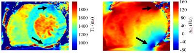
T1 Errors from Off-Resonance Effects for MOLLI at 3T: Experience in a Clinical Study
Justin Grenier, Natasha Wiebe, Stephanie Thompson, Scott Klarenbach, Marcello Tonelli, Paolo Raggi, Richard Thompson
The widely used MOLLI (MOdified Look-Locker Inversion recovery) T1 mapping approach underestimates T1 values as a function of several factors, including off-resonance frequency.  Native T1 and matched off-resonance frequency (Δω) maps were acquired in 24 subjects as part of a study of patients with kidney disease (3T field strength).  Δω within individuals had an average range 133±43Hz, and Δω was significantly correlated with underestimation of native T1, in good agreement with Bloch equation simulations. ~50% of slices had relatively large (75-150 Hz) off-resonance frequencies, for which T1 errors ranged from -25ms to -150ms.


2729.  
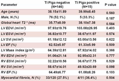
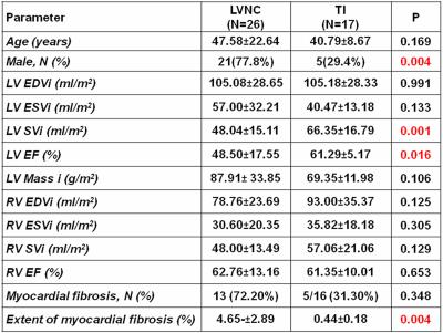
The prognostic role of hypertrabeculation  by cardiac magnetic resonance in thalassemia intermedia patients
Antonella Meloni, Francesca Macaione, Vincenzo Positano, Andrea Barison, Laura Pistoia, Salvatore Novo, Pasquale Assennato, Alessia Pepe
We prospectively assessed whether the Piga’s criterion for left ventricle non-compaction (LVNC) (NC/C ratio threshold of >2.5) had a  prognostic role for adverse cardiovascular outcomes in thalassemia intermedia patients. We found out that patients with Piga’s positive criterion had a significant higher risk of developing cardiac complications globally considered and arrhythmias.


2736.   
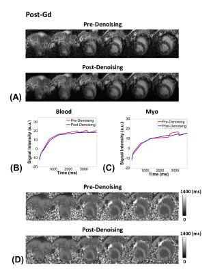
Multi-Slice GRE-MOLLI at 3T using Denoising with Low-Rank and Sparsity Constraints
Paul Han, Chao Ma, Nicolas Guehl, Nathaniel Alpert, Marc Normandin, Georges El Fakhri
Modified Look-Locker inversion recovery (MOLLI) uses bSSFP readout due to its high SNR, however, bSSFP is sensitive to off-resonance effects which result in banding artifacts. Recently, GRE has been proposed as an alternative readout for MOLLI, however, the low SNR efficiency of GRE-MOLLI is still a major problem. In this work, we propose to use a denoising reconstruction framework with low-rank and sparsity constraints to improve the low SNR of GRE-MOLLI. The proposed denoising method improved the low SNR of GRE-MOLLI, and multi-slice GRE-MOLLI is feasible for artifact-free T1 mapping with wider spatial coverage at high magnetic fields ($$$\geq 3T$$$).


2737.   
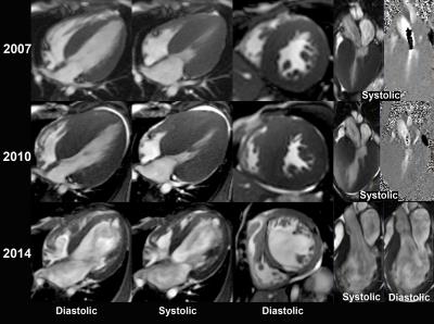
Prognostic value of cardiac MR imaging for end-stage phase of hypertrophic cardiomyopathy patients with or without adverse ventricular remodeling
Sainan Cheng, Chen Cui, Gang Yin, Lu Li, Shihua Zhao
Cardiac morphology as well as LGE extent proved to be particularly heterogeneous in ES phase of HCM patients. For ES phase of HCM patients with adverse ventricular remodeling, LGE was a significant predictor of poor outcomes. But for ES phase of HCM patients without ventricular dilatation, biatrial enlargement, extremely diastolic dysfunction and higher incidences of AF may contribute more to adverse prognosis.


2730.  
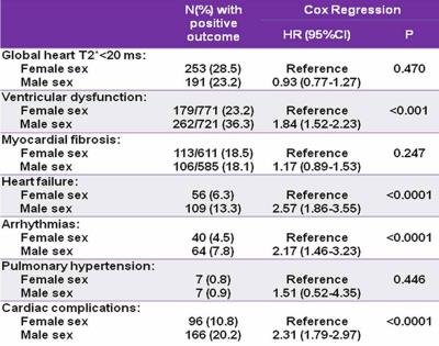
Gender-based optimization of cardiac follow up in thalassemia major patients
Antonella Meloni, Laura Pistoia, Silvia Maffei, Giuseppe Peritore, Valentina Vinci, Massimiliano Missere, Vincenzo Positano, Massimo Allň, Antonella Massa, Alessia Pepe
Females seem to tolerate iron toxicity better, possibly as an effect of reduced sensitivity to chronic oxidative stress. Based on our data about the significantly different risk in developing cardiac complications, in females older than 20 years the FU may be performed every 24 months, thus optimizing health care costs.


2738.  
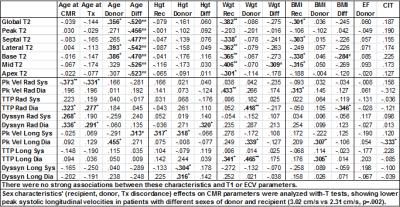
CMR-Derived Regional T2, T1/ECV, Myocardial Velocities, and Dyssynchrony Influenced by Donor and Recipient Characteristics after Heart Transplantation
Ryan Dolan, Amir Rahsepar, Julie Blaisdell, Allen Anderson, Kambiz Ghafourian, Esther Vorovich, Jonathan Rich, Jane Wilcox, Clyde Yancy, Jeremy Collins, Michael Markl, James Carr
Cardiac MRI is increasingly being used for cardiac allograft surveillance following transplantation, so it is important to investigate which recipient and donor characteristics influence several CMR parameters: global ventricular function, myocardial velocities, dyssynchrony, T2, native T1, and ECV.  Notable associations with T2 included donor age, normalized recipient-donor age difference, and recipient weight.  Peak diastolic longitudinal velocity was associated with donor age and cold ischemic time.


2739.  
Non-contrast T1 mapping can detect myocardial fibrosis in hypertrophic cardiomyopathy without gadolinium. Is native T1 a superior alternative to late gadolinium enhancement?
Yoshiaki Morita, Naoaki Yamada, Teruo Noguchi, Yoshiaki Watanabe, Tatsuya Nishii, Atsushi Kono, Tetsuya Fukuda
Native T1 mapping is a novel cardiac magnetic resonance technique for myocardial tissue characterization without contrast administration. Native T1-mapping in hypertrophic cardiomyopathy was correlated with T1 map-based extracellular volume fraction, and native T1 of apparently late gadolinium enhancement (LGE)-negative segments were significantly longer than normal myocardium. Therefore, native T1 mapping has the potential to quantify the volume of interstitial space without gadolinium, which would be useful particularly in patients who are limited in use of gadolinium. Furthermore, native T1 would be a useful biomarker for the detection of diffuse myocardial damage difficult to evaluate using conventional LGE alone.


2743.  
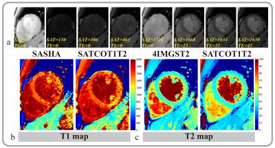
A 2D combined myocardium T1 and T2 mapping
Rui Guo, Zhensen Chen, Jianfeng zhang, Jianwen Luo, Haiyan Ding
In this study, we developed a 2D combined myocardial T1 and T2 mapping sequence that uses a combination of saturation pulse and T2 preparation pulse and allows simultaneously obtaining T1 and T2 map with acceptable breath holding time (12 heartbeats). High quality multiple T1 and T2 weighted images were obtained in the other cardiac cycle between the saturation pulse and T2 preparation pulse. Phantom experiment showed that T1 and T2 measured by proposed method highly correlated with reference methods. In vivo experiment showed that the proposed sequence can yield comparable myocardium T1 and T2 values with the conventional separated T1 and T2 mapping sequences.


2744.   
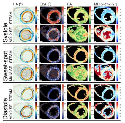
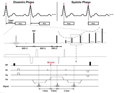
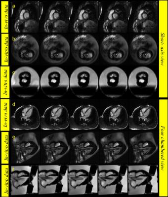
An in-vivo comparison of STEAM and 2nd order motion compensated spin-echo imaging in multi-phase cardiac DTI at 3T
Andrew Scott, Sonia Nielles-Vallespin, Pedro Ferreira, Zohya Khalique, Dudley Pennell, David Firmin
Cardiac diffusion tensor imaging (cDTI) is a novel non-invasive method of interrogating myocardial microstructure that has seen a recent surge in interest. Many of the most interesting clinical results were obtained using stimulated echo acquisition mode (STEAM) imaging at multiple cardiac phases. Recently however, spin-echo cDTI with second order motion compensated diffusion gradients (M012-SE) was proposed. In this study we report results of a comparison of M012-SE and STEAM imaging in multiple cardiac phases at 3T in 15 healthy subjects with matched sequence parameters. While M012-SE provides comparable quality data in systole, STEAM is the more reliable technique in diastole.


2745.   
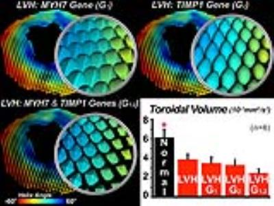
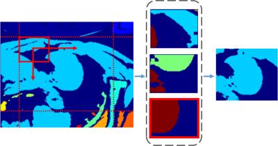
Integrated Analysis of Cardiac Genetic and Structural Alterations in Left Ventricular Hypertrophy using the Supertoroidal Model
Choukri Mekkaoui, Howard Chen, Iris Chen, Ronglih Liao, William Kostis, Timothy Reese, Marcel Jackowski, David Sosnovik
Response to disease occurs over many scales ranging from individual gene expression to whole organ physiology. We employed the supertoroidal model of the diffusion tensor to study the interaction between gene expression and microstructure of the heart. Left ventricular hypertrophy (LVH) was induced in C57Bl6 mice through aortic banding, and characterization of the cardiac microstructure was performed in vivo with DTI. The supertoroidal model was constrained by both diffusion information and gene expression data related to cardiomyocyte hypertrophy and myofiber orientation. Our model enabled further characterization of LVH by unifying information at different scales and across domains.


2746.   
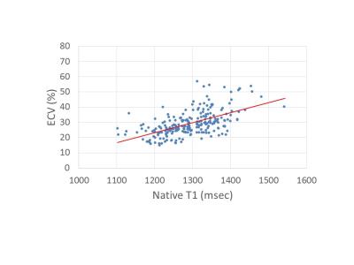
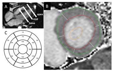
Segment and sexual variation of myocardium in T1 mapping and extracellular volume fraction with cardiovascular magnetic resonance in healthy volunteers
Yukun Cao, Yue Cui, Xiangchuang Kong, Shan Zhang, Xu Yan, Heshui Shi
     This study aimed to evaluate sex and segment differences of myocardium in native T1 value and ECV in healthy volunteers. We measured native T1 value of 425 segments and ECV of 424 segments. The results showed that myocardial ECV of females was higher than that of males in basal, middle and apical segments. The ECV of the apical segments was higher than in the basal and middle segments. The mean native T1 value and ECV were not associated with age, ejection function(EF), end diastolic volume(EDV), end systolic volume(ESV), and stroke volume(SV).


2747.   
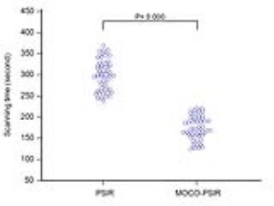
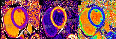
Role of free-breathing motion-corrected phase-sensitive inversion recovery (MOCO-PSIR) imaging technique for the assessment of late gadolinium enhancement
Yinyin Chen, Shan Yang, Hong Yun, Xiaoming Bi, Caixia Fu, Hang Jin, Mengsu Zeng

We aimed to investigate the role of motion-corrected phase-sensitive inversion recovery (MOCO-PSIR) technology for the evaluation of LGE. LGE imaging was conducted in 55 patients with MOCO-PSIR and conventional breath-hold PSIR sequences ‎successively. Image quality was scored using a four-point scale. Compared with conventional PSIR, MOCO-PSIR showed better image quality and detected larger LGE volumes in nonischemic cardiomyopathy. Free-breathing motion-corrected PSIR method is a promising alternative to conventional PSIR sequence.


2748.   
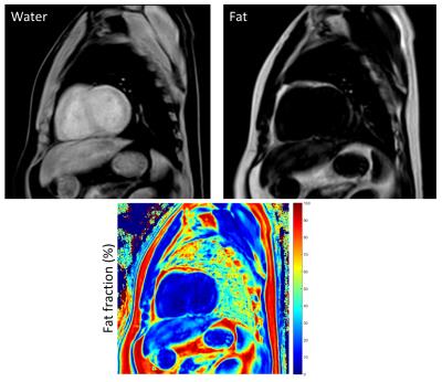
Myocardial fat quantification of normal subjects via 7-peaks mDixon model
Yu-Fen Huang, Feng Mao Chiu, Queenie Chan, Ya-Wen Shen, Chao-Jung Wei, Chih-Miang Chiang
Cardiac magnetic resonance imaging provides a lot of physiological information about myocardial pathology, and the mDixon technique also has been developed on cardiac application recently. The fat deposition in myocardium is possibly related with cardiomyopathy, so it is important to determine the fat composition of myocardium. The mDixon provides an easy way to acquire such this information within one breath hold, and it has a consistent result with magnetic resonance spectroscopy from the previous study. Also it has been reported that multipeak fat spectrum model gives more robustness, and 7-peaks model is used in this study. The aim of this study is to evaluate fat fraction of myocardium with 7-peaks model.


2749.   
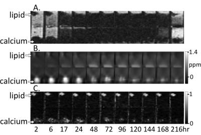
Reperfusion Hemorrhage Following Prolonged Myocardial Ischemia Leads to Fatty Degeneration of Myocardial Infarctions via Iron-Mediated, Self-Perpetuating Loop of Foam Cell and Ceroid Accumulation
Ivan Cokic, Avinash Kali, Hsin-Jung Yang, Richard Tang, Joseph Francis, Rohan Dharmakumar
Fatty infiltration within chronic myocardial infarctions (MI) is a common finding. It is typically observed in the peri-infarct border zone of old scars and has been linked to adverse outcomes in the chronic post-MI setting. To date, the trigger for fat deposition within old MI is unknown. Recent reports showed that iron deposits from hemorrhagic MI drive the recruitment of new monocytes/macrophages into the infarcted territory throughout the chronic phase after MI. Since iron-laden macrophages (siderophages) are prone to transforming into foam cells, we hypothesized that fatty degeneration of hemorrhagic myocardial infarctions has its origin in iron-driven foam cell formation. 


2750.   
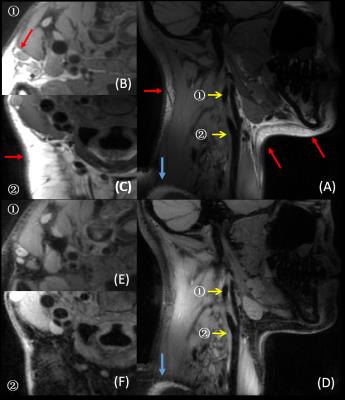
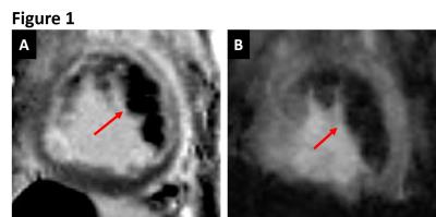
Cardiomyopathy in later-onset Fabry disease: a correlative study of T1 mapping on MR and histology
Jian-Ling Chen, Liang-Wei Chen, Sheng-Che Hung, Hsien-Tzu Liu, Mei-Han Wu, Fu-Pang Chang, An-Hung Yang, Ting-Rong Hsu, Dau-Ming Niu, Ming-Ting Wu, Chui-Mei Tiu, Chien-Yuan Lin
Fabry disease is a rare and X-linked disorder characterized by accumulation of glycosphingolipid within lysosomes and resultant multiple organ damage including heart. Since lipid is known to shorten the MRI parameter T1, non-contrast T1 mapping has emerged as key imaging modality to assess Fabry cardiomyopathy and early detection of lipid deposition. This study provides a histologic validation of 7 male patients of untreated later-onset Fabry disease to demonstrate the negative correlation between native myocardial T1 value and severity of lipid deposition (correlation coefficient, -0.771; p, 0.042) and justifies the application of T1 mapping as a noninvasive predictor of surveillance strategy.


2751.  
Cardiac MR Derived Extracellular Volume Measurements Using Different Contrast Agents
Amir Ali Rahsepar, Ahmadreza Ghasemiesfe, Monica Korell, Jeremy Collins, James Carr
T1 and ECV values calculated from gadoterate meglumine enhanced CMR are comparable to more routinely used gadopentetate dimeglumine and gadobutrol CMR measurements. 


2752.  
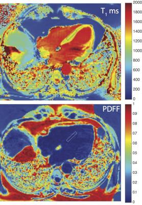
Single Breath-hold Measurement of T2 Corrected Myocardial Proton Density Fat Fraction in Humans at 3T
Ronald Ouwerkerk, Ranganath Muniyappa, Monica Skarulis, Ahmed Gharib
Localized 1H-MRS was used to determine T2s for water and lipid signals in the human heart in a single breath hold and derive relaxation corrected proton density fat fractions.


2740.  
Assessment of myocardial inflammation in cardiac sarcoidosis using early gadolinium enhancement
Yoshiaki Morita, Naoaki Yamada, Teruo Noguchi, Yoshiaki Watanabe, Tatsuya Nishii, Atsushi Kono, Tetsuya Fukuda
In patients with cardiac sarcoidosis, assessment of the activity of myocardial inflammation is as crucial as the choice of therapeutic strategy and monitoring of therapeutic effects. It is known that early gadolinium enhancement (at 2–5 minutes after gadolinium administration) can visualize myocardial inflammation and/or edema such as in acute myocarditis and acute myocardial infarction. In this study, images of gadolinium enhancement at 2 minutes delay were significantly associated with findings regarding active inflammation, suggesting that early gadolinium enhancement has the potential to act as a marker of inflammation activity in cardiac sarcoidosis.


2753.   
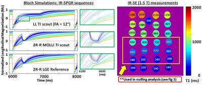
A Versatile MOLLI-based Inversion Time (TI) scout sequence for Myocardial Nulling Determination in emerging Late Gadolinium Enhanced CMR Variants: A Phantom Study
Keigo Kawaji, Adam Hasse, Amita Singh, Akhil Narang, Hui Wang, Timothy Carroll, Amit Patel
A Look-Locker scout is used for optimal inversion time (TI) determination of myocardial nulling in Late Gadolinium Enhancement (LGE) Cardiac Magnetic Resonance. Recently, novel LGE technique variants such as the Wideband approach have been proposed, and these approaches can lead to variability in the signal evolution between the Look-Locker TI-scout and LGE. In this study, we propose a Modified Look-Locker Inversion (MOLLI)-based TI-scout that closely matches its signal evolution to that of any LGE variant.  Bloch simulation and  phantom evaluation are performed to compare the null-times from the conventional LL-based and proposed MOLLI-based approaches against the LGE protocol over a range of clinically relevant T1 values observed in viability imaging. 


2741.  
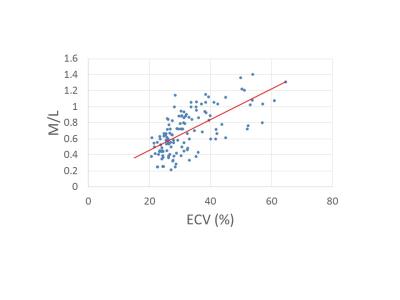
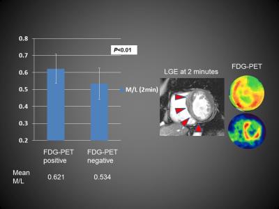
Simple quantification using the myocardium-to-lumen signal ratio in diffuse myocardial fibrosis of non-ischemic cardiomyopathies: Correlation with T1 mapping derived ECV
Yoshiaki Morita, Naoaki Yamada, Teruo Noguchi, Yoshiaki Watanabe, Tatsuya Nishii, Atsushi Kono, Tetsuya Fukuda
Myocardial T1 mapping has recently been applied to the quantification of extracellular volume fraction (ECV) and has shown potential for the detection of myocardial fibrosis. However, for the measurement of ECV, additional scans of pre- and post-contrast T1 mapping are necessary, and the post-processing procedure is time consuming. In this study, the myocardium-to-lumen signal ratio (M/L) in conventional late gadolinium enhancement images with fixed inversion time showed acceptable levels of correlation with ECV obtained by the T1 mapping in non-ischemic cardiomyopathy, suggesting that M/L has the potential to allow for simple quantification of the fibrotic change in non-ischemic cardiomyopathies.


2754.  
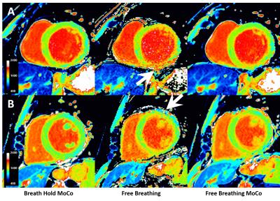
Free-breathing native cardiac T1 and T2 mapping with flexible elastic image registration: Preliminary Clinical Result
Shuo Zhang, Tomoyuki Okuaki, Sven Kabus, Bao Ru Leong, Yiying Han, Yi Hui Hung, Ping Wang, Ru San Tan
Current myocardial T1 and T2 mapping based on inversion-recovery and single-shot readout techniques require generally at least 7 to 10 seconds breath hold. Improper and difficulties of breath hold is one of the main sources of error and reduced reproducibility. To mitigate the dependence of mapping on breath holds and also to increase patient comfort, we report free-breathing T1 and T2 mapping using different acquisition schemes using flexible elastic image registration. We demonstrate the feasibility of this approach with motion correction for increased image quality and quantification accuracy.


2755.   
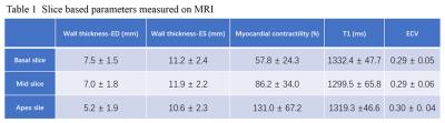
The Correlation of Clinical and Image of Acromegaly Patients Based on 3.0T Cardiac Magnetic Resonance Quantitative Analysis of Myocardial T1 and Extracellular Volume
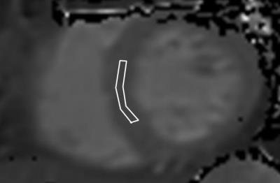
Jian Cao, Peijun Liu, Lu Lin, Xiao Li, Xiaopeng Guo, Jing An, Bing Xing, Yining Wang
The aim of this study was to find out if there might be some correlation between clinical information and image measurements for acromegaly patients. And it found that for acromegaly patients, the basal slice of heart might be the most involvemented position, and its contractility had a positive correlation with GH burden, and both T1 and ECV had a negative correlation with IGF-1. And we need to enlarge the sample size and compare the changes before and after the surgery in order to give more informations to the clinicians.


2756.  
SMART1Map: Accuracy and Influencing Imaging Parameters For Cardiac T1 Mapping.
Malek MAKKI, Barbara Burkhardt, Emanuela Valsangiacomo
SMART1Map sequence was performed on 6 phantoms with different T1 values. Sixteen schemes were prescribed: 4 heart rates (60, 80, 100, 120 bpm), 2 spatial resolutions (FOV = 30 cm and FOV = 48 cm) and 2 cardiac phases (systole and diastole). The results show that SMART1Map underestimates long T1 (native T1 myocardium) at any scheme. We also found an increase in error with reduced FOV (all other parameters being identical), and an influence of the cardiac phases on T1 at the levels of native and post-contrast blood and myocardium


2757.  
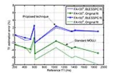
An alternative T1 estimation algorithm with a higher flip angle improves T1 mapping accuracy and precision of the MOLLI sequence
Jiaxin Shao, Kim-Lien Nguyen, Peng Hu
Good T1 estimation accuracy and precision is important for clinical application of the myocardial T1 mapping technique. Although the widely used T1 mapping technique, modified Look-Locker inversion-recovery (MOLLI), has good precision and reproducibility, it is not accurate. Several T1 estimation algorithms have been proposed to improve the accuracy for MOLLI. These algorithms however, often have reduced T1 estimation precision. We developed an improved MOLLI technique to achieve better accuracy and precision than the standard MOLLI sequence. Based on phantom and volunteer studies, a higher flip angle MOLLI with Bloch equation simulation and slice profile correction (BLESSPC) is used for T1 estimation.


2758.   
Coronary artery disease is not related to pathologic epicardial fat volumes, left ventricular strain or T1-relaxation times in hypertensive patients
Rami Homsi, Michael Meier-Schroers, Alois Sprinkart, Juergen Gieseke, Daniel Kuetting, Stefan Fischer, Darius Dabir, Julian Luetkens, Christian Marx, Hans Schild, Daniel Thomas
Hypertension is related to increased amounts of epicardial fat, to myocardial fibrosis and to left-ventricular contractility disturbances despite a normal systolic left-ventricular ejection fraction. However, the presence of stable coronary atherosclerotic disease in general - and with no cardiac damage due to prior coronary pathologies - does not additionally affect these parameters.


2759.  
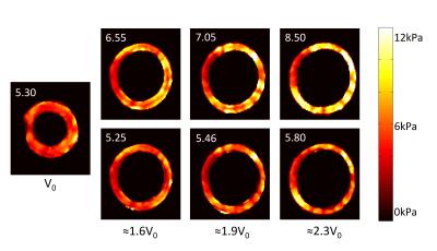
3D High Frequency Cardiac Magnetic Resonance Elastography Quantitatively Differentiates Myocardial Stiffness in Patients with HFPEF and Healthy Volunteers
Shivaram Poigai Arunachalam, Arvin Arani, Ian Chang, Yi Sui, Phillip Rossman, Kevin Glaser, Joshua Trzasko, Kiaran McGee, Armando Manduca, Barry Borlaug, Richard Ehman, Philip Araoz
Increased myocardial stiffness in patients with heart failure with preserved ejection fraction (HFpEF) is known to affect diastolic filling. The purpose of this work was to determine if 3D high frequency cardiac MR elastography (MRE) can quantitatively differentiate increased myocardial stiffness in HFpEF patients compared to healthy volunteers. Two patients with clinical diagnosis for HFpEF and 47 healthy volunteers were studied. The myocardial stiffness of HFpEF patients (mean: 10.57 kPa) was found to be significantly stiffer (p < 0.05) than healthy controls (mean: 7.79 kPa). Recruitment of more HFpEF patients is underway for further validation of this finding.


2742.  
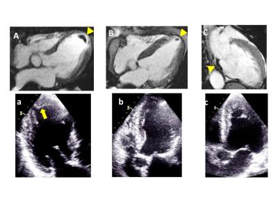
Assessment and detection of left ventricular thrombus in patients with acute ischemic stroke using cardiac MR (ADVENT study)
Yoshiaki Morita, Junji Takasugi, Naoaki Yamada, Teruo Noguchi, Yoshiaki Watanabe, Tatsuya Nishii, Atsushi Kono, Tetsuya Fukuda
In patients with acute ischemic stroke (AIS), the assessment of left ventricular thrombus (LVT) is essential. In this study, we showed the utility of the early phase of gadolinium enhancement for detecting LVT in AIS patients, which conventional TTE hardly identified. CE-CMR, when using the early phase of gadolinium enhancement, should be performed on acute ischemic stroke patients, especially those with prior myocardial infarction or LV dysfunction and without definitive stroke etiologies.


2760.  
Reconstructing Inherent Stiffness in a Deforming Heart Phantom using MR Elastography
Myrianthi Hadjicharalambous, Adela Capilnasiu, Daniel Fovargue, Ayse Dokumaci, Stefan Hoelzl, Jack Lee, Ralph Sinkus, David Nordsletten
Cardiac MR-Elastography has significant potential to provide a non-invasive measure of myocardial tissue health. A core challenge in its application is the substantial motion of the heart over the cardiac cycle, which introduces a bias in the quantified apparent stiffness. In this work we apply a reconstruction technique for MRE designed to correct changes in apparent stiffness due to deformation. The reconstruction method is tested on anatomically accurate homogeneous and heterogeneous heart phantoms that are inflated to mimic diastolic function. The results demonstrate the ability of the reconstruction technique to retrieve intrinsic stiffness values, even under substantial inflation.    


2761.   
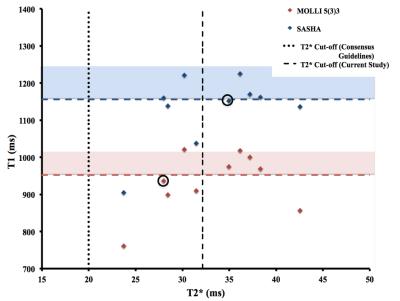
Comparison of SASHA and MOLLI sequences for Iron Quantification in the Myocardium.
Sarah McElroy, Torben Schneider, Daniel Sado, Tarique Hussain, Amedeo Chiribiri, Sarah Peel
Development of T1 mapping in the myocardium in recent years has demonstrated that the technique can add information to inform the diagnosis or management of patients in a number of pathologies.  Sequences have been developed to enable T1 mapping of the myocardium within a single breath-hold and the limitations of these sequences have been well characterized. Recent studies have investigated myocardial T1 mapping in iron overload patients using variants of the MOLLI sequence. These studies have concluded that T1 mapping could be more sensitive and reproducible than T2* mapping for the measurement of iron overload in the myocardium. The current study aims to determine the optimal T1 mapping sequence for iron overload. Simulations, phantom studies and in-vivo studies were carried out to investigate MOLLI and SASHA sequences used for T1 mapping of low T1 and T2 tissues (comparable to iron overload in the myocardium).


2762.  
Optimization of MOLLI reconstruction for free-breathing myocardial T1 imaging
Yun-Wen Wang, Yi-Fu Tsai, Teng-Yi Huang
The free-breathing MOLLI (FB-MOLLI) presented in our previous study allowed T1 mapping in vivo without breath-hold. In this study, we attempted to implement unsupervised reconstruction for FB-MOLLI data sets and used a deformable method for image registration to improve the reliability of free-breathing T1 mapping. The results supported that the method improved the image alignments of the FB-MOLLI data sets and thus increased the quality of the T1 map. The variations of the repeated T1 measurements were significantly reduced in the anterolateral of the LV walls.


2763.   
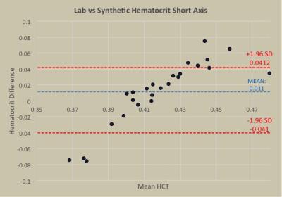
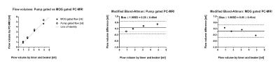
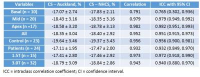
Derivation and Validation of Synthetic Hematocrit Calculation from Blood Pool T1 values at multiple different cardiac blood pools, in both high and low flow states on 3T MRI
Ozair Rahman, Kelvin Chow, James Carr, Jeremy Collins
Comparison of the relationship between blood R1 and venous derived hematocrit in the left ventricle, left atrium, descending thoracic aorta, and short axis (apex, mid, and base) blood pools.


2764.   
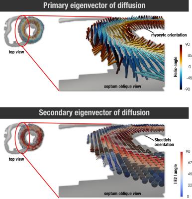
Dual identity of the interventricular septum with in vivo diffusion tensor imaging
Pedro Ferreira, Sonia Nielles-Vallespin, Andrew Scott, Zohya Khalique, Dudley Pennell, David Firmin
Heart muscle has a complex cellular helical arrangement, where bundles of myocytes, known as sheetlets interleave with collagen-lined shear layers. The right and left ventricle form separately in early stages of cardiogenesis, resulting in a dual inter-ventricular septum. In this work we compare the myocyte and sheetlet microstructure in the septum and LV free-wall in healthy and HCM hearts with in vivo diffusion tensor imaging. Results show that the right-side of the interventricular-septum has myocyte orientations not seen in the free-wall, and also lower sheetlet angles, which may indicate an RV identity in this region.


2765.   
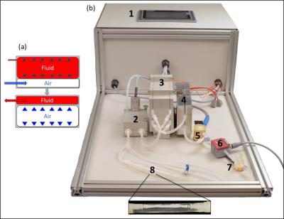
A Cost-effective 3D Printed Cardiac MR Phantom
Shivaprasad Chikop, Amaresh Konar, Nimitha Reddy, Nithin Vajuvalli, Darshan Keelara, Ashwini Kumnoor, Ramesh Venkatesan, Sairam Geethanath
Cardiac phantoms have been employed as testing and validating tools for newly developed techniques focused on sampling and reconstruction strategies. In this work, a 3D printed cardiac phantom was built to mimic the human heart. This was achieved through the integration of a peristaltic pump.  Results depict the structural and functional behavior of the cardiac phantom, based on MR imaging on 1.5T scanners from 2 vendors. Ongoing work involves implementing the post-processing pipeline to correlate UI parameters with those derived from images. Future work focuses on employing PVA for preparing the heart model employing the 3D printing heart mold.


2766.   
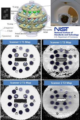
Inter-Scanner T1 and T2 Measurement Variability Evaluation on Two 3T Scanners with Identical Hardware and Software Configuration
Anshuman Panda, Clinton Jokerst, Kristopher Cummings, Prasad Panse
Inter-scanner T1 and T2 measurement variability was evaluated on two 3T scanners with identical hardware and software configuration to identify the most robust combination of sequence, parameters and post-processing that produces accurate measurement and to establish confidence intervals for T1 and T2 measurements for in-vivo studies by incorporating “native” inter-scanner variability. 


2767.   
Cardiac T1 Mapping Using True Hybrid Inversion and Saturation Recovery
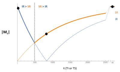
Glenn Slavin, Anne Menini, Haonan Wang, Anja Brau
Despite limited accuracy, MOLLI remains popular for T1 mapping due to high precision and visual quality of the maps. This is due primarily to the large dynamic range afforded by the inversion-recovery (IR) acquisition. Methods using saturation-recovery (SR) have better T1 accuracy but have relatively poor precision compared with MOLLI. This work presents a true hybrid IR/SR acquisition that targets the optimal regions of both the IR and SR relaxation curves, employs a novel method for maximizing dynamic range, and uses an improved sampling strategy. The proposed hybrid method combines the accuracy of single-point SR with the precision of IR.
 
Atherosclerosis Imaging
Traditional Poster
Cardiovascular

 
Thursday, 27 April 2017
Exhibition Hall  08:15 - 10:15


2768.   
Identification of Intrapalque Hemorrhage in Carotid Artery by Simultaneous Non-contrast Angiography and intraPlaque hemorrhage (SNAP) Imaging: A Comparison Study with MP-RAGE
Xihai Zhao, Dongye Li, Huilin Zhao, Xiaoyi Chen, Huiyu Qiao, Le He, Rui Li, Jianrong Xu, Chun Yuan
It is important to accurately identify carotid artery intraplaque hemorrhage (IPH) due to its significant association with ischemic stroke. MP-RAGE sequence has been demonstrated to be the best approach to detect carotid IPH. Recently, investigators proposed SNAP imaging technique which allows non-contrast MR angiography and identifying IPH simultaneously. This study sought to investigate the performance of SNAP imaging in detecting carotid IPH by comparing with MP-RAGE. We found that SNAP imaging detected more IPHs and showed larger IPH size than MP-RAGE, suggesting SNAP imaging might be a sensitive imaging tool to detect IPH.


2769.   
Predicting Procedure Successful Rate after Endovascular Recanalization for Chronic Carotid Artery Occlusion by 3D Vessel Wall Imaging
Huilin Zhao, Jianrong Xu, Xiaosheng Liu, Beibei Sun, Jieqing Wan, Weibo Chen, Xihai Zhao, Chun Yuan
Endovascular recanalization in patients with carotid chronic total occlusion (CTO) has been reported to be technically challenging. In this study, we retrospectively analyzed the 3D vessel wall imaging in patients with carotid CTO and its relationship to technical success rates. A total of 15 consecutive carotid CTO recanalization attempts were performed with overall technical success rate 60.0%. Compared with failure group, patients with an occlusion length <50mm, distal true lumen visibility in C1 segment on MR vessel wall images had a higher rate of successful recanalization. 3D vessel wall MR imaging may play a role in patient selection of endovascular recanalization in patients with carotid CTOs.


2770.   
Distribution and burden of atherosclerosis in patients with anterior circulation cerebral ischemic events: Characterization using combined extracranial and intracranial vessel wall MRI
Haifeng Gao, Jie Sun, Niranjan Balu, Dongxiang Xu, Daniel Hippe, Chun Yuan, Thomas Hatsukami
Both extracranial and intracranial atherosclerosis may be implicated in large-artery atherothrombotic stroke. This proof-of-concept study characterized the distribution and burden of atherosclerosis in thirteen patients with anterior circulation cerebral ischemic events using combined extracranial and intracranial vessel wall MRI. We found that atherosclerotic plaques were highly prevalent in both extracranial and intracranial carotid arteries. Larger plaque burden measured as plaque index on black-blood vessel wall MRI, rather than luminal stenosis on time-of-flight MRA, was significantly associated with clinical symptoms. Black-blood vessel wall MRI may be useful in identifying the culprit plaque in patients with suspected large-artery atherothrombotic stroke.


2771.   
Reproducibility of Semi-automatic Carotid Intraplaque Hemorrhage Quantification using SNAP MRI
Jin Liu, Jie Sun, Thomas Hatsukami, Marina Ferguson, Niranjan Balu, William Kerwin, Daniel Hippe, Amy Wang, Chun Yuan
Intraplaque hemorrhage (IPH), a characteristic feature of high-risk atherosclerosis, can be identified as hyperintensity areas on T1-weighted MRI. Simultaneous Non-contrast Angiography and intraPlaque hemorrhage (SNAP) MRI has been shown to provide comparable IPH detection as MPRAGE with additional benefit of lumen assessment in the same scan. In this study, we developed a semi-automatic method to detect and quantify IPH on SNAP MRI with histology confirmation. An objective IPH detection threshold was identified and high scan-rescan reproducibility was obtained for semi-automatic IPH volume measurement and maximum IPH intensity.


2772.   
MR Imaging of Vasa Vasorum During Lipid-Lowering Therapy to Carotid Unstable Plaque: A Prospective Study in Chinese Patients
Cui Bao, Cui Jinguo, Cai Jianming
Carotid plaque vasa vasorum  angiogenesis from adventitia may be influenced by inflammation, could promote intraplaque hemorrhage and decrease the thickness of fibrous cap, finally, the cap is prone to rupture. Drug therapy and assessment to the vulnerable plaque, in vivo, would be of major clinical interest. To investigate whether lipid therapy leads to changes in vasa vasorum characteristics of carotid unstable plaque in chinese patients, as measured by using dynamic contrast-enhanced (DCE) MRI. Focus on inflammation to monitor the early beneficial therapy using vasa vasorum MR imaging, biomarker in vivo.


2773.   
Assessment of aortic pulse wave velocity in patients with peripheral artery disease
Erin Englund, Michael Langham, Emile Mohler, Thomas Floyd, Felix Wehrli
Pulse wave velocity (PWV) provides a measure of arterial stiffness by quantifying the propagation velocity of the systolic pressure wave along the arterial wall. Prior studies have reported that PWV is elevated in patients with peripheral artery disease (PAD), and here we sought to determine whether the elevation in PWV is correlated with PAD disease severity as measured by the ankle-brachial index (ABI). No correlation was observed between ABI and PWV, suggesting that the systemic nature of atherosclerosis may be contributing to the increase in PWV more than the specific lesions that result in a reduction in ABI. 


2774.   
3D black-blood DCE-MRI using radial stack-of-stars acquisition and CS reconstruction: application in carotid and femoral arteries
Jasper Schoormans, Kang Zheng, Erik Stroes, Gustav Strijkers, Aart Nederveen, Bram Coolen
We implemented a high temporal resolution 3D black-blood protocol for DCE imaging of atherosclerotic plaques using a combination of a radial stack-of-stars sampling scheme, motion-sensitised (iMSDE) blood suppression and CS reconstruction. Using this approach, 3D black-blood DCE images with a temporal resolution of 12s could be obtained. In this work, we show the application of our method in patients with femoral and carotid artery plaques. 


2775.   
Robust fat suppressed direct thrombus? imaging (MPRAGE) sequence with a large field-of-view at 3T
Nobuyuki Toyonari, Masami Yoneyama, Seiichiro Noda, Yukari Horino, Kazuhiro Katahira
MR plaque imaging is promising for characterizing atherosclerotic plaque. We proposed direct thrombus imaging (MPRAGE), based on mDIXON turbo field-echo sequence, could provide more robust and stable black-blood imaging with a large field-of-view compared with conventional sequences.


2776.   
Molecular Magnetic Resonance Imaging of Vascular Myeloperoxidase Activity for the Identification of Unstable Atherosclerotic Plaque
Imran Rashid, David Cheng, Jihan Talib, Ghassan Maghzal, Andre Bongers, Ren Minqin, Roland Stocker
Non-invasive imaging of unstable atherosclerotic plaques prone to rupture remains an unmet need in clinical cardiology. This is because current imaging modalities provide information on plaque burden, luminal stenosis and calcification, whereas they have limited ability to discern unstable from stable plaques. We used the tandem stenosis mouse model of unstable plaque in conjunction with MRI and a sensor (bis-5-hydroxytryptamide-DTPA-Gd) for the activity of the inflammatory enzyme myeloperoxidase. Our results reveal sustained and greater enhancement of unstable experimental plaque with the targeted sensor compared with its non-targeted analog (DTPA-Gd), and these results were confirmed by separate biochemical and histological analyses.


2777.   
Motion Insensitive 3D Multiple Echo Inversion Recovery Stack of Star(ME IR SOS) technique for measurement of T2* in Intraplaque Hemorrhage(IPH)
Seong-Eun Kim, J Rock Hadley, J Scott McNally, Bradley Bolster, Jr. , Gerald Treiman, Dennis Parker
The purpose of this work was to develop a 3D ME IR SOS technique to reduce off resonance blurring and allow measurement of T2* in Atherosclerotic plaque. In a prior study we observed lower ADC values in symptomatic IPH. We hypothesized that the T2* measurements from 3D ME IR SOS would help identify IPH and further characterize symptomatic and asymptomatic IPH. The 3D ME IR SOS can provide motion insensitive IPH visualization and T2* values simultaneously which may provide important clinical information to detect the plaque progression.


2778.   
3D visualization of Vascular Cell Adhesion Molecule-1 (VCAM-1) specific Ultrasmall Superparamagnetic Iron Oxide (USPIO) nanoparticles in the atherosclerotic mouse with accelerated self-navigated radial 4D-MRI.
Kristina Andelovic, Patrick Winter, Thomas Kampf, Volker Herold, Peter Jakob, Wolfgang Bauer
USPIO-based, functionalized contrast agents targeting VCAM-1 enable the visualization of inflamed areas in the vessel wall during early atherogenesis. We present a novel 3D technique for coverage of the whole aortic arch at high spatial resolution and a time-resolved detection of nanoparticles with an ECG-free flow-compensated radial 3D-Cine acquisition. T2* weighted 3D-Cines were acquired using two different gradient echoes to provide phase difference maps. The results indicate a reliable detection of the nanoparticles with the new method due to a true 3D coverage of the aorta and the additional phase information.


2779.   
Gradient Echo Derived Multi-Contrast Imaging for Carotid Atherosclerosis Assessment
Zechen Zhou, Shuo Chen, Xihai Zhao, Rui Li, Chun Yuan
In this work, we developed a post processing framework to generate multi-contrast images from a single three dimensional MR scan of multi-echo gradient echo sequence, which can alleviate the registration problem and further improve the scan efficiency with a total scan time of 3min22sec for carotid atherosclerosis imaging. The initial experiments has demonstrated its feasibility for vessel wall delineation and its potential for plaque component characterization.


2780.   
Quantitative characterization of calcified and lipid-laden blood clot in vitro at 3T
Spencer Christiansen, Junmin Liu, Trevor Wade, Joy Dunmore-Buyze, Michael Boffa, Luciano Sposato, Maria Drangova
Thrombus composition in embolic occlusion, particularly the presence of thrombolysis-resistant components such as calcium and fat, can significantly influence treatment efficacy, yet current MR methods for inferring composition are qualitative and sensitive only to red blood cells. We examined the ability of novel post-processing algorithms applied to a tailored GRE acquisition to discriminate and quantify important components within in vitro blood clots of varied hematocrit over a nine-day ageing period. Calcium and lard were readily discernable throughout the experiment, while clots were differentiable from one another between two to six days, demonstrating this protocol's potential for thrombus characterization in vivo.


2781.   
Optimization of whole-brain intracranial arterial wall imaging sequence using Bloch equation simulation
Lei Zhang, Chao Zou, Lijie Ren, Dong Liang, Xin Liu
T1 weighted SPACE has gain popularity in intracranial wall imaging, but its long scan time makes it impractical and its multiple parameters make it very difficult to be optimized. In this study, we looked into the effect of various imaging parameters of T1w SPACE on the signal-to-noise (SNR) and contrast ratio (CR) between vessel wall and cerebrospinal fluid using Bloch equation simulation. An optimized whole-brain intracranial artery wall imaging protocol which had comparable higher CR and SNR within 8min and 0.55mm iso resolution was found. The simulation results were verified by the volunteer study and matched the experiments well. 


2782.   
Assessment of Local Pulse-Wave-Velocity Distribution in Mice using k-t BLAST MRI with Semi-Automatic Area Segmentation
Volker Herold, Patrick Winter, Stefan Herz, Fabian Gutjahr, Kristina Andelovic, Wolfgang Bauer, Peter Jakob
Arterial stiffness (AS) assessed by pulse wave velocity (PWV) has been shown to represent a valuable biomarker of cardiovascular disease risk. Local elastic properties of the murine aortic vessels such as the pulse-wave-velocity (PWV) can be examined using PC-MRI [1]. In the present study we provide an accelerated method based on a k-t BLAST undersampling scheme combined with a new semi-automatic segmentation algorithm to quantify the distribution of the local PWV along the aortic vessel.


2783.   
Improvement of fat separation for simultaneous carotid and intracranial vessel wall imaging by dual-echo fat-water imaging
Chao Zou, Lei Zhang, Chuanli Cheng, Xin Liu, Hairong Zheng
In this study, we used dual-echo fat-water imaging technique in variable flip angle turbo spin echo sequence for simultaneous carotid and intracranial artery imaging to obtain fat-free vessel wall images. Results show that the technique was promising for simultaneous carotid and intracranial vessel wall imaging to improve the SNR and better delineation of carotid vessel wall.


2784.   
3D Golden-Angle Spiral Sparse Parallel-Imaging for Lumen Area Measurements of the Entire Proximal and Mid-Segments of the Coronary Arteries in a Breath-Hold
Michael Schär, Gabriele Bonanno, Allison Hays, Robert Weiss
Coronary artery endothelial function (CEF) could historically only be measured with invasive catheterization-based testing. Recently, 2-dimensional cine MRI in combination with isometric handgrip exercise was introduced to quantify CEF noninvasively. However, the MRI technique only assesses CEF at one or two locations in an artery and does not allow assessment of regional heterogeneity present throughout diseased vessels. The aim of this work was to develop high-resolution isotropic 3-dimensional cine coronary MRI that enables measures of coronary lumen area along the entire proximal and mid segments of the coronary arteries in a single breath-hold, a pre-requisite for future 3-dimensional CEF measurements.


2785.   
Identification of Carotid Lipid-Rich Necrotic Core by Three-Dimensional Magnetization-Prepared Rapid Acquisition Gradient-Echo Imaging
Huiyu Qiao, Dongxiang Xu, Feiyu Li, William Kerwin, Lars Johansson, Chun Yuan, Xihai Zhao
Carotid atherosclerotic lipid-rich necrotic core (LRNC) is associated with ischemic cerebrovascular events. Currently, LRNC was mainly characterized by contrast-enhanced T1 weighted (CE-T1W) or T2 weighted (T2W) imaging. However, CE-T1W imaging needs gadolinium application and T2W imaging is challenging to identify LRNCs with long-T2 components. This study investigated the usefulness of MP-RAGE in identifying carotid LRNC by comparing with CE-T1W and T2W imaging. We found that LRNC had the lowest relative signal intensity against muscle or fibrous tissue on MP-RAGE images among three sequences (P<0.05), indicating that MP-RAGE might be a better non-contrast imaging tool to identify LRNC than T2W imaging.


2786.   
Comparison of black-blood T2 mapping sequences in carotid artery at 3T
Jianmin Yuan, Andrew Patterson, Scott Reid, Martin Graves, Jonathan Gillard
Carotid black-blood quantitative T2 mapping sequences have the potential to help plaque component segmentation and are more suitable for multi-centre studies across different MRI systems. The purpose of this study is to compare the accuracy of three different black-blood quantitative T2 sequences in the phantom and healthy volunteers at 3T.


2787.   
Optimization of 3D black-blood multi-echo T2* weighted sequence in carotid artery
Jianmin Yuan, Ammara Usman, Pascal Ruetten, Andrew Patterson, Andrew Priest, Martin Graves, Jonathan Gillard
Carotid T2* weighted images have multiple applications, including differentiating plaque components and detecting ultrasmall superparamagnetic iron oxide (USPIO) contrast agents. The current work develops and optimizes the three-dimensional black-blood multi-echo T2* weighted sequence through Bloch simulation and volunteer scans.


2788.   
Three-dimensional T1-Weighted Black-Blood Turbo Spin-Echo with Navigator Respiratory Gating for the Vessel Wall Imaging: A Novel Approach to Evaluate Plaque in the Aortic Arch
Kenichi Nakagawa, Noriyoshi Morimoto, Sachi Fukushima, Takashi Tabuchi
Complex plaques in the aortic arch on transthoracic echocardiography (TEE) can cause brain embolism. However, TEE, which is associated with a number of complications and contraindications, is not suitable for all patients. Therefore, we focused on contrast evaluation of MRI with 3D T1w black-blood TSE as an adjunct to diagnosis of complex plaques. The aim of this study is to clarify the optimal scan method for 3D T1w black-blood TSE. Our results suggest that 3D T1w black-blood TSE in the aortic arch can reduce the amount of artifact by using the optimal trigger timing by ECG-gating and navigator respiratory gating.


2789.   
Comparison of Two Different Implementations for the Simultaneous Non-Contrast Angiography and Intraplaque Hemorrhage (SNAP) Sequence
M Louis Lauzon, Niranjan Balu, Chun Yuan, Richard Frayne
We compare, using Bloch equation simulation, the relative signal in intraplaque hemorrhage (IPH), arterial wall (Wall) and Lumen (i.e., blood), along with the IPH–Wall contrast for two different implementations of the SNAP sequence. The first implementation is the original version (Ver1) that uses a linear phase-encoding scheme post-IR, whereas the second version (Ver2) uses centric slice-encodes post-IR. We show that, although Ver2 appears to be less time-efficient, it actually provides increased signal and contrast for the same acquisition time.


2790.   
Accelerated multi-contrast high isotropic resolution 3D intracranial vessel wall MRI using a tailored k-space undersampling and partially parallel reconstruction strategy.
Niranjan Balu, Zechen Zhou, Thomas Hatsukami, Mahmud Mossa-Basha, Chun Yuan
Identification and differentiation of intracranial vessel wall pathologies requires multi-contrast high isotropic resolution 3D vessel wall MRI. However scan times for multi-contrast intracranial vessel wall imaging (IVWI) are too long to be tolerated by patients. We have developed a tailored k-space undersampling and partially parallel reconstruction strategy to accelerate 0.5mm 3D isotropic multi-contrast IVWI sequences to five minutes each. We demonstrate an accelerated multi-contrast IVWI protocol that provides similar vessel delineation as fully sampled sequences. Protocol performance was assessed in patients with intracranial atherosclerotic disease and showed clear delineation of vessel wall and atherosclerotic plaque.


2791.   
Quantitative 3D Dynamic Contrast Enhanced (DCE) Imaging of Carotid Vessel Wall by Fast T1 Mapping
Nan Wang, Anthony Christodoulou, Yibin Xie, Debiao Li
Dynamic contrast enhanced (DCE) MRI is a promising technique to quantitatively evaluate the inflammatory status of atherosclerosis noninvasively. However, its demanding sampling requirement leads to sacrifices in slide resolution, coverage, and/or temporal resolution in the applications to vessel wall imaging. In this work we designed accelerated dynamic T1-mapping technique using Low Rank Tensor (LRT) framework to achieve 3D high-resolution quantitative DCE of the carotid arteries.
 
MR Angiography
Traditional Poster
Cardiovascular

 
Thursday, 27 April 2017
Exhibition Hall  08:15 - 10:15


2792.   
Clinical outcomes after magnetic resonance angiography (MRA) compared to computed tomographic angiography (CTA) for pulmonary embolism evaluation
Scott Nagle, Michael Repplinger, John Harringa, Aimee Broman, Christopher Lindholm, Christopher Francois, Thomas Grist, Scott Reeder, Mark Schiebler
In this retrospective case-control study of 1173 subjects with suspected pulmonary embolism, the 6-month rate of major adverse events (venous thromboembolic, major bleeding, or death) was lower following contrast-enhanced magnetic resonance angiography (MRA) (5.4%) than following computed tomographic angiography (CTA) (13.4%, p < 0.001). The technical success rate of MRA (92.6%) and CTA (90.5%) performed in a routine clinical setting did not differ significantly (p = 0.41). MRA is a clinically effective imaging exam for the primary evaluation of pulmonary embolism.


2822.  
Direct and Indirect Findings of Pulmonary Embolism Using Contrast Enhanced Magnetic Resonance Angiography (CE-MRA)
Donald Benson, Scott Nagle, Christopher Francois, Scott Reeder, Thomas Grist, Michael Repplinger, Mark Schiebler
Contrast enhanced pulmonary MRA is an important modality for detecting PE in those situations where there are concerns about excess exposure to ionizing radiation or contraindications to iodinated contrast.  While most radiologists are experienced in interpreting CT angiographic studies for pulmonary embolus, many are unfamiliar with CE-MRA for the diagnosis of PE.  It is important for interpreting physicians to understand both the direct and indirect findings associated with PE.  In our retrospective study of 682 patients, we found 136 PE in 61 patients.  The prevalence of both direct and indirect findings associated with PE were reviewed in this study.


2793.   
Does TWIST with iterative reconstruction improve diagnostics of AVM of the hand?
Claudia Fellner, Walter Wohlgemuth, Michaela Schmidt, Christoph Forman, Christian Stroszczynski, Wibke Uller
Highly accelerated TWIST MRA with standard and iterative reconstruction was compared to a standard TWIST technique in 11 patients with AVM of the hand and/or fingers. Qualitative and quantitative analysis revealed significant advantages for the accelerated protocol with iterative reconstruction: Separation of arterial and venous phase as well as delineation of central and peripheral arterial feeders were significantly improved due to higher temporal resolution compared with our standard protocol while maintaining high spatial resolution.


2794.   
Precision and Reproducibility of Cardiac Phase Resolved Ventricular Volumetry using 4D MUSIC MRI in Congenital Heart Disease
Takegawa Yoshida, Kim-Lien Nguyen, Maxine Tang, Fei Han, Peng Hu, Paul Finn
Precise and reproducible quantification of ventricular function and volume, particularly right ventricular volumetry, is important in congenital heart disease. Accurate quantification is highly dependent on accurate delineation of myocardial borders. Ferumoxytol-enhanced MUSIC provides multiphase, high resolution, dynamic 3D imaging of the whole heart. We evaluated the precision and reproducibility of ventricular function and volumetric measurements using a commercial 3D image design and modeling software whose segmentation algorithm takes into account partial volume effects (Mimics, Materialise). We demonstrate that quantification of ventricular function and volumetry for 4D MUSIC images using Mimics is reproducible with high intra-class correlation coefficient.


2795.   
Quantification of Cerebrovascular Tortuosity of the SCD Patients using Angiography
Shayan Farzad, Adam Bush, Damini Dey, Natasha Lepore, Thomas Coates, John Wood, Julie Coloigner, Matthew Borzage
Stroke is a critical complication of Sickle Cell Disease (SCD) and is predicted by increased transcranial Doppler velocities. Recent computational fluid dynamic studies demonstrate a combination of high flow and vessel tortuosity are responsible for high TCD velocities. We determined the predictors of vessel tortuosity in control subjects and SCD patients without known vasculopathy. We applied three different tortuosity metrics (distance measure, inflection count, and sum of angles method) to middle cerebral artery segments measured in 19 SCD patients and 13 controls. Both distance and inflection count metrics were increased in SCD patients but the distance metric was more discriminatory. Age and hemoglobin levels were the strongest predictors of tortuosity in SCD patients. Both terms were retained in multivariate analysis, suggesting that chronic anemia exacerbates the normal increase in vessel tortuosity with age. 


2796.   
Comparison of Simultaneous Non-Contrast Angiography and intraPlaque Hemorrhage (SNAP) and 3D Time of Flight (TOF) for Non-Contrast Intracranial MR Angiography (MRA)
Qiang Zhang, Jia Ning, Shuo Chen, Zhensen Chen, Xihai Zhao, Chun Yuan, Huijun Chen
The purpose of this study is to compare the MRA image contrast of SNAP and TOF sequences in in-vivo cerebral artery images and explore the explanation of the observation by theoretical simulation. We found TOF MRA showed better artery-background contrast than SNAP MRA in proximal large cerebral artery for all cases, but worse in distal small cerebral artery for most of the cases. The theoretical simulation considering the blood velocity and the blood travel distance in imaging slab further validated this observation, suggesting the contrast difference between SNAP and TOF were related to blood velocity and travel distance.


2797.   
Unenhanced Peripheral MRA with Robust Background Suppression using Chemical-Shift-Encoded Single-Slab 3D GRASE: Decomposition of Angiogram and Fatty Backgrounds
Byungjae Hwang, Hahnsung Kim, Seong-Gi Kim, Jaeseok Park
Unenhanced peripheral MRA, which is based on cardiac-gated fresh blood imaging, exploits a pulsatile nature of arterial blood flow to differentiate arteries from veins and stationary backgrounds by subtracting two sets of images in cardiac diastole and systole, respectively. However, background signals remain substantial after subtraction due to either varying R-R intervals in between the two cardiac phases or subject motion, potentially obscuring the delineation of angiograms. Under the hypothesis that most of bright background signals result from fatty tissues, we develop a novel, unenhanced peripheral MRA method with robust background suppression using chemical-shift-encoded single-slab 3D GRASE to decompose angiograms and fatty background tissues directly from the subtracted, undersampled k-space data between the dual cardiac phases under the framework of compressed sensing.


2798.   
Diagnostic Accuracy Of 3D Head And Neck Joint Black-Blood Vessel Wall Imaging In Patients With Carotid Artery StenosisComparison with DSA
Zhenjia Wang, liu wen, yu wei
Joint head and neck vessel wall imaging technology with variable flip angle turbo spin echo (SPACE)1 has recently been introduced as a promising MRI method for simultaneous evaluation of extra-cranial and intra-cranial vessel wall. However, this technique is yet to be validated with established imaging techniques. We aim to evaluate the accuracy of this technique using DSA as reference in assessing carotid artery stenosis and plaque morphology.


2799.   
Coronary MRI allows Assessment and Monitoring of Coronary Patency and Blood Flow Velocity Quantification in Patients treated with Bioresorbable Vascular Scaffolds
Simon Reiss, Axel Krafft, Marius Menza, Lisa Besch, Timo Heidt, Christoph Bode, Constantin von zur Mühlen, Michael Bock
Bioresorbable Vascular Scaffolds (BVS) provide a new and rapidly evolving alternative to drug eluting metal stents (DES) in the treatment of coronary artery disease. Besides potential advantages over DES in the restoration of the vessel function and reduction of post-interventional angina BVS allow for artifact-free coronary MRI of scaffolded arteries. In this study, we demonstrate that MRI for non-invasive monitoring of coronary arteries after BVS implantation is feasible by assessing coronary patency in a group of 11 patients initially and one year post-intervention.


2800.   
Improving visualization of 4D Flow MRI with four-dimensional angiographic data
Mariana Bustamante, Vikas Gupta, Carl-Johan Carlhäll, Tino Ebbers
We present a technique for the generation of four-dimensional angiographic data from 4D Flow MRI.  Using registration between the timeframes of the 4D Flow MRI, the method concentrates information from the entire cardiac cycle into an angiographic dataset at one timeframe. This step is followed by another set of registrations to generate a time-resolved three-dimensional angiography (4D). Visual comparison of the generated data versus conventional techniques resulted in higher scores in all the regions evaluated. The resulting data allow for visualization of the cardiovascular anatomy throughout the cardiac cycle, facilitating anatomical orientation and enhancing visualization of 4D Flow MRI data.


2801.   
Objective Measurement of Carotid Lumen Using Non-Contrast SNAP MRA
Haining Liu, Jie Sun, Niranjan Balu, Daniel Hippe, Thomas Hatsukami, Chun Yuan
Maximum intensity projection (MIP) is a commonly used tool to measure luminal stenosis. MIP images may underestimate stenosis. Directly measuring lumen size represents an alternative solution, but manual segmentation is laborious and sensitive to flow artifacts and bias. Simultaneous non-contrast angiography and intraplaque hemorrhage (SNAP) imaging affords a novel 3D non-contrast MRA approach. The polarity map available with SNAP makes it less sensitive to flow artifacts and facilitates objective lumen boundary definition. In this study, objective lumen measurements on SNAP and 3D-TOF are compared using CE-MRA as a reference. Results showed good agreement between SNAP and CE-MRA on lumen assessment.


2802.   
Novel ASL-based flow imaging for cardiovascular hemodynamics and valvular function visualization: comparison with a feasibility study
Yasuhiro Goto, Michinobu Nagao, Kenji Fukushima, Masami Yoneyama, Hitoshi Tadenuma, Mamoru Takeyama, Shuji Sakai
We developed a novel ASL-based flow imaging (ASL-Flow) with modified flow-sensitive alternating inversion recovery technique and Look-Locker sequence to evaluate cardiovascular hemodynamics and valvular dysfunction. We validate that time-intensity curves of pulmonary artery form ASL-Flow matches for time-velocity curves from 2D phase-contrast MRI. ASL-Flow can visualize more clearly regurgitateion and stenotic jet in valvular diseases than standard cine imaging. The scan time for ASL-Flow is short (15 seconds/slice), and the image reconstruction needs no special software; therefore, this is easy to clinically use. We promise that ASL-Flow is a feasible non-contrast imaging to detect hemodynamics abnormality in structural heart disease.


2803.   
Transluminal Attenuation Gradient in Non-contrast Whole Heart Magnetic Resonance Coronary Angiography (MRA TAG) – Noninvasive Methods for Assessment of Coronary Flow: A Comparison with Coronary Computed Tomography Angiography TAG
Yuki Ohmoto-Sekine, Junji Takahashi, Takashi Yoshida, Rieko Ishimura, Makiko Ishihara, Yasuji Arase, Mitsue Miyazaki
CT coronary angiography (CTA) and non-contrast coronary MRA are established non-invasive methods for anatomical assessment of coronary artery stenosis; however, functional information is limited. Transluminal attenuation gradient (TAG) of CTA predicts a functional significant stenosis. The aim of this study was to access TAG using non-contrast whole heart coronary MRA (MRA TAG) as compared to TAG using CTA (CTA TAG). Preliminary evaluation of MRA TAG provides acceptable prediction of invasive FFR. Further study is required to determine an appropriate prediction of MRA TAG.


2804.   
Validation and Error Quantification of Pulmonary Artery 4D Flow-MRI in a digital broadband 3T MR setup
Thekla Oechtering, Clara Berlin, Malte Sieren, Daniel Droemann, Joerg Barkhausen, Alex Frydrychowicz
4D Flow-MRI offers unique possibilities for the diagnosis of pulmonary hypertension. A validation of the sequence on a digital broadband 3T MR system is essential before introduction into clinical routine. This study compared 4D Flow–MRI derived stroke volume (SV) and maximal flow velocity (Vmax) in the pulmonary arteries of 23 healthy volunteers to 2D phase contrast MRI, right ventricular stroke volume (RVSV), conservation of mass-analysis (COM), and to a static phantom for additional error analysis. Results revealed clinically acceptable differences with a trend of phantom correction to improve results except for the COM-analysis.


2805.   
Breath-hold and Free-Breathing Single-Shot Radial Quiescent-Interval Slice-Selective (QISS) MRA for Evaluation of the Pulmonary Arteries
Robert Edelman, Shivraman Giri, Robert Silvers, Kiran Thakrar, Ioannis Koktzoglou
For suspected pulmonary embolism, CTA is the first-line imaging test, with CEMRA a potential alternative.  Disadvantages including exposure to ionizing radiation and iodine contrast for CTA, while CEMRA is sensitive to respiratory motion and requires a gadolinium-based contrast agent.  As a nonenhanced alternative, we tested breath-hold and free-breathing versions of single-shot radial QISS.  Compared with the reference standard, all pulmonary artery branches down to the segmental level were visualized by breath-hold and navigator-gated QISS.  With further optimization and validation, radial QISS has the potential to provide a nonenhanced alternative to CTA and CEMRA for the evaluation of suspected pulmonary embolism.


2806.   
Improved 3D phase contrast MR angiogram calculation using 3D CINE bSSFP and 4D flow MRI
Kelly Jarvis, Susanne Schnell, Alex Barker, Shivraman Giri, Nivedita Naresh, James Carr, Jeremy Collins, Michael Markl
Improvements in the 3D PC-MRA calculated from 4D flow MRI are needed for better vessel wall depiction and assessment of vascular dimensions. Balanced steady state free precession (bSSFP) is a promising imaging method to combine with 4D flow for an improved depiction of cardiac anatomy and blood-tissue contrast. This study of 10 healthy subjects compares multiple PC-MRA algorithms using bSSFP with 4D flow magnitude and flow images.


2809.   
A novel method for Background Suppression in Continuous Arterial Spin Labeling (cASL) Angiography
Jianxun Qu, Bing Wu, Zhenyu Zhou
In this work, we implemented background suppression for continuous arterial spin labeling based MR angiography to suppress the noise resulting from the influence of magnetic transfer effect. Pulsed labeling is also incorporated in the background suppression scheme to address the flow void effect in continuous labeling. With the proposed method, the overall vessel clarity was improved without prolonging the scan time.


2807.   
Multi-echo sliding interleaved projection reconstruction (SLIPR) imaging of the carotid artery
Jason Mendes, John Roberts, Bradley Bolster, Jr., Seong-Eun Kim, J Scott McNally, Gerald Treiman, Dennis Parker
An optimized 3D stack of stars with Sliding Interleaved Projection Reconstruction (SLIPR) provides improved carotid MRA over conventional Time of Flight (TOF) techniques. The stack of stars SLIPR technique takes better advantage of the inflow effect to maximize contrast to noise between blood and tissue. In addition, a multi-echo radial readout minimizes off resonance blurring, allows for fat water separation and shortens acquisition time compared with non-sliding 3D TOF techniques.


2808.   
Non-Contrast-Enhanced MRA for Hepatic Vasculature Imaging using Off-Resonance-Robust Velocity-Selective Pulse Train
Lyu Li, Qin Qin, Weibo Chen, Ting Li, Kangan Li, Hua Guo
Preoperative hepatic vascular evaluation is of great importance in a variety of liver surgeries, including tumor resection and transplantation. An accurate depiction of hepatic vascular anatomy can help to prevent the complications and decrease the morbidity and mortality in liver surgeries. However, few studies have reported good image quality in hepatic vasculature imaging using non-contrast-enhanced MR angiography (NCE-MRA) because of the motion and complex structure. In recent years, some studies reported the potential of NCE-MRA with velocity-selective pulse train (VSMRA). In this study, we demonstrated the feasibility of VSMRA for imaging the hepatic vasculature with an off-resonance-robust velocity-selective pulse train at 3T.


2810.   
The Use of Flip Angle Modulation in Zero TE ASL MRA for Improved Angiogram Quality
Jianxun Qu, Bing Wu, Yu Kang, Zhenyu Zhou
Time resolved MRA is valuable in study of cerebrovascular disorders. ASL based MRA permits visualization of blood flow without injection of contrast agent. Continuous labeling combined with zero TE readout features maximal labeling efficiency and high angiogram fidelity. However, the constant small flip angle excitation scheme limits its applicability in time resolved visualization. This work performed variable flip angle modulation to improve the quality of angiography in zTE-ASL MRA.


2811.  
Can non-contrast Fresh Blood Imaging (FBI) provide sufficient pretreatment information of peripheral artery disease (PAD)?
Takashi Okigawa, Takashi Fukunaga, Akihiko Arakawa, Hirohumi Wada, Takeshi Oota, Kouichi Nakao, Joji Urata, Katsumi Nakamura, Mitsue Miyazaki
 Diagnostic accuracy of nonenhanced Fresh Blood Imaging (FBI) was compared with digital subtraction angiography (DSA) or digital angiography (DA), which was performed at percutaneous transluminal angioplasty (PTA), in the evaluation of the peripheral lower-extremity arteries. In the evaluation of 173 segments in 25 patients with peripheral arterial occlusive disease, the sensitivity, specificity, and accuracy of FBI of a consensus reading for detection of 50% or greater stenotic lesions were 100%, 83%, and 88%, respectively. FBI achieves high diagnostic accuracy for the detection of peripheral arterial disease (PAD) in comparison with DSA.


2812.   
Non-contrast enhanced MR angiography of lower extremity in patients with diabetes: initial experience of using TRANCE technique at 3.0 T
Zhang Lan, Xin Liu
There is a clinical need for implementing a Non-contrast enhanced imaging technique for patients with peripheral arterial disease or diabetes and contraindications for the use of contrast medium, especially patients with decline in kidney function or renal artery stenosis. We optimized TRANCE technique combined with VISTA for best performance of NCE-MRA for lower extremity artery at 3.0T MRI.  


2813.  
A Novel Technique for 4D Time-of-Flight MR Angiography using Double Adiabatic Inversion Recovery Pulses
Susumu Takano, Tetsuo Ogino, Shuhei Shibukawa, Tomohiko Horie, Isao Muro, Nao Kajihara, Toshiki Saito, Tetsu Niwa, Toshiki Kazama, Yutaka Imai
Although previously developed 4D time-of-flight (4D-TOF) at a 3T MR system for evaluating cerebral hemodynamics, this method did not fit at a 1.5T MR system in order to early recovery the brain tissue signals. We proposed a novel 4D-TOF technique using double adiabatic inversion recovery pulse (DIR 4D-TOF) for suppressing background recovery and depiction of intracranial artery at a 1.5T MR system. The results presented show that DIR 4D-TOF with long interval between the first and the second adiabatic inversion recovery pulses could effectively delay T1 recovery of the brain tissue and improved visibility of intracranial artery. 


2814.  
Optimization of signal-to-noise ratio and signal contrast in time-resolved single-echo Dixon imaging
Eric Stinson, Joshua Trzasko, Stephen Riederer
Dixon imaging has recently been shown to be useful in contrast-enhanced MR angiography due to motion robustness and improved signal-to-noise ratio. Single-echo Dixon mostly avoids the time penalty associated with multi-echo Dixon imaging, but may incur some time penalty when choosing an echo time to give optimal signal-to-noise ratio (SNR). For dynamic scans, it is beneficial to optimize the imaging parameters for speed, even at the expense of SNR.  Additionally, signal contrast can be optimized to further suppress the background water signal.  The purpose of this work is to describe optimization of SNR and signal contrast for single-echo Dixon imaging.


2815.   
Magnetic resonance imaging discriminates organizing from non-organizing area in deep vein thrombus.
Yasuyoshi Kuroiwa, Atsushi Yamashita, Eriko Nakamura, Tosiaki Miyati, Auxeisu Fukumi, Masaji Maeda, Yasushi Kihara, Takuroh Imamura, Yujiro Asada
Deep vein thrombus (DVT) is gradually replaced by fibrous tissue, and is called as organizing reaction. We examined magnetic resonance image (MRI) findings of the organizing DVT in vitro using a 1.5T MR system in autopsy cases with DVT. The organizing area of DVT showed low signal intensity (SI) on T1 weighted images, in contrast to high SI on T1 weighted images in non-organizing area. Both areas showed iso to low SI on T2 weighted image. T1 SI positively correlated with the erythrocyte content in the thrombus. MRI could discriminate organizing from non-organizing area in DVT. 


2816.   
3D MR black-blood thrombus imaging for the diagnosis of acute deep vein thrombosis at 1.5 T: a feasibility study
Guoxi Xie, Hanwei Chen, Xueping He, Yufeng Ye, Zhuonan He, Wei Deng, Jianke Liang, Debiao Li, Xin Liu, Zhaoyang Fan
Accurate diagnosis of acute deep vein thrombosis (DVT) is relevant for appropriate treatment and avoiding life-threatening events. Recently proposed MR black-blood thrombus imaging (BTI) has demonstrated the potential to provide high sensitive and specific diagnosis of DVT. However, previous studies were performed at 3T and patients investigated were all in subacute to chronic phases. It remains unclear that if BTI could be used for the diagnosis of acute DVT and could work at 1.5T. Therefore, we sought to prospectively assess whether BTI is feasible at 1.5T for diagnosis of acute DVT, using contrast-enhanced MR venography as the standard reference. 


2817.   
The Diagnostic Value of Sampling Perfection Application with Contrast Optimized Using Variable Flip Angle Evolutions (SPACE MR) in Evaluating Lower Extremity Deep Venous Thrombus
Gang Wu, Xu Yan, Xiaolei Zhu, Tianjing Zhang
The study evaluate the diagnostic performance of SPACE MR sequence in detecting lower extremity deep venous thrombosis (DVT) and evaluating clot burden, with comparison to ultrasound (US). The result showed that SPACE MR is highly accurate in detecting lower extremity DVT and reliable in the evaluation of clot burden, thus could be considered as an important alternative for patients in whom US cannot be performed.


2818.   
Clinical utility of the pulmonary artery to ascending aortic ratio by cardiac magnetic resonance in patients with pulmonary hypertension.
Noriko Oyama-Manabe, Osamu Manabe, Ichizo Tsujino, Hiroshi Ohira, Tadao Aikawa, Kohsuke Kudo, Noriko Oyama-Manabe
Thirty-three patients with pulmonary hypertension (PH) and age-, sex-matched 15 controls were retrospectively evaluated. The pulmonary artery to ascending aortic ratio (PA-A ratio) measured by cardiac MRI was significantly higher in PH patients than that in controls. The patients with pulmonary arterial hypertension showed a significantly higher PA-A ratio compared to patients with other causes of PH. Increased PA-A ratio showed significant correlations with right ventricular (RV) dilatation and decreased RVEF. The PA-A ratio showed high sensitivity and specificity for detection of PH. The PA-A ratio using cardiac MRI is an easy surrogate marker for detection of RV dysfunction and PH.


2819.   
Estimation of Circulating Blood Volume using Ferumoxytol
Rajiv Ramasawmy, Miguel Alcantar, Jaffar Khan, Adrienne Campbell-Washburn, Anthony Faranesh, Robert Lederman
Clinical assessment of total blood volume is significant for management of patients with decompensated chronic heart failure. We investigate the feasibility of measuring circulating blood volume as part of an interventional cardiovascular MR exam by measuring T1 changes due to the presence of Ferumoxytol - an intravascular, FDA-approved, iron supplement. Three pigs were scanned prior and twenty minutes post Ferumoxytol administration, from which a mean blood volume of 81.6 ± 1.1 mL/kg was estimated, which approximately overestimates by 15-30% from the literature. This technique has promise as a non-ionizing and non-toxic alternative to measuring patient volume. 


2820.   
Visibility of the draining location of the thoracic duct to the venous system on balanced turbo field echo with extended k-space sampling
Tetsu Niwa, Takakiyo Nomura, Shuhei Shibukawa, Yutaka Imai
The knowledge regarding the visibility of the thoracic duct draining to the venous system has remained sparse. We assessed the visibility of draining location of the thoracic duct to the venous system in the subclavian region using balanced turbo field echo (bTFE) with extended k-space sampling. As a result, a relatively good visibility was achieved in the thoracic duct and the venous branches in the region. This sequence may be useful to grasp the draining location of the thoracic duct to the venous system at the subclavian region.


2821.  
Evaluation of Pediatric Tracheobronchial Anomalies with congenital heart disease using Three-dimensional Turbo Field Echo Magnetic Resonance Imaging Sequence
Yu-min Zhong, Ai-min Sun, Qian Wang, Li-Wei Hu, Shi-Yu Wang, Qiao-Ru Hou, Min Zhu
Tracheobronchial anomalies are common in congenital heart disease (CHD). Cardiovascular anomaly is the principal extrinsic lesion causing tracheobronchial stenosis. MSCT remains an ionizing procedure even though can demonstrate tracheobronchial tree clearly. MRI has the advantage of being non-ionizing and providing excellent soft tissue contrast for the diagnosis of CHD and tracheobronchial anomalies. Spin echo (SE) sequence can demonstrate the tracheobronchial tree but this typically a 2D sequence and therefore difficult to depict the entire tracheobronchial tree optimally. Three-dimensional turbo field echo (3D-TFE) can delineate the entire tracheobronchial tree clearly through post-processing. 


2823.  
Cardiac manifestations of diffuse lung disease: A pictorial review of cardiac magnetic resonance imaging findings
Donald Benson, Scott Nagle, Mark Schiebler, Christopher Francois
Many disease processes associated with diffuse lung disease also have cardiac abnormalities. These cardiac abnormalities can easily be overlooked if one focuses too heavily on the lung ?ndings. The purpose of this educational exhibit is to review the characteristic imaging ?ndings associated with the cardiac manifestations of di?use lung disease.
 
Flow & Velocity
Traditional Poster
Cardiovascular

 
Thursday, 27 April 2017
Exhibition Hall  08:15 - 10:15


2824.  
Voxel-by-Voxel 4D Flow Based Assessment of Retrograde Flow in the Aorta
Xin Shen, Kenichiro Suwa, Alex Barker, Susanne Schnell, Jeremy Collins, James Carr, Michael Markl
Retrograde fraction stratifies the degree of aortic regurgitation (AR) in patients with aortic valve disease. The purpose of this study was to develop 4D flow based voxel-by-voxel regional analysis of forward flow, reverse flow, and retrograde fraction with full volumetric coverage of the aorta. In a study with 10 subjects (5 healthy controls, 5 patients with AR), reverse flow and retrograde fraction maps were used to systematically analyze regional flow patterns and assess differences between standard (plane-wise) and voxel-by-voxel quantification of AR.  


2825.   
Temporal variation of cerebrovascular transit time measured by BOLD-based time lag mapping
Toshihiko Aso, Shin-ichi Urayama
A pilot study of perfusion-related signal component in fMRI signal, demonstrates possibility of measuring blood transit time variation by tracking BOLD lag through the vascular structure. Effect of short breath-holding and mild hyperventilation on the time-shift analysis of whole-brain BOLD signals were tested. Temporal variation of the relative BOLD transit time up to ±15% on average were found to be negatively correlated with the global BOLD signal change, which is consistent with the Central Volume Principle under the condition of small CBV change. This relative BOLD transit time is likely to reflect the global CBF/CBV dynamics with high temporal resolution.


2826.   
An automatic method to estimate 3D Pulse Wave Velocity from 4D-flow MRI data
Joaquin Mura, Julio Sotelo, Cristian Montalba, Bram Ruijsink, David Nordsletten, Reza Razavi, Pablo Irarrazaval, Cristian Tejos, Marcelo Andia, Sergio Uribe
One of the most common and well-accepted biomarkers for Cardiovascular diseases is the Pulse Wave Velocity (PWV), related with the time-shift observed in pressure or flow waveforms along the artery. Some drawbacks in its application can be summarized as data often collected by catheterization or separated 2D flow planes, centerlines do not necessarily coincide with the path followed by wavefronts and, the methods are user-dependent. We propose a novel method for PWV avoiding centerlines by evaluating flows over wavefronts to improve results. A systematic analysis of phantom scans and to a set of volunteers and patients show promising results.


2827.   
Phase-Contrast MRI with Hybrid One- and Two-sided Flow-Encodings and Velocity Spectrum De-aliasing (HOTDEAL) using Low Spatial Resolution Reference Four-point Phase-Contrast MRI
Da Wang, Ziwu Zhou, Peng Hu
The proposed technique, phase-contrast MRI (PC-MRI) technique with hybrid one- and two-sided flow-encoding and velocity spectrum de-aliasing (HOTDEAL) using low spatial resolution reference four-point PC-MRI for accelerated blood flow and velocity measurement, is a novel M1-space (gradient first moment space) under-sampling technique for accurate Fourier velocity spectra separation.


2828.   
Evaluate Systolic and Diastolic Wall Shear Stress of Aorta for Marfan Syndrome by 4D Flow MRI
Pin-Chen Chen, Hsin-Hui Chiu, Wen-Yih Isaac Tseng, Hsu-Hsia Peng
We aim to reveal the abnormality of plane-wise and segmental WSS in different cardiac phases of patients with Marfan syndrome (MFS) with dilated aorta. MFS group presented lower plane-wise axial WSS than normal group at ascending aorta and descending aorta during systole. In respect of segmental WSS, either in the whole cardiac cycle or during systole, MFS patients demonstrated reduced values at almost all segments in proximal ascending aorta. In conclusion, the quantitative indices of plane-wise and regional WSS clearly differentiate MFS patients from normal controls. Therefore, quantification of WSS can provide promising approaches for patient managements in the future.


2829.   
Independent phantom validation of Metric Optimized Gating for fetal cardiovascular phase-contrast flow imaging and application in a second center
Sebastian Bidhult, Johannes Töger, Einar Heiberg, Erik Hedström, Anthony Aletras
Fetal cardiovascular MRI may enhance clinical flow measurements. The lack of fetal ECG for gating however makes phase contrast (PC) flow quantification challenging. Metric Optimized Gating (MOG) overcomes this limitation, but is currently applied only in a single center. We provide independent pulsatile-flow phantom validation of MOG PC-MRI for fetal flow volumes and heart rates, and measurements in human fetuses in a second center. Flow by MOG agrees with timer/beaker and gated flow quantification, and gives pulsatile fetal flow curves in a second center, suggesting MOG PC-MRI as a reliable tool for fetal flow quantification in more centers.


2830.   
A novel platform to study hemodynamics and morphology in an ex vivo carotid artery model under pulsatile flow conditions
Kristina Andelovic, Patrick Winter, Thomas Kampf, Volker Herold, Sebastian Schuerlein, Jan Hansmann, Peter Jakob, Wolfgang Bauer
Biological artery models, cultured in a bioreactor-platform with adjustable pulsatile flow conditions, represent a potential in vitro test system for atherosclerosis research and provide a suitable tool for the development of new flow quantification techniques as well as studies of arterial elasticity and flow dynamics ex vivo and in vitro. A major requirement for these studies is viable motion synchronization in order to achieve time-resolved flow measurements. We present a new platform that uses self-navigation instead of external trigger signals for measurements of 2D- and 4D flow dynamics, vessel wall morphology and quantifications of arterial pulse-wave-velocity and wall shear stress.


2831.  
Distribution of 4D flow MRI-derived wall shear stress and oscillatory shear index and its relation with the ascending aorta dilation in bicuspid valve patients.
Lydia Dux-Santoy , Andrea Guala, José F. Rodríguez-Palomares , Julio Sotelo , Daniel E. Hurtado , Sergio Uribe , Arturo Evangelista
Elevated WSS is atheroprotective but implies greater medial degradation. Lower WSS degenerates endothelium and determines aneurysm area with lower dilation and rupture risk. OSI distinguishes aneurysm for rupture. Through 4D-flow MRI we analyze WSS and OSI ascending aortic regional differences in 27 BAV patients and 11 controls. Despite average WSS and OSI do not different among TAV and BAV, RL-BAV and RN-BAV and dilated and non-dilated BAV, regional differences have been found. Of most interest, dilated BAVs have partially overlapping regions of lower WSS and high OSI in the distal-anterior ascending aorta which may explain ascending aorta dilation morphotype


2832.   
The feasibility of correcting for through-plane heart motion on phase contrast aortic blood flow measurements using feature tracking cine-MRI
Alexander Johansson, Frida Svensson, Ĺse Johnsson, Kerstin Lagerstrand
Phase contrast measurements are prone to velocity offsets due to through-plane motion of the heart. Here we present a promising method for correction of through-plane heart motion on phase contrast aortic blood flow measurements. The method, which utilizes the inherent information in conventional cine MR images for tracking of the aortic valve, was shown to be both robust and accurate. The reliability, but also the availability of the method makes it an attractive tool for correction of velocity offsets due to through-plane heart motion.


2833.  
4D Flow MRI and lumped parameter modelling for subject-specific assessment of cardiovascular function
Belen Casas, Jonas Lantz, Federica Viola, Ann Bolger, Carl-Johan Carlhäll, Matts Karlsson, Tino Ebbers
Lumped-parameter models of the cardiovascular system can improve the understanding of cardiovascular function and assist treatment planning. The clinical applicability of these models improves when they are subject-specific. This work proposes an approach to personalizing a model of the heart and the systemic circulation using exclusively non-invasive measurements from routine cardiovascular MRI and 4D Flow MRI. Personalized models were constructed for eight healthy volunteers. The model-based pressures and flows agreed well with the in-vivo measurements for each subject. The proposed approach can be used to synthesize medical data into clinically relevant information and estimate parameters that cannot be measured clinically.


2834.   
Evaluation of Aortic Hemodynamics in Patients with Sievers Type II BAV
Carmen Blanken, Ozair Rahman, Alex Barker, Kenichiro Suwa, Michael Rose, James Carr, Jeremy Collins, Michael Markl
Sievers type II bicuspid aortic valve (BAV) is a rare disease that has been associated with more severe aortopathy than the more common type I BAV. To study the relation between aortic valve (AV) morphology and altered aortic hemodynamics in this disease, 4D flow MRI data from 32 type II BAV patients with different AV fusion types were analyzed. The helicity direction of blood flow in the ascending aorta was found to be influenced by AV morphology. Additional research with a larger cohort and comparison to type I BAV should render further insights into the pathophysiologic mechanism underlying BAV aortopathy.


2835.   
Peak ventricular transit time with first pass perfusion on cardiac MRI is a new marker of right ventricular dysfunction and pulmonary hypertension
Noriko Oyama-Manabe, Osamu Manabe, Ichizo Tsujino, Hiroshi Ohira, Tadao Aikawa, Kohsuke Kudo
Thirty-three patients with pulmonary hypertension (PH) and 16 controls were retrospectively evaluated. The right-to-left peak ventricular transit time (PVTT) determined using cardiac first-pass perfusion MRI was prolonged in patients with PH without left ventricular dysfunction. The degree of PVTT prolongation showed a modest association with right ventricular dilatation and dysfunction. Significant positive correlations were observed between PVTT and pulmonary artery pressures. The PVTT showed moderate sensitivity (65%) and high specificity (90%) for detection of PH. PVTT may be an additional simple tool to evaluate right ventricular dysfunction and pulmonary artery pressure in patients with PH.  


2836.  
Quantification of helical flow and aortic tortuosity using 4D Flow MRI
Filippa Gustafsson, Magnus Ziegler, Martin Welander, Marcus Lindenberger, Niclas Bjarnegĺrd, Tino Ebbers, Toste Länne, Petter Dyverfeldt
Due to the complex anatomy of the heart, its valves, and the aorta, the blood flow in the aorta is similarly complex and can exhibit a swirling, or helical flow pattern. Previous studies have shown that aortic geometry changes with age. As the shape of the aorta is complex and evolves over time, the aim of this study is to examine the relationship between the shape of the aorta and helical flow. The results show that the aorta gets more tortuous with age, and that the increased tortuosity is associated with increased helicity.


2837.   
Magnetic Resonance Imaging Reveals Elevated Aortic Pulse Wave Velocity in Overweight Adolescents
Jessica Caterini, Laura Banks, Greg Wells, Brian McCrindle, Mike Seed
This study provides a comparison of four commonly-used estimates of PWV to evaluate aortic stiffness, and compares these estimates of PWV with factors indicating cardiometabolic risk in obese youth. Hemodynamic parameters and phase-contrast CMR of the aortic arch at 3.0T were measured in 19 participants (12 obese, 7 normal-weight). Four methods of estimating PWV were used (cross-correlation, foot-to-foot, half-peak, area under the curve). There was an association between increasing measures of inflammation (C-reactive protein) and BMI, indicating that aortic PWV may be associated with changes in arterial stiffness in adolescents with subclinical biomarkers of cardiovascular disease. Careful consideration should be given to the method of PWV estimation due to measurement bias.


2838.   
Comprehensive Noninvasive Hemodynamics Using High Temporal Resolution Phase Contrast MR Imaging
Michael Quail, Rebekah Short, Bejal Pandya, Jennifer Steeden, Abbas Khushnood, Andrew Taylor, Patrick Segers, Vivek Muthurangu
Implementation of an imaging based technique to perform a non-invasive, comprehensive hemodynamic assessment, using high temporal resolution phase contrast imaging. 


2839.  
Influence of the k-t Principal Component Analysis acceleration factor on the accuracy of flow measurement in 4D PC-MRI
Gwenaël Pagé, Jérémie Bettoni, Anne-Virginie Salsac, Olivier Balédent
The main purpose of this phantom study is to assess the influence of k-t Principal Component Analysis reconstruction technique on the 4D PC-MRI velocity measurements through millimetrics vessels. 4D PC-MRI sequence is repeated 4 times with a k-t PCA acceleration factor set at 0, 2, 4 and 8. Flow curves are reconstructed and their mean flow and amplitude are compared. Results shows it is possible to reduce the acquisition time and still maintain a good mean flow measurement accuracy. However, the k-t PCA acceleration factor causes loss of information, resulting in a decrease of the amplitude of the flow curve.


2840.  
Assessment of global cerebral flow: comparing 2D and 4D flow strategies
Karin Markenroth Bloch, Jonas Svensson, Mariam Al-Mashat, Marcus Carlsson, Danielle van Westen
Quantifying the total blood supply to the brain and relating that to brain tissue mass gives a measure of global cerebral blood flow (CBFglo). The cerebral blood flow can be measured using phase contrast MRI in the supplying arteries. For accuracy, separate 2D flow measurements for each vessel has been recommended. An alternative is to use one 4D flow measurement that covers all vessels of interest and allows flexible definition of measurement planes. The aim of this work is to compare the flow results obtained from a single 2D flow measurement to those from a 4D flow scan.


2841.   
Real-time monitoring of Exercise Stress using Spiral Flow MRI
Rajiv Ramasawmy, Anthony Faranesh, Jaffar Khan, Toby Rogers, Robert Lederman, Michael Hansen, Adrienne Campbell-Washburn
Interventional cardiovascular MR exams can augment catheter procedures with simultaneous functional measurements. This preliminary work implements an accelerated spiral acquisition to measure real-time, beat-to-beat cardiac output changes during exercise stress induced using a MR-compatible ergometer. At rest, the beat-to-beat variation in cardiac output had a coefficient of variation of 19.1 ± 16.8%. Using this high temporal resolution sequence, a significant increase in cardiac output (5.23 L/min to 8.39 L/min) was measured between rest and exercise sustained at 160-180 W.


2842.  
Improved Visualization of Common Iliac Artery 3D-cine Phase Contrast Magnetic Resonance Imaging Using Selective Water Excitation
Sachi Fukushima, Kenichi Nakagawa, Masami Yoneyama, Noriyoshi Morimoto, Masayuki Kumashiro, Takashi Tabuchi
The motion artifacts in the pelvic area using 3D-cine phase contrast (4D PC) MRI may be provide the severity results. The purpose of this study was to demonstrate the usefulness of fat suppression for the common iliac artery (CIA).  The study protocol compared 4D PC MRI date without fat suppression to with principle of selective excitation technique (ProSet). We evaluated the streamline visualization, SNR of magnitude images and blood flow volume. ProSet can reduce respiration artifacts. Extending of TR by using ProSet increases SNR. Thus ProSet1-1 is the optimal fat suppression technique to improve vascular visualization.


2843.  
Comprehensive Hemodynamics of Living Donor Liver Transplantion Using MRI-based In-Vitro Experiments and Computational Simulation
David Rutkowski, Scott Reeder, Alejandro Roldán-Alzate
The purpose of this study was to develop a patient specific experimental methodology to complement computational surgical planning models for living donor liver transplant.   4D flow MRI was performed on one healthy liver donor before and after partial hepatectomy. Physical models of the donor vasculature were created, and experiments were conducted to measure flow and pressure throughout the system. Experimental results were compared with 4D flow measurements and simulation results.  Flow measurements were significantly similar between imaging and simulation, and between simulation and experiment.  This methodology will be used to improve boundary value assumptions in surgical planning models.


2844.  
An outlier rejection scheme to improve background phase correction in PC-MRI
Aaron Pruitt, Ning Jin, Orlando Simonetti, Yingmin Liu, Rizwan Ahmad
The accuracy of flow quantification in phase contrast MRI (PC-MRI) is limited by the presence of eddy current-induced background phase. A widely reported method to correct background phase relies on polynomial fitting of the pixels within regions of static tissue. However, separating regions with steady flow from static tissue can be challenging because such regions lack temporal variations that are often used to identify and eliminate pixels that are not static. In this work, we present and validate a processing method that identifies and eliminates outliers such as pixels belonging to venous flow and thus improves flow quantification.


2845.   
Improving Non-Contrast 4D Flow MRI Using Multiple-Thin-Slab Dual-Venc Acquisition
Fatih Hafalir, Peng Lai, Ana Beatriz Solana, Anja Brau, Malek Makki, Axel Haase, Martin Janich
Dual-venc 4D flow MRI can improve velocity-to-noise ratio (VNR) using a low-venc velocity map to provide a good VNR and a high-venc velocity map to correct the velocity aliasing. However, dual-venc 4D flow MRI significantly lengthens the scan duration to longer than the 10min delayed enhancement wait time and therefore it is difficult to take advantage of T1 shortening immediately after contrast injection. On the other hand, whole volume acquisition saturates blood signal and decreases SNR for 4D flow MRI, especially without contrast agent. Multi Thin Slab (MSLAB) 4D flow MRI acquisition technique provides bright blood imaging with improved SNR and motion robustness. Therefore, by combining dual-venc with MSLAB we propose a more accurate 4D flow method without requirement of contrast agent. In this study, we analyzed our proposed method in a pulsatile flow phantom in terms of VNR and performance in a volunteer.         


2846.  
Volumetric Segmentation-Free Method for Quantitative Visualization of Cardiovascular Wall Shear Stress Using 4D Flow MRI
Evan Masutani, Joseph Cheng, Marcus Alley, Shreyas Vasanawala, Albert Hsiao
We demonstrate a volumetric technique for calculation and visualization of great vessel wall shear stress (WSS) from 4D Flow MRI data.  Traditional methods for WSS have relied on planar sections of the data followed by explicit manual segmentation of vessel boundaries, which can be labor-intensive to perform.  We propose a volumetric strategy for computation and visualization of WSS, which may facilitate its clinical translation.


2847.  
Feasibility of non-contrast-enhanced cardiovascular 4D flow MRI using a balanced SSFP approach
Christopher Sandino, Marcus Alley, Joseph Cheng, Brian Hargreaves, Shreyas Vasanawala
Balanced steady state free precession (bSSFP) phase-contrast sequences have desirable tissue contrast properties that allow for non-contrast-enhanced cardiovascular flow exams. However, they are susceptible to flow-related signal dephasing especially in regions with highly accelerating flow like the heart. To address this, we propose a variable-density radial view-ordered bSSFP 4D flow sequence. Acquired images show reasonable visualization of cardiac anatomy, and similar velocity measurements in aortic regions. Further sequence modifications are suggested to improve its robustness. If proven to be a viable alternative to the standard exam, bSSFP 4D flow would reduce exam costs and greatly improve patient experience.


2848.   
Evaluation of three-dimensional flow characteristics behind the prosthetic mechanical valve under subvalvular pannus formation; in-vitro 4D flow MRI
Hyung Kyu Huh, Hojin Ha, Sang Joon Lee, Namkug Kim, Donghyun Yang
A pannus formation is a growth of abnormal tissues around the heart valve, which is often found in patients whom underwent heart valve replacement surgery. Significant hemodynamic changes caused by the formation of the pannus may contribute to the failure of the valves even without direct contact to the leaflets. In current study, flow in the sinus if Valsalva under subvalvular pannus formation was analyzed using in vitro phantom and 4D flow MRI. Symmetric pannus causes imperfect opening the valve, which might lead to the heart failure.


2849.   
Quantification of cerebrospinal fluid flow through the cerebral aqueduct using 7T MRI
JM Spijkerman, JCW Siero, LJ Geurts, MJ Donahue, J Hendrikse, PR Luijten, JJM Zwanenburg
In this work the feasibility of recording high spatial resolution quantitative flow (Qflow) measurements in the cerebral aqueduct at 7T was investigated. Five volunteers were scanned, two encoding velocities (venc) were used: 5 and 13 cm/s. Repeated measurements were performed (venc=5cm/s: n=4; venc=13 cm/s: n=5). Mean net CSF flow through the aqueduct (directed towards the spine) was (mean±std) 332±128 ml/day for venc=5cm/s and 441±149 ml/day for venc=13cm/s, which is in accordance with values found in literature. Reasonable reproducibility of the measurements was found.


2850.   
The Magnitude of Static Phantom Corrections for Velocity Encoded Phase Contrast Cine MRI and Clinical Importance
Evan Nelson, Li-Yueh Hsu, Anna Noreuil, Sara Berger, Marcus Chen, W. Bandettini, Sujata Shanbhag, Shahryar Saba, Giancarlo Serafini, Christine Mancini, Vandana Sachdev, Andrew Arai
The purpose of this study was to investigate the magnitude of ex vivo static phantom corrections affecting Qp, Qs, and the Qp-to-Qs ratio measurements in diagnosing  patients with intracardiac shunts. In the patients with Cath as the reference standard, phantom correction improved both the bias as well as the limits of agreement for Qp and the Qp:Qs ratio.  For Qs, phantom correction improved the bias but did not significantly change the limits of agreement.  While phantom correction appears to improve phase contrast MRI measurements, the main benefit is limited to patients with Qp:Qs close to the important diagnostic threshold selected.  


2851.  
Image-based Background Phase Error Correction in 4D Flow MRI
Julia Busch, Daniel Giese, Sebastian Kozerke
Background phase errors occurring in 4D Flow MRI are analyzed with respect to their spatial order. Results demonstrate that background errors can range from first up to third spatial order requiring correction with appropriate polynomial orders. While higher order corrections perform well even for low SNR, they are highly sensitive to the amount of stationary tissue present for background phase estimation requiring at least 25%, 60% and 75% of stationary tissue for systems with first, second and third order offsets The amount of stationary tissue available in-vivo, however, limits the use of higher order polynomial models for background phase correction.


2852.   
4D flow MR measurements for functional assessment of idiopathic pulmonary arterial hypertension: comparison between patients and healthy volunteers
Xiaole Wang, Yunlong Yue, Lei Pan, Hong Jiang, Yunduo Li, Rui Li
We applied parameter analysis in 4D flow imaging for pulmonary artery hypertension (PAH) and compare the MR parameters between five patients with iPAH and five healthy volunteers. Compared with healthy volunteers, patients with idiopathic PAH have more retrograde flow in MPA, enlarger area of MPA, higher peak acceleration. And Plane-to-plane variations of these parameters were evaluated using the Bland-Altman comparisons, some parameters have relatively low variations which means they can be calculated through 2D flow imaging. In conclusion, MRI results show that the effect of increased pulmonary artery pressure and resistance has influence the flow in MPA.


2853.   
Phase Contrast MRI with Automatic Inline Second Order Background Phase Correction
Gary McNeal, Timothy Slesnick, Sassan Hashemi, Mehmet Gulsun, Andreas Greiser, Ning Jin
This work presents an automatic inline method for correction of background phase errors in 2D PC MRI. Flow images were automatically processed during image reconstruction for background phase correction. Uncorrected and corrected images were compared qualitatively and quantitatively.  Quantitative flow measurements were performed by two experienced observers using a fully-manual method and a semi-automated method. Mean Qp/Qs of the patient cohort was used as a metric to evaluate the proposed second order background phase correction method.


2854.   
4D-flow-MRI in patients with Fontan circulation for evaluation of pulmonary artery blood distribution
Harald Kramer, Anja Lehner, Adrian Curta, Robert Dalla-Pozza
4D-flow-MRI can serve as a non-invasive imaging modality to monitor patients with congenital heart disease and status post surgical repair. Patients with Fontan-circulation need to be followed up regularly to identify the right time for re-intervention to avoid pulmonary hypertension or atrophy due to hyper- or hypoperfusion, respectively and the development of protein-losing enteropathy.


2855.   
4D-Flow enables depictions and quantitative analysis of the characteristic flow fluctuations in the infrarenal aorta and diastolic suction flow in renal arteries
Masataka Sugiyama, Yasuo Takehara, Marcus Alley, Tetsuya Wakayama, Atsushi Nozaki, Hiroyuki Kabasawa, Takasuke Ushio, Yohei Ito, Harumi Sakahara
4D-Flow can be used to visually and quantitatively evaluate characteristic retrograde flow within the infrarenal abdominal aorta and the diastolic suction flow within the RAs, which might be the initiation factor of degradation of abdominal aortic wall followed by fatal aortic disease.


2856.  
MRI-based Fluid Structure Interaction Simulation of the Bicuspid Aortic Valve using Native Non-linear Valve Properties
Alex Barker*, Anvar Gilmanov*, Henryk Stolarski, Fotis Sotiropoulos
Limitations of temporal and spatial resolution prevent MRI from visualizing boundary layers and fluid structure interaction (FSI) occurring immediately adjacent to the valve leaflets as well as at the aortic wall downstream from the valve. We make use of CMR image-based patient specific anatomy and boundary conditions to perform computational fluid dynamic (CFD) analysis which incorporates moving boundary conditions. The numerical approach is shown to elucidate complex and dynamic blood and leaflet behavior previously not seen with CMR alone.


2857.   
Analysis of WSS variability subjected to changes of 4D flow parameters using a realistic aortic phantom.
Cristian Montalba, Julio Sotelo, Jesús Urbina, Marcelo Andia, Cristian Tejos, Pablo Irarrázaval, Israel Valverde, Sergio Uribe
The purpose of this work was to study the accuracy of the estimation of WSS calculated from 4D flow data acquired at different spatial and temporal resolutions. The data was acquired using a realistic thoracic aortic phantom in nine different hemodynamic conditions. We conclude that WSS measurements are more sensitive to changes in spatial resolution than in temporal resolution.


2858.  
Left Ventricle Strain Estimation using flow MRI
Hernán Mella, Joquín Mura, Julio Sotelo, Cristian Montalba, Sergio Uribe
The stiffness in the myocardium represents a reliable biomarker of cardiac dysfunction or diseases, such as atherosclerosis or cardiac infarction. Actually, there exist methods to retrieve this indicator using MRI, however, they can be very time-consuming, especially for volumetric evaluations. In this work, we propose a novel method to estimate strains using Phase-Contrast or 4D-flow data, where velocity fields are transformed into strain tensors in a numerically simple but robust manner. Preliminary results are promising and we expect to validate our method in volunteers and patients.


2859.   
Quantification of blood flow in the great veins and right heart using 4D flow MRI in Chronic Obstructive Pulmonary Disease: A pilot study in the MESA COPD Study
Ozair Rahman, Carmen Blanken, Pallavi Balte, Bharath Venkatesh, Martin Prince, David Bluemke, Oliver Wieben, Joao Lima, Stephen Dashnaw, James Carr, Graham Barr, Michael Markl
Chronic Obstructive Pulmonary Disease is the third leading cause of death in the United States, and affects 24 million Americans with over 65 million people affected world-wide. Up-to 58% of patients develop exertional pulmonary hypertension and right ventricular volume changes. Understanding this phenomenon, known as cor pulmonale can help us gain insight into the complex pathophysiology involved. This pilot study attempts to apply 4D flow MRI in patients with varying degrees of COPD, and assess the feasibility, reproducibility and accuracy of this technique.


2860.  
Kinetic energy and vorticity in the pulmonary artery in pediatric patients with repaired tetralogy of Fallot using 4D flow MRI
Julio Garcia, Silvia Hidalgo Tobon, Benito de Celis Alonso, Manuel Obregon, Porfirio Ibanez, Julio Erdmenger, Pilar Dies-Suarez
Flow alterations in the pulmonary artery (PA) of patients with repaired tetralogy of Fallot (rTOF) may be link with elevated kinetic energy (KE). 4D flow MRI allows for the non-invasive volumetric assessment of flow hemodynamics, vorticity, and KE in pediatric patients with rTOF in the pulmonary (PA). Thus, the aim was to investigate the impact of flow alterations in the PA and its association with KE and vorticity.


2861.   
Visualization of atrial septal defects and shunt quantification at multiple locations using 4D Flow MRI in a multicenter pilot study
Raluca Chelu, Michael Horowitz, Dominika Sucha, Shreyas Vasanawala, Koen Nieman, Jean-Francois Paul, Albert Hsiao
4D Flow shows clinical promise for a wide variety of cardiovascular applications. We hypothesized that flow measurements may be valuable across a variety of scanning parameters and field strengths typically used in clinical practice.  We therefore established a multicenter clinical study at 4 sites using MRI in patients with atrial septal defects. We investigated the consistency of blood flow measurements obtained along the systemic and pulmonary vasculature. Quantitative data appear robust across multiple observers and measurement locations, from data obtained across multiple sites, representing a step forward in the implementation of 4D Flow in clinical practice.
 
Function
Traditional Poster
Cardiovascular

 
Thursday, 27 April 2017
Exhibition Hall  08:15 - 10:15


2862.  
Automated Cardiac Resting Phase Detection in 2D cine MR images for Acquisition Window Selection in High-Resolution Coronary MRI
Davide Piccini, Robin Demesmaeker, Gabriella Vincenti, Tobias Kober, Matthias Stuber
In MR coronary angiography, the synchronization of the ECG-triggered imaging sequence with periods of minimal diastolic/systolic myocardial motion (resting phases) is essential. The selection of the resting phases is usually performed manually by an expert user. Here, automated detection of the period of minimal myocardial motion is described and tested in 30 cine patient datasets. After normalization of the cine image series, a 1D curve representative of the overall amount of motion for each cine frame is extracted. Frames belonging to diastolic/systolic resting phases are selected from such curve using peak detection and threshold-based region-growing. Testing is performed in comparison to manually expert-selected resting phases.


2863.   
On the link between cardiac action and magnetic field dynamics observed with NMR sensors
Simon Gross, Christoph Barmet, Benjamin Dietrich, Julia Busch, Sebastian Kozerke, Klaas Pruessmann
Magnetic field dynamics recorded with NMR field sensors located close to beating heart provide detailed access to cardio-vascular action. We present a realistic model that examines the physical pathways which translate mechanical heart activity into observable magnetic field dynamics. It is based on a 4D tissue model obtained from cardiac cine MR images. We confirmed that the observed signals are mainly caused by displacements of blood and muscle tissue. The presented modelling approach is expected to advance the physiological interpretation of the observed signals.


2864.  
The diagnostic accuracy of cardiac magnetic resonance imaging for the differential diagnosis between the left ventricular non-compaction and the negative heart remodeling in thalassemia intermedia patients
Antonella Meloni, Francesca Macaione, Vincenzo Positano, Andrea Barison, Laura Pistoia, Salvatore Novo, Pasquale Assennato, Alessia Pepe
The Grothoff's criteria should be used in the clinical practice in order to improve the specificity of  the diagnosis of LVNC and distinguish LVNC and negative heart remodeling in β-TI.


2865.   
Determining prolate spheroidal modes of cardiac deformation directly from tagged heart images
Walter O'Dell, Shruti Siva Kumar
An approach was developed to compute modes of left ventricular wall deformation directly from tagged MR images via generation of simulated tagged images undergoing modes of deformation expressed in a prolate spheroidal coordinate system (PSCS). This eliminates the laborious pre-processing step of detecting tags and/or points along tags while employing an efficient and accurate representation of LV wall motion in terms of PSCS modes. The method was tested with simulated and low-quality human image data sets with acceptable initial results. It is hoped that this advancement will help rekindle application of quantitative MR tagging in the clinical and research arenas.


2866.   
Three-dimensional Bi-ventricular Myocardial Feature Tracking for Congenital Heart Disease Using Standard Cardiac Cine MRI with Interpolation Technique Based on Moving Gradients
Masateru Kawakubo, Yuzo Yamasaki, Hiroshi Akamine, Michinobu Nagao
Patients with congenital heart disease (CHD) often have complicated ventricular motion and configuration.  Myocardial feature tracking (MFT) MRI can quantitatively analyze 2-dimensional myocardial motion. Although three-dimensional (3D) MFT is required for the patients with CHD, standard cine MRI is limited the setting of thin slice thickness attributed to increasing scan time. In this study, we analyzed bi-ventricular function with 3D MFT using only standard cine MRI datasets reconstructed by moving gradients based image interpolation technique. As the result, 3D MFT is useful to evaluate bi-ventricular function for patients with CHD, and can be easily applied to routine clinical MR examination.


2867.  
Real-Time Cardiac Functional Imaging Using an Arrhythmia-Robust Radial Imaging Sequence
André Fischer, Peng Lai, James Holmes, Ty Cashen, Kevin Johnson, Anne Menini, Christopher Francois, Anja Brau, El-Sayed Ibrahim
Cardiac cine datasets are difficult to obtain in the presence of arrhythmia or poor gating signal. Here, completely ungated radial real-time imaging may help. Cartesian real-time imaging usually offers compromised spatial resolution to maintain sufficient temporal resolution. Hence, we propose a radial bSSFP-based real-time approach with Golden Angle increment which enables view sharing of temporally adjacent projections. To minimize temporal blurring, a narrow tornado shaped filter, followed by subsequent iterative SENSE reconstruction, was used. Remaining streaking artifacts were reduced by a principal component analysis based technique. Results show good agreement in terms of image quality to a standard Cartesian cine dataset.


2868.   
Left Atrial Enlargement and Systolic Failure Measured by Cardiac MRI in Severe Isolated Mitral Regurgitation with Preserved Left Ventricular Ejection Fraction
Xiaoxia Zhang, Steven Lloyd, Himanshu Gupta, Nouha Salibi, James Davies, Louis Dell'Italia, Thomas Denney
Mitral regurgitation (MR) from degeneration of the mitral valve (MV) results in a relatively   low-pressure form of volume overload caused by excess volume being ejected through a secondary ejection pathway into the left atrium (LA).  Optimal timing for MV repair is under debate because a spuriously normal left ventricular (LV) ejection fraction belies severe myocardial damage and that the onset of symptom has increased risk for LV dysfunction post MV repair. LA function measured by cMRI may be an important indicator for timing of MV repair before the onset of symptoms in patients with well-preserved LV systolic function.


2869.  
One Minute Free-Breathing 3D Cardiac Cine Imaging with Adaptive Respiratory Self-Gating Efficiencies
Jing Liu, Li Feng, Karen Ordovas, David Saloner
Cardiac cine imaging has become the standard for cardiac functional measurements. However, a series of breath-holds are required to acquire 2D cine images covering the whole heart. The capability of children or sick patients to perform consistent breath-holds is limited and often results in non-diagnostic images. We aim to develop a fast and reliable 3D imaging technique for cardiac functional assessment, which only requires one minute of scan time during free breathing. To compensate for respiratory motion, which varies substantially among subjects, we propose to apply adaptive respiratory self-gating efficiencies to generate reliable image quality for 3D cardiac cine imaging.


2870.  
Evaluation of left ventricular (LV) geometric models for estimating LV volumes in 980 children using cardiac cine magnetic resonance imaging
Jiming Zhang, Carlo Uribe, Benjamin Cheong, Amol Pednekar, Paolo Angelini, Raja Muthupillai
The results from this study that included cine SSFP images of the LV from 980 children show that LV volumes computed using commonly used bi-plane and tri-plane ellipsoidal models deviate significantly when compared to LV volumes estimated from a stack of short axis slices.  A cut-cone+cone model of the LV proposed in this manuscript can estimate metrics describing LV function (EDV, ESV, and EF) with just two projections that are comparable to that obtained from an entire stack of short-axis slices.


2871.  
Feature-Tracking for Volume and Strain with Subtly Tagged SSFP
Eric Schrauben, Andreas Greiser, Brett Cowan, Alistair Young
A novel subtly tagged cardiac acquisition coupled with non-rigid registration is developed and tested in healthy volunteers. The technique produces myocardial strain characterization and ventricular volumetrics within a single scan. 


2872.  
Inter-center reproducibility of left ventricular circumferential strain analysis with spatial modulation of magnetization imaging analysis in healthy and repaired Tetralogy of Fallot patients
Xiaodan Zhao, Kathleen Gilbert, Hua Zou, Ru San Tan, Wen Ruan, Ju Le Tan, Alistair Young, Liang Zhong
The aim of this study was to examine inter-center reproducibility of tagging magnetic resonance imaging (MRI) analysis in human patients undergoing either 1.5T or 3.0T MR examinations. A total of 20 subjects, 10 heathy subjects and 10 patients with repaired tetralogy of Fallot (rTOF), aged between 18 and 69 years, underwent either 1.5T or 3.0T MR scans. Circumferential strain (CS) from base, mid, and apex regions were analysed using CIM v8.4 at two centres (NHCS Singapore and University of Auckland). Reproducibility was moderate at the base (ICC 0.765), and excellent at the mid (0.979) and apex (0.982) regions. Overall reproducibility was excellent in both healthy controls and rTOF patients (ICC: 0.956 and 0.932), and at both 1.5T and 3.0T (0.972 and 0.940).


2873.  
Cardiovascular Magnetic Resonance Imaging Evaluation of Right Ventricular Efficiency in Health and Disease: State of the Art
Christopher Francois, Niti Aggarwal, Alan McMillan, Mark Schiebler
Assessing RV function is complex, particularly in patients with RV dysfunction. Traditional indices, such as RV volumes and ejection fraction, incompletely describe the alterations that occur with changes in RV volume or afterload. New CMR methods of assessing RV efficiency based on strain and energetics have the potential to detect dysfunction earlier than currently used approaches.


2874.  
Improved Time Efficiency and Workflow for Fully Self-Gated Non-Contrast 5D Imaging of the Heart
Jerome Yerly, Davide Piccini, Lorenzo Di Sopra, Jessica Bastiaansen, Simone Coppo, Matthias Stuber
Current solutions for cardiac and respiratory motion resolved whole-heart MR imaging rely on ECG signal to synchronize data acquisition, sub-optimal strategies for fat suppression which interrupts steady-state magnetization, or contrast agent for anatomical differentiation. To address these hurdles, we present a self-gated framework with bSSFP contrast and binomial spectrally selective excitation pulse which suppresses epicardial fat signal without interrupting steady-state. When compared to existing sequences, the proposed framework reduces energy deposition and acquisition time while preserving or even improving the final image quality.


2875.   
Global and regional wall motion abnormalities detected using strain-encoded MRI in comparison with late gadolinium enhancement in patients with sarcoidosis
Osamu Manabe, Noriko Oyama-Manabe, Hiroshi Ohira, Masanao Naya, Tadao Aikawa, Nagara Tamaki
We investigated global and regional myocardial wall motion abnormalities using strain-encoded (SENC) MRI in comparison with late gadolinium enhancement (LGE) in patients with systemic sarcoidosis. Fourteen patients were retrospectively evaluated. We found that global strain assessed using SENC MRI correlated well with global left ventricular (LV) dysfunction and the extent of LGE. In addition, regional longitudinal strain significantly decreased in segments with >10% LGE. SENC MRI has the potential to detect global and regional LV dysfunction and to predict the extent of LGE.


2876.   
3D left atrial strain imaging based on multi-slice radial cine and feature tracking
Chenxi Hu, Nripesh Parulji, Haiying Lu, Xenophon Papademetris, James Duncan, Dana Peters
Left atrial (LA) strain is an important marker of left atrial remodeling.  Two-dimensional atrial strain has been studied using echocardiography and MRI cine.  Here we develop a 3D strain method, using volumetric cine data sets at multiple phases.  The volumes were registered and 3D strain maps were generated, demonstrating patterns of regional strain in the LA. 


2877.   
Respiratory variation in left ventricular cardiac function with 3D double-golden-angle whole-heart cine imaging
Karen Holst, Martin Ugander, Andreas Sigfridsson
Respiratory variation in ventricular volume can potentially provide clinically important information in cardiac diseases involving left ventricular stiffness changes, and can be measured using respiratory resolved imaging. 3D double-golden-angle radial data were acquired during free breathing, and respiratory self-gating signals were extracted from k-space. All radial spokes were sorted into 8 respiratory phases and one end-diastolic cardiac phase and left ventricular end-diastolic volume (EDV) was segmented from a short-axis image stack. Respiratory induced changes in EDV were demonstrated and changed 9.0±2.3 % with respect to maximum expiratory EDV. 


2878.   
3D-Cine Whole-heart Magnetic Resonance Imaging Using a Novel Prospective Respiratory Self-Gating Technique
Mehdi Hedjazi Moghari, Ashita Barthur, Matteo Gazzola, Andrew Powell
We developed a novel prospective respiratory motion compensation algorithm, Heart-NAV, for free-breathing retrospective electrocardiogram (ECG)-gated 3D-cine steady-state free precession whole-heart magnetic resonance imaging. In 10 patients, there was good agreement between the 3D-cine and conventional breath-hold 2D-cine imaging measurements of ventricular volumes. The mean scan time for the 3D-cine acquisition was 5.9±2.7 minutes. Advantages of the Heart-NAV approach include real-time motion correction allowing for immediate in-line image reconstruction, compatibility with a variety of k-space filling approaches, and utilization of standard scanner hardware/software. Such a 3D-cine approach eliminates the need for breath-holding and simplifies planning for ventricular function assessment.


2879.  
3D Stack-of-Stars Cardiac Cine MRI: Free-Breathing vs. Respiratory Gated Reconstruction
Jan Paul, Yangyang Qu, Volker Rasche
3D cardiac cine MRI provides consistent data for complete LV analysis in a single scan. As this acquisition is too long for a breathhold, respiratory gating is typically applied.

The aim of this study is to investigate whether respiratory gating is also required for 3D cardiac cine MRI when using a Stack-of-Stars acquisition.

Ungated and Self-Gated reconstructions are compared visually and for LV function parameters.

As no motion artifacts are apparent even in the ungated cine data, and LV function results are comparable to the gated images, ungated reconstruction seems to be feasible for LV analysis.


2880.   
Improvements in Cardiac MR Elastography Using Reduced FOV Techniques
Yi Sui, Arvin Forghanian-Arani, Joshua Trzasko, Shiv Arunachalam, Kevin Glaser, David Lake, Kiaran McGee, Armando Manduca, Phillip Rossman, Richard Ehman, Philip Araoz
Single-shot spin-echo EPI (SS-SE-EPI) has been utilized for cardiac MR Elastography (cMRE) due to its fast imaging speed and insensitivity to cardiac motion. However, the long echo train in SS-EPI makes it vulnerable to image distortion, which can be prominent in cMRE due to  the large susceptibility gradient present at the heart-lung interface. In this study, a reduced phase FOV (rFOV) technique combined with a 2D selective excitation has been implemented in the cMRE. And the results showed that the MRE image quality was improved with reduced image distortions and higher SNR. 


2881.   
Application of optimized TPAT technique in evaluating arrythmia patients' cardiac function
Hui Chen, Xiaohai Ma, Lei Zhao, Xiaoyong Zhang, Guoxi Xie, Tianjing Zhang, Zhanming Fan
Traditional CMR sequences are useful tools for assessing cardiac structure and function, However, the poor image quality and motion artifacts caused by arrhythmia may hamper the diagnostic quality of CMR images. We hypothesized that the optimized temporal parallel acquisition  technique (TPAT) may improve this situation by allowing free-breathing of subjects and meanwhile provides a similar diagnostic values for radiologists .


2882.  
Cardiovascular Magnetic Resonance (CMR) imaging quanti?cation of right and left ventricular strain in repaired Tetralogy of Fallot: Preliminary results in 10 patients and 10 normal volunteers
Donald Benson, Mark Schiebler, Zach Borden, Christopher Francois
Patients with repaired Tetralogy of Fallot (rTOF) have worsening right and left ventricular function due to their lack of a functional pulmonary valve and the resulting pulmonary insufficiency. Determining the severity of this dysfunction is important for deciding when interventions like pulmonary valve replacement become necessary. Radial and longitudinal myocardial strain is a functional parameter than can aid in this clinical decision. In this small study, myocardial strain was calculated using tissue tracking software applied to axial cine balanced steady-state free precession (bSSFP) images. Signi?cant di?erences in RV and LV radial and longitudinal strain were found between patients with rTOF and healthy volunteers.


2883.  
Left Ventricle Circumferential Strain from Radially Tagged Images through CIRCOME combined by SinMod
Fatemeh Rastegar Jouybari, Elham Mohammadi, Abbas Moghaddam
Left ventricle function can be evaluated by measuring circumferential strain. CIRcumferential COMpression Encoding (CIRCOME), as a technique for quantification of circumferential strain, needs local frequency and displacement maps during deformation. This study aims to use sine wave modeling approach for estimation of local spatial frequency and displacement maps used in CIRCOME for radially tagged series of images. Circumferential strain computed from proposed method shows expected results in agreement with previous studies.


2884.  
Reducing Specific Energy Deposition in Free Breathing Balanced Steady-State Free Precession (bSSFP) Cine Imaging using Cardio-respiratory Synchronization
Amol Pednekar, Benjamin Cheong, Janie Swaab, Melissa Andrews, Debra Dees, Raja Muthupillai
Free breathing cine bSSFP sequences with minimal compromises in spatio-temporal resolution requirements are highly desirable in patients who can not hold their breath, e.g., sedated patients, patients with poor cardiopulmonary reserve, patients with arrhythmia etc.  The results from this prospective clinical study of 14 clinical patients shows that a cardiorespiratory synchronized (CS) sequence can reduce specific energy deposition (0.68 ± 0.24 kJ/kg) by 40% compared to a respiratory gated (RG) sequence (1.2 ± 0.36 kJ/kg) without compromising spatial, temporal, and contrast resolution thus permitting the prescription of bSSFP cine sequences with higher spatial or temporal resolution, and coverage.   


2885.  
A Comparison Study between the 2D Breath-holding and 3D Free-breathing approaches to in vivo Cardiac Quantitative Susceptibility Mapping
Yan Wen, Thanh Nguyen, Pascal Spincemaille, Jiwon Kim, Jonathan W. Weinsaft, Yi Wang
Our previous study showed that high quality in vivo cardiac susceptibility maps (QSM) can be obtain using a 2D breath-hold (2DBH) sequence. However, the 2DBH approach is vulnerable to slice misregistration caused by inconsistent breath-holds, which will cause artifacts in the resulting susceptibility maps. Here we introduce a 3D navigator gated sequence (3DNAV) which allows for free-breathing during acquisition, and compare it to the 2DBH approach.


2886.  
Quantification of Left Ventricular Dyssynchrony and Action Time in Patients with Fabry Disease by Magnetic Resonance Tissue Phase Mapping
Bo-Yan Chuang, Ming-Ting Wu, Marius Menza, Mao-Yuan Su, Hsu-Hsia Peng
Recent studies showed that LV dyssynchrony and deceleration time can be recognized as an important predictor of poor outcome in patients. However, the regional myocardial function investigated by MRI is less discussed. In this study, we applied MR tissue phase mapping (TPM) and aim to investigate the myocardial dyssynchrony and action time in LV for Fabry disease (FD). We observed increased longitudinal dyssynchrony and lower systolic action time in FD group. In conclusion, the quantification of LV myocardial dyssynchrony and action time may provide useful information to comprehend the impaired cardiac manifestation and diastolic dysfunction in patients with FD.


2887.  
Interaction of Pulmonary Regurgitation Flow and Myocardial Motion Velocity in Patients with Repaired Tetralogy of Fallot
Chin-Wei Liu, Ming-Ting Wu, Mao-Yuan Su, Hsu-Hsia Peng
In this study, we measured pulmonary regurgitation fraction and three-directional myocardial wall motion velocities to establish indices for characterizing cardiac function in patients with repaired tetralogy of Fallot (rTOF). We aim to reveal the interaction of pulmonary regurgitation fraction and myocardial motion velocity and thereby to speculate the myocardial damage in rTOF patients. In conclusion, the pulmonary RF displayed negative correlation with Vz and positive correlation with Vr in the right ventricle of rTOF patients. The analyses of pulmonary flow and myocardial motion velocity can help to comprehend the interaction between pulmonary regurgitation flow and impaired myocardium in rTOF patients.
 

The International Society for Magnetic Resonance in Medicine is accredited by the Accreditation Council for
Continuing Medical Education to provide continuing medical education for physicians.