|
Electronic Poster Session
Neuro |
Monday, 18 June 2018
Electronic PosterNeuro
3123 -3146 Image Analysis for Neuroimaging
3147 -3170 Emerging Methods
3171 -3194 Neuroimaging: Pulse Sequences & Reconstruction
3243 -3266 Brain Morphometry
3267 -3290 Functional & Structural Connectivity in the Brain
3291 -3314 Neuroimaging at Ultra-High Fields |
| |
Image Analysis for Neuroimaging
Electronic Poster
Neuro
Monday, 18 June 2018
| Exhibition Hall |
08:15 - 09:15 |
| |
|
Computer # |
|
3123.
 |
49 |
 Atlas-based brain extraction for bias field-affected structural imaging demonstrated using MPRAGE in marmoset Atlas-based brain extraction for bias field-affected structural imaging demonstrated using MPRAGE in marmoset
Isaac Huen, Krishna Kanth Chitta, Kuan Jin Lee, Philip Lee, Kheng Choon Lim, Lisa F. P. Ng, Bhanu Prakash KN
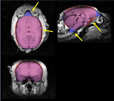
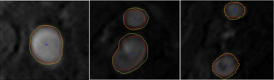
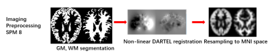
Changes in volume and shape of structures under an intervention or disease state can be measured in many subjects by automatic segmentation of structural imaging. However, unwanted intensity variation (bias field artefact), such as observed using a surface coil, can compromise such segmentation. We investigated how this artefact could be resolved using post-processing, to yield accurate brain extraction from MPRAGE acquisition in a marmoset. This atlas-based method (ASM) significantly improved brain extraction, correcting multiple inaccuracies of the initial FSL-based method (FSM) such as exclusion of the olfactory bulb and inferior cerebellar structures, and is robust to bias field artefact.
|
|
3124.
 |
50 |
Fast and robust unsupervised identification of MS lesion change using statistical detection of changes (SDC) algorithm
Thanh Nguyen, Shun Zhang, Ajay Gupta, Susan Gauthier, Yi Wang
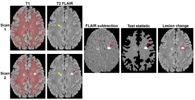
The objective of this study was to develop a robust automated lesion change detection algorithm for MS. Our preliminary results in 30 patients show that our SDC algorithm achieves much higher sensitivity and specificity (99%/76%) compared to that obtained with off-the-shelf LPA algorithm (76%/27%).
|
|
3125.
 |
51 |
Clinical feasibility of Quantitative Susceptibility Mapping with Automatic Uniform Cerebrospinal Fluid Zero Reference
Shun Zhang, Zhe Liu, Yihao Yao, Thanh Nguyen, Pascal Spincemaille, Yi Wang
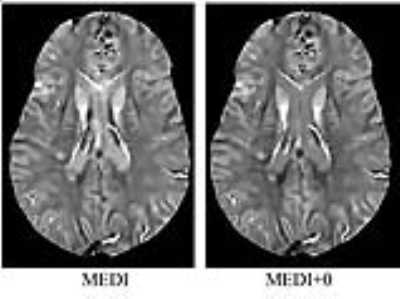
Longitudinal and cross-center studies using conventional quantitative susceptibility mapping (QSM) methods require the choice of a reference tissue, its manual segmentation and subtraction of its average. In this work, we report our initial clinical experience with a fully automated zero-referenced Morphology Enabled Dipole Inversion (MEDI+0) method that uses the ventricular cerebrospinal fluid (CSF) as zero reference in 393 consecutive patients. In 92.62% of cases, excellent agreement of image quality between MEDI+0 and MEDI was observed with high correlation of lesion susceptibility in a combined glioma, ischemic stroke and multiple sclerosis subset of patients.
|
|
3126.
 |
52 |
Thalamus in schizophrenia revisited: a partial-volume estimation study
Philipp Baumann, Elena Najdenovska, Mário João Fartaria, Alessandra Griffa, Timo Roine, Yasser Alemán-Gómez, Emeline Mullier, Philippe Golay, Zita Rovo, Patric Hagmann, Kim Do, Philippe Conus, Pascal Steullet, Meritxell Bach Cuadra
The thalamus has a central role in the pathophysiology of schizophrenia. Formed by several nuclei, it is mainly constituted by a mixture of grey and white matter and, thus, its MR signal is heavily affected by the partial volume (PV) effect. We hypothesize that tissue segmentation based on a PV model will better depict subtle changes in schizophrenia patients than total thalamus volume or local tissue volume measurements that do not consider PV. Results show statistically significant changes in gray matter and white matter average concentration from PV model within the thalamus in schizophrenia patients (SCHZ) compared to healthy controls (HC).
|
|
3127.
 |
53 |
Comparison study of Visualization of Low Intensity Spots between Susceptibility Weighted Imaging and Quantitative Susceptibility Mapping Created from the Same Raw Phase
Yasutaka Fushimi, Tomohisa Okada, Yuta Urushibata, Takuya Hinoda, Takayuki Yamamoto, Hikaru Fukutomi, Yusuke Yokota, Sonoko Oshima, Akira Yamamoto, Tsutomu Okada, Kaori Togashi
This study was conducted to evaluate the contrast of microbleeds between SWI and QSM created from the identical raw phase of SWI. QSM was created by using STI Suite. The contrast between low intensity spot on SWI and the surrounding brain parenchyma was significantly higher in QSM than SWI.
|
|
3128.
 |
54 |
Regional Differences in Cortical Diffusion MRI Measurements Based on the HCP Dataset
Video Permission Withheld
Zifei Liang, Di Wang, Tanzil Arefin, Yulin Ge, Jiangyang Zhang
Diffusion MRI (dMRI) is widely used for investigation of microstructural properties of white matter structures, but its application in cortical gray matter structures has only emerged recently due to limited spatial resolution. Little is known of the normal dMRI measurements among different cortical gray matter structures, their variations, and regional differences. In this work, we present the normative values of healthy volunteers for dMRI metrics across 74 parcellated cortical regions based on well-characterized high-resolution human connectome project (HCP) dataset. We found regional differences in several dMRI metrics among these cortical regions to be mainly in mean diffusivity and kurtosis.
|
|
3129.
 |
55 |
PROBABILISTIC MODEL-BASED FUNCTIONAL PARCELLATION OF THE PRIMARY OLFACTORY CORTEX
Prasanna Karunanayaka, Jiaming Lu, Christopher Sica, Bing Zhang, Qing Yang, Paul Eslinger, Ronald Janssen
The primary olfactory cortex (POC) is the largest recipient of olfactory bulb projections. It has a functionally versatile organization with extensive reciprocal connections to several higher-order cortical regions likely resulting in specific brain signals. Here, we propose a Bayesian model-based clustering approach, applied solely to resting state functional MRI time courses, to identify intrinsic POC functional parcellations. Results of this study suggest that multiple regions within the POC, with clear inter-hemispheric correspondence and functional relevance, can be identified using resting state fMRI data.
|
|
3130.
 |
56 |
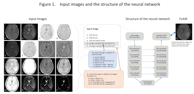
A Deep Learning Approach to Synthesize FLAIR Image from T1WI and T2WI
Takashi Abe, Noriko Salamon
We synthewized FLAIR images of the brain from T1WI and T2WI by using autoencoder, which is one of the state of the art deep-learning technology. Autoencoder compresses the input information and reproduces the information therefrom. We used T1WI and T2WI as an input and synthewized FLAIR image with high accuracy. This method could be applicable to other body part other than the brain and might synthewize of other MR imaging sequences. This technology seems to be useful to improve clinical diagnosis and computer-aided diagnosis.
|
|
3131.
 |
57 |
Automatic quality assessment of high-resolution T2-weighted images used in hippocampus volumetry and functional studies – a tool developed as part of DZNE-DELCODE study.
Arturo Cardenas-Blanco, Yi Chen, Jose Pedro Valdes-Herrera, Laura Dobisch, Renat Yakupov, Klaus Fliessbach, Michael Wagner, Annika Spottke, Stefan Teipel, Katharina Buerger, Anja Schneider, Oliver Peters, Peter Nestor, Josef Priller, Jens Wiltfang, Christoph Laske, Frank Jessen, Emrah Duezel
This abstract presents a processing pipeline developed to automatically assess the quality of specific structural T2-weighted images typically acquired in the study of the hippocampus. By combining existing neuroimaging tools, the presented pipeline generates descriptive information about the signal properties in different tissue classes of the T2-weighted image. This information could subsequently be used to detect sub-optimal volumes due to noise or motion artifacts. Similarly as it measures the angulation of the T2-weighted slices with respect to the HC, it could also be used to automatically determine whether the field of view angulation follows protocol.
|
|
3132.
 |
58 |
Large Vessel Filtering of In Vivo High-Resolution Mouse CBV Map Using Expectation-Maximization Gaussian Mixture Model
Jia Guo, Scott Small
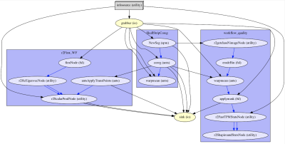
When examining relative cerebral blood volume (rCBV) between mice, values from the unwanted large vessels should be excluded, otherwise, they may result in findings that are not associated with the basal metabolism mapped by microvascular blood volume. Our proposed method provides an automated and robust approach to estimate the rCBV distribution in large vessels with the maximum likelihood estimation using expectation-maximization Gaussian mixture model and to help filter out this unwanted confounding. In research related to rCBV analysis, we suggest applying this as one preprocessing step, which may help improve both sensitivity and specificity when comparing rCBV between subjects.
|
|
3133.
 |
59 |
Regression of Quasi-Periodic patterns diminishes BOLD functional connectivity and reveals hidden dynamic correlations
Michaël Belloy, Behnaz Yousefi, Anzar Abbas, Annemie van Der Linden, Georgios A. Keliris, Marleen Verhoye, Shella Keilholz
Quasi-Periodic patterns (QPPs) represent large-scale recurring patterns in the brain, which appear as promising contributors to low frequency BOLD fluctuations. To assess the impact of QPPs on functional connectivity (FC), we used a general linear model approach to regress their contribution out of the functional images of a group of wild-type mice. We show that QPP regression diminished FC in co-active regions within the QPP, while anti-correlated regions became correlated. By calculating FC on QPP-scrubbed images, we highlight that these effects are not solely an artifact of regression. These results suggest that QPPs orchestrate dynamic correlations between resting state networks.
|
|
3134.
 |
60 |
Preliminary investigation of QSM stability across sites
Renat Yakupov, Arturo Cardenas-Blanco, Klaus Fliessbach, Michael Wagner, Annika Spottke, Stefan Teipel, Katharina Bürger, Anja Schneider, Oliver Peters, Peter Nestor, Josef Priller, Jens Wiltfang, Christoph Laske, Frank Jessen, Emrah Duezel, Julio Acosta-Cabronero
We compared quantitative susceptibility mapping (QSM) from two groups of subjects scanned at two different sites on different 3T Siemens MRI systems running different Syngo MR software versions. Qualitative whole-brain inspection of summary statistics and a statistical test comparing data from both sites revealed no major QSM offsets. The present results are only preliminary but they suggest QSM might be well suited for multi-site studies.
|
|
3135.
 |
61 |
Quantifying cortico-cerebellar structural covariance
Christopher Steele, Sejal Patel, Jurgen Germann, Gabriel Devenyi, Mallar Chakravarty
Invasive tract-tracing in primates has shown that cerebellar regions (lobules) are differentially connected to the cerebral cortex. As diffusion tractography to/from the cerebellum is problematic in living humans, we propose a simple non-invasive approach to identify the patterns of grey-matter structural covariance between cerebellar lobules and the cerebral cortex as a proxy for anatomical connectivity. We performed vertex-wise linear regressions between lobular volumes and cortical thickness/surface area. We then clustered lobule-wise correlations to identify similar spatial patterns of cortico-cerebellar covariance. We identified differential patterns that may reflect the underlying connectivity between the cerebellum and cortex.
|
|
3136.
 |
62 |
Carotid Artery Segmentation Based on Level Set Method in MR Images
Video Permission Withheld
Lian Luo, Fei Shang, Xinyu Tong, Shuai Liu, Xihai Zhao
Segmentation of blood vessels is the first step of visualization of vascular geometry and morphological analysis in cardiovascular diagnosis. However, the manual segmentation of blood vessels is time consuming. This study applied the level set method to automatic segmentation for carotid artery morphology on time-of-flight MR angiography images. The brightness weighting term is added to optimize the model, and the optimal setting of the parameters of the carotid artery images is discussed in detail. The results showed that the model with the brightness weighting term was more stable, accurate and efficient, which might be used to automatic segmentation of carotid arteries.
|
|
3137.
 |
63 |
Spatio-temporal features of brain's iron accumulation across the life span.
Did Not Present
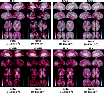
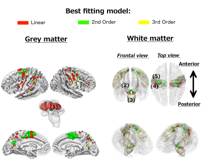
Vyacheslav Karolis, Thomas Hope, Nikolaus Weiskopf, Martina Callaghan, Geraint Rees, Marinella Cappelletti
In this study we characterised the spatiotemporal features of age-related iron accumulation marked by R2* maps in 97 participants distributed over the whole range of ages between 20 and 75 years old. The analyses showed a broad spectrum of linear and non-linear patterns of age-related differences in R2*. These were predominantly confined to striatum, motor, pre-motor and parietal regions of grey matter as well as subgyral pre- and postcentral regions of white matter. The spatial distribution of R2* changes seems to be related to a higher prevalence of long-distance white matter connections originating from a region.
|
|
3138.
 |
64 |
Quantitative Assessment of the Intracranial Vasculature of an Elderly Population using the intraCranial Artery Feature Extraction (iCafe) Technique
Li Chen, Quan Yuan, Niranjan Balu, Isabelle Yuan, Daniel Hippe , Jie Sun, Xihai Zhao, Rui Li, Le He, Jenq-Neng Hwang, Chun Yuan
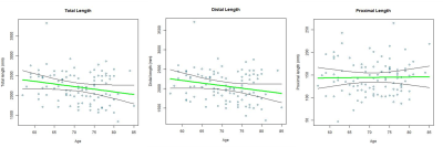
iCafe is a novel technique which semi-automatically traces intracranial arteries from 3D magnetic resonance angiography (MRA) and computes corresponding quantitative morphometry and intensity features. MRA images of 100 healthy subjects (age 57-85) were processed and 8 representative features were extracted. We found significant decreases in total artery length (p=0.026), distal artery length (p=0.025), and number of branches (p=0.005) with increasing age using linear regression. These results suggest reduced vascularity with age, consistent with prior results showing cerebral blood flow with age. iCafe may be a useful tool to generalize systemic quantitative measurements of intracranial vascular structures.
|
|
3139.
 |
65 |
Prediction of Alzheimer’s disease by using deep learning 3D-Convolutional Neural Networks
Did Not Present
Na Sang, Francisco Garcia, Wanshun Wei, Huabing Li, Tao Ma, Silun Wang
We analyzed the T1 structural MRI by using deep learning 3D-CNN method. The results indicate that deep learning models can accurately predict AD patients with diagnostic accuracy of 96%. This can be achieved using raw MRI data, with a minimum of processing necessary to generate an accurate AD prediction. Our model shows highly sensitivity and negative predictive value and thus appropriate for use for screening testing in population study. Currently model has the potential to be used as a screen biomarker to investigate the neurodegeneration, brain aging and associated brain diseases.
|
|
3140.
 |
66 |
Automated MR-based volumetry of basal ganglia and thalamus at the chronic phase of cortical stroke
Cindy Baudat, Bénédicte Maréchal, Ricardo Corredor-Jerez, Patric Hagmann, Philippe Maeder, Vincent Dunet
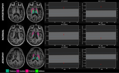
We investigated the potential of automated T1-MPRAGE-based brain segmentation to assess individual basal ganglia and thalamus atrophy in nineteen patients with cortical stroke at the chronic phase. The basal ganglia and thalamus volumes z-scores were compared to the initial stroke ipsilateraly and contralateraly, and the relationship with the stroke volume was assessed. Except caudate nucleus, basal ganglia and thalamus, atrophy ipsilateraly to the stroke was observed and negatively correlated with the stroke volume in the territory of the middle cerebral artery. This suggests a potential role for automated MRI volumetry to assess brain plasticity after stroke at the individual level.
|
|
3141.
 |
67 |
Role of MRI Texture Analysis in Differentiating Post-treatment Changes from Tumour Recurrence in Patients with High Grade Glioma
Did Not Present
Fahad Essbaiheem, Rebecca Thornhill, Gregory Cron, John Woulfe, Mario Kontolemos, Beckie Manouchehri, Nader Zakhari, Andrew Boivin, Thanh Binh Nguyen
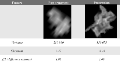
In this prospective study, we have identified a combination of textural features from contrast-enhanced T1-weighted images which can help in differentiating tumour recurrence from post-treatment changes in patients with high grade gliomas. The diagnostic accuracy of textural analysis was similar or slightly higher than that of two neuroradiologists who performed visual assessment.
|
|
3142.
 |
68 |
Deep Learning approach for Automatic Segmentation and characterization of Traumatic Brain Injury using Multi-parametric MRI
Krishna Kanth Chitta, Abdalla Mohamed, Fatima Nasrallah, Bhanu Prakash KN
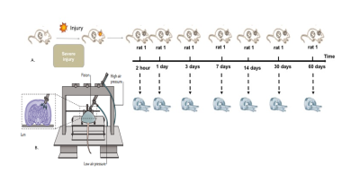
Automatic and accurate segmentation of Traumatic brain injury is vital to improve assessment of pathophysiology, plan treatment methods and enable large cohort studies. In this work we propose a framework based on 3D CNN and FCM to perform automatic segmentation of whole TBI volume and its sub-regions. The proposed framework utilizes multiple MRI contrasts and has shown high accuracy in delineating injury and sub-regions

|
|
3143.
 |
69 |
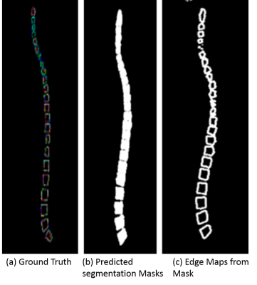
A Deep Learning Based Solution for Vertebrae Segmentation of Whole Spine MR Images: A Step Closer to Automated Whole Spine Labeling
Kavitha Manickam, Jignesh Dholakia, Vignesh Singh
Any reporting on an MR spine scans involves labeling of the vertebrae. Hence, providing labeled spine images for reading can save significant time for radiologists. First step of an automated labeling is reliable segmentation of vertebral bodies. Most of the studies provide methods only for the segmentation and labeling of only a part of the spine. Here, we have used a variant of U-Net based Deep Learning architecture for segmenting vertebrae of Whole Spine. The network was trained with 165 datasets of whole spine images and tested with 8 datasets. We achieved average DICE score of 0.921.
|
|
3144.
 |
70 |
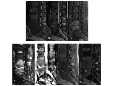
Deciphering predictive models for differentiating vertebral lesions using multiparametric MRI
Durgesh Dwivedi, Anit Parihar, Rashi Rathore, Neera Kohli, Alok Dwivedi, Anil Chandra
Conventional MR imaging has high sensitivity but limited specificity in differentiating various vertebral lesions. We aimed to assess the ability of multiparametric MR imaging in differentiating spinal vertebral lesions and to develop statistical models for predicting the probability of malignant vertebral lesions. On the basis of the mean ADC and signal intensity ratio, we established automated statistical models that would be helpful in differentiating vertebral lesions. Our study shows that multiparametric MRI differentiates various vertebral lesions, and we established prediction models for the same.
|
|
3145.
 |
71 |
Evaluation of errors in image registration and diffusion measurement due to the cavum septum pellucidum
Chen-Hsiang Weng, Yung-Chin Hsu, Wen-Yih Isaac Tseng
To determine the registration errors caused by the cavum septum pellucidum (CSP), Dice coefficient and generalized fractional anisotropy (GFA) were assessed in brain using T1-weighted imaging and diffusion spectrum imaging (DSI), respectively. The subjects included 30 subjects with the CSP and 30 subjects without the CSP. Comparing with the subjects without the CSP, the subjects who had enlarged CSP showed significantly decreased Dice coefficient and significantly different GFA values in nine tracts. Our findings indicate that the existence of the CSP leads to errors in image registration and diffusion index calculation, and that the size of the CSP should be taken into consideration as a covariate in statistical analysis.
|
|
3146.
 |
72 |
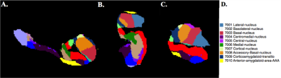
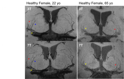
Automated Segmentation and Volumetric Analysis of the Amygdala Nuclei in Epilepsy Patients at 7 Tesla
John Rutland, Rebecca Feldman, Lara Marcuse, Madeline Fields, Bradley Delman, Prantik Kundu , Priti Balchandani
The present study employs ultra-high field MRI (7 Tesla) to perform structural imaging on a group of 19 epilepsy patients and 9 healthy controls. We use automated segmentation of the amygdala to derive volumes of constituent sub-nuclei. When comparing epilepsy patients and controls, we found that the volume of the right lateral nucleus was reduced compared with controls. We also found that the anterior-amygdaloid-area and the whole right amygdala approach significance for reduced volume. These are the first in vivo findings that indicate particular nuclei are affected in epilepsy patients.
|
|
Emerging Methods
Electronic Poster
Neuro
Monday, 18 June 2018
| Exhibition Hall |
08:15 - 09:15 |
| |
|
Computer # |
|
3147.
 |
73 |
Magnetic susceptibility in subcortical gray matter is associated with age-related neuropathologies: An ex-vivo QSM and pathology investigation in a community cohort of older adults
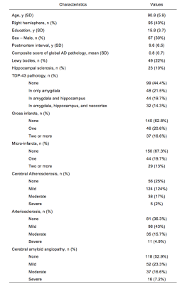
Arnold Evia, Ashish Tamhane, Aikaterini Kotrotsou, Robert Dawe, Sue Leurgans, Julie Schneider, David Bennett, Konstantinos Arfanakis
To understand the associations of quantitative susceptibility mapping with age-related neuropathologies, it is essential to combine QSM with direct assessments of age-related brain pathologies on the same individuals. Using ex-vivo QSM for this purpose may be more advantageous than in-vivo QSM, since ex-vivo QSM overcomes several of the obstacles that complicate MRI-pathology investigations and can provide magnetic susceptibility measurements that are linked to those collected in-vivo. Therefore, our goal was to investigate the associations of magnetic susceptibility with the pathology of multiple age-related diseases by combining ex-vivo QSM with histology.
|
|
3148.
 |
74 |
Imaging the effect of high-protein diet on the brain Glutaryl-CoA dehydrogenase deficient mice using GluCEST MRI
Puneet Bagga, Damodara Reddy, Delia Talos, Kimberly Sansalone, Leah Jacobs, Gaurav Verma, Hari Hariharan, Ravinder Reddy
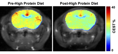
Autosomal recessive inheritance of Glutaryl-CoA dehydrogenase (GCDH) deficiency or Glutaric Acidemia-1 is one of the common inherited metabolic disorders affecting many children worldwide. We applied GluCEST MRI to image the metabolic changes in the brain of GCDH-/-mice following the exposure to high-protein diet. GluCEST contrast was found to be significantly reduced in the striatum and cortex of the GCDH-/- mice following high-protein diet exposure. 1H MRS data corroborated with GluCEST results showing a significant reduction in the striatal glutamate level. GluCEST MRI may be used as an imaging biomarker for assessing the neurovascular damage caused by high-protein diet in GCDH-/- subjects.
|
|
3149.
 |
75 |
On Resonance Variable Delay Multiple Pulse (onVDMP) CEST MRI Detects Transient Lactate Depletion during Experimental Autoimmune Encephalomyelitis Progression
Aline Thomas, Jiadi Xu, Peter Calabresi, Peter van Zijl, Jeff Bulte
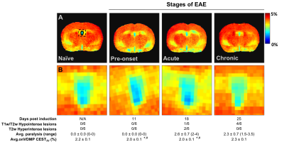
On resonance variable delay multiple pulse CEST MRI detected a transient reduction in lactate levels in the cerebrospinal fluid during the disease course of a mouse model of multiple sclerosis, which was validated using magnetic resonance spectroscopy.
|
|
3150.
 |
76 |
On Resonance Variable Delay Multiple Pulse (onVDMP) CEST MRI for Monitoring Stem Cell Therapy in Experimental Autoimmune Encephalomyelitis
Aline Thomas, Jiadi Xu, Shen Li, Piotr Walczak, Peter Calabresi, Peter van Zijl, Jeff Bulte
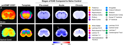
In mice with experimental autoimmune encephalomyelitis, spatiotemporal signal changes were detected with on resonance variable delay multiple pulse (onVDMP) CEST MRI for the brain and spinal cord. Transplantation of glial-restricted precursor cells resulted in normalization of those signal changes during the pre-onset stage of the disease.
|
|
3151.
 |
77 |
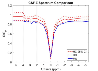
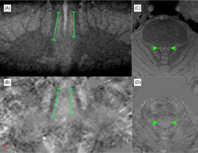
APT CEST in cerebrospinal fluid in patients with multiple sclerosis at 3T
Richard Lawless, Quinn Weinberg, Bailey Box, Samantha By, Francesca Bagnato, Seth Smith
There is currently no clinically available MRI method capable of monitoring chronic, systemic inflammation in the central nervous system as a confirmatory biomarker. Chemical exchange saturation transfer (CEST) is a novel MR technique sensitive to low concentration, exchangeable mobile solutes. Amide proton transfer (APT) CEST provides information about concentration of proteins/peptides with amide backbones. Our results show that APT CEST of CSF in the spinal cord demonstrates significant changes when compared between the control and MS cohort.
|
|
3152.
 |
78 |
MT and QSM of the Locus Coeruleus and Substantia Nigra on human healthy subjects
Catarina Rua, Luca Passamonti, Claire O’Callaghan, James Rowe, Adrian Carpenter, Guy Williams
The Locus Coeruleus (LC) and the Substantia Nigra (SN) are located in the mid-brain are known to show severe cell loss in several neurodegenerative diseases (e.g., Alzheimer’s disease). Developing new imaging techniques to quantify the signal changes in these regions is of high clinical relevance. In this pilot study of five healthy adults from different age groups, the LC and SN were evaluated for neuromelanin and iron content with an Magnetisation Transfer-weighted sequence and with Quantitative Susceptibility Mapping, respectively.
|
|
3153.
 |
79 |
Susceptibility weighted imaging with Compressed-SENSE: Quantitative and Clinical evaluation
Jaladhar Neelavalli, Rakesh Kumar Gupta, Jakob Meineke, Rupsa Bhattacharjee, Suthambhara Nagaraj, Tejas Shah, Ulrich Katscher, Indrajit Saha
The current work evaluates the influence of a combined SENSE and compressed sensing algorithm, Compressed-SENSE, on the quantitative and qualitative aspects of SWI data. Quantitative evaluation is done using quantitative susceptibility maps. Clinical qualitative assessment is done in patients with cerebral glioma using ITSS score as the metric for image diagnostic quality.
|
|
3154.
 |
80 |
Functional MRI reveals resting state network alterations upon DREADD-induced silencing of the right dorsomedial prefrontal cortex in mice.
Lore Peeters, Rukun Hinz, Stephan Missault, Marleen Verhoye, Annemie Van der Linden, Georgios A. Keliris
Combining chemogenetics with non-invasive functional MRI (fMRI) allows establishing a link between the activity of selected populations of neurons with large-scale network activity. Here, we show that Kappa Opioid Receptor (KOR) DREADD-induced decreases in neural activity result in network alterations that can be picked up by pharmacological and resting state fMRI. In particular, inhibition of the right dorsomedial prefrontal cortex (dmPFC), a core region of the attention network in rodents, induces functional connectivity changes between other regions of the attentional network and between regions of distinct sensory networks (e.g. the visual network).
|
|
3155.
 |
81 |
Cross-Platform Comparison of Regional Brain Stiffness
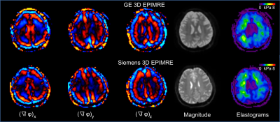
Jun Chen, Matthew Murphy, Kevin Glaser, Curtis Johnson, Arvin Forghanian-Arani, Yuan Le, David Lake, Roger Grimm, John III Huston, Richard Ehman
Brain MR Elastography (MRE) has been shown to add value to neurological imaging. Brain biomechanical properties are correlated with age, gender, exercise and memory in normal volunteers. In patients with neurological diseases, such as multiple sclerosis, dementia and intracranial tumors, global and regional brain mechanical properties may be sensitive biomarkers. Characterization of the cross-platform performance of regional stiffness measurements using 3D brain MRE is important for future multiplatform, multicenter brain MRE studies. The purpose of our study was to compare regional 3D brain MRE in normal volunteers using two vendor platforms.
|
|
3156.
 |
82 |
Magnetization Transfer Ratio based Metric for APTw or CESTw MRI Suppressing Signal from Fluid Compartments - Initial Application to Glioblastoma Assessment
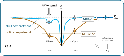
Jochen Keupp, Osamu Togao
Amide proton transfer weighted (APTw) MRI has been shown to allow assessment of the tumor aggressiveness/tumor grade with high sensitivity and specificity. Compartments with significant fluid content, like cysts, haemorrhage or necrosis may complicate reading. A metric for APTw MRI is developed, applicable for chemical exchange saturation transfer (CEST) MRI in general, which suppresses fluids based on the spectral shape of the background magnetization transfer ratio (MTR). It is shown to essentially conserve numerical values of APTw MRI in solid tissues. Furthermore, no extra acquisition time is needed, because the metric can be computed from the minimum Z-spectral data also required for standard CEST MRI. The novel metric is explored in an intial study on N=12 gliobastoma cases.
|
|
3157.
 |
83 |
Longitudinal Change in the Developmental Rat Brain using in vivo Glutamate Chemical Exchange Saturation Transfer
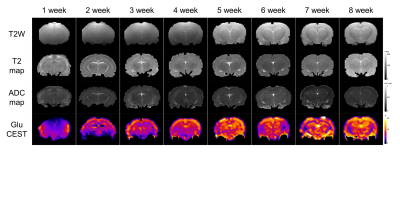
Wooyul Paik, Dong Cheol Woo, Ho Sung Kim
Our study results and analyses show the trend in glutamate concentration and the decreases in T2 and ADC values influenced by myelination, neuronal change, and the water and macromolecule content of the developing rat brain, and also provide evidence indicating the time point where a neurochemical balance is achieved. Our results demonstrating changes in glutamate concentration up to the eighth postnatal week should provide valuable reference data for other studies of the developing healthy rat brain, and should be useful for comparisons with diseased rat brain in further developmental studies.
|
|
3158.
 |
84 |
Optimizing Brain MR Elastography with Multiple Motion Encoding Gradient Cycles
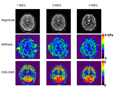
Yuan Le, Kevin Glaser, Matthew Murphy, Yuxiang Zhou, Jun Chen, William Pavlicek, Joseph Hoxworth, Bradley Bolster Jr., John Huston III., Joel Felmlee, Richard Ehman
A spin-echo EPI MR Elastography sequence was optimized so that multiple motion encoding gradient (MEG) cycles can be added to increase the motion sensitivity. Volunteer tests showed that comparing with the original one MEG version, optimized two or three MEG cycles provided higher Octahedral Shear Strain Signal-to-Noise Ratio (OSS-SNR), which means higher stiffness measurement precision. Global brain Images acquired with 2 and 3 MEG cycles are in most cases comparable in stiffness and OSS-SNR while images with 1 MEG tend to have a slightly lower OSS-SNR.
|
|
3159.
 |
85 |
Regional Changes in Brain Stiffness with Age Assessed with MR Elastography
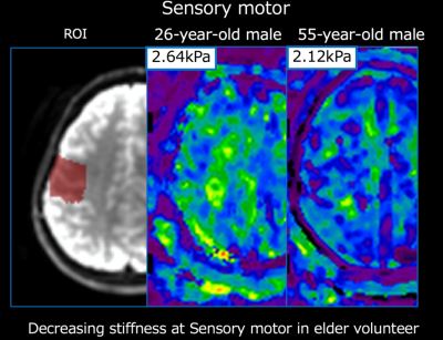
Yu Sasaki, Utaroh Motosugi, Tomohiro Takamura, Kevin Glaser, Richard Ehman, Hiroshi Kumagai, Takashi Kakegawa, Hiroshi Onishi
Various MR-assessed quantities change with age in the brain, including T2, diffusion, and fractional anisotropy. MR elastography (MRE) is a new technique that can measure the stiffness of tissue and is now widely used in the clinic for assessing hepatic fibrosis. Recently, it has become feasible to perform brain MRE as well. In this study, we showed that the stiffness of the brain, measured using MRE, decreased with age, especially in the parietal lobes and the sensory-motor area.
|
|
3160.
 |
86 |
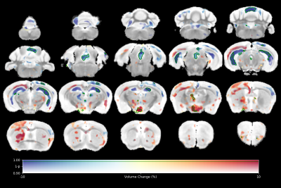
The impact of age and sex on mouse brain stiffness measured with Magnetic Resonance Elastography
Katharina Schregel, Miklos Palotai, Navid Nazari, Julie Merchant, Walter Taylor, Charles Guttmann, Ralph Sinkus, Tracy Young-Pearse, Samuel Patz
Aging is accompanied by neurodegeneration, which affects the cerebral biomechanical properties. We investigated the impact of age and sex on mouse brain stiffness using magnetic resonance elastography (MRE). Repeated MRI and MRE exams were performed on 5 male and 5 female healthy C57BL/6 mice over 14 months. A significant decrease of the viscoelastic modulus |G*| was observed, while the phase angle Y remained unaltered. Grey and white matter exhibited significant differences in |G*| and Y. Sex differences were observed in the cortex at 11 months. This is relevant for future cerebral MRE studies on mice.
|
|
3161.
 |
87 |
Brain stiffness changes due to Alzheimer’s disease in cortical-centric regions
Matthew Murphy, David Jones, Clifford Jack, Kevin Glaser, Matthew Senjem, Armando Manduca, Joel Felmlee, Richard Ehman, John Huston
Brain stiffness is known to decrease in subjects with Alzheimer’s disease (AD). However, previously reported stiffness estimates were heavily weighted toward white matter. Here we investigate the sensitivity of cortical-centric stiffness measurements for detecting AD pathophysiology, given that the cortex is the primary site of pathology. Using a neural network-based inversion algorithm, cortical-centric measurements are highly repeatable with test-retest errors of less than 2% on average. With respect to AD, the medial temporal lobe region of interest is found to best discriminate those with dementia from cognitively normal subjects, and performs better than previously reported methods.
|
|
3162.
 |
88 |
Acute changes in rat brain metabolism after intravenous administration of alcohol, cocaine, and nicotine: A simultaneous PET/MR study with dynamic 1H-MRS and continuous infusion 18FDG.
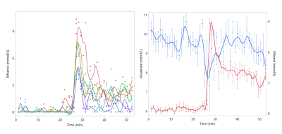
Bart de Laat, Akila Weerasekera, Gwen Schroyen, Cesar Molinos, Uwe Himmelreich, Cindy Casteels, Koen Van Laere, Willy Gsell
We have combined PET imaging with continuous 18F-deoxy-glucose PET and dynamic 1H-MRS focused on the prefrontal cortex in a rodent model of substance abuse. During imaging, animals received an intravenous injection of saline, alcohol, cocaine, or nicotine. Cocaine administration reduced the regional cerebral metabolic rate of glucose (rCMRGlu) as measured by PET, but increased prefrontal glucose and creatine levels as measured with dynamic MRS. Furthermore, alcohol administration significantly influenced the prefrontal concentration of ethanol, glucose, creatine, and glutamate. Finally, our data show that alcohol induces a transient decrease in prefrontal glutamate coinciding with the peak in ethanol concentration.
|
|
3163.
 |
89 |
Assessment of cerebral infarction due to intracranial arterial stenosis in the human brain using hyperpolarized xenon-129 MRI
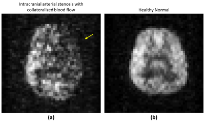
Madhwesha Rao, Graham Norquay, Neil Stewart, Nigel Hoggard, Paul Griffiths, Jim Wild
In this pilot study we evaluate the feasibility of hyperpolarised 129Xe brain perfusion MR imaging to evaluate brain tissue perfusion patho-physiology in a subject with established intracranial arterial stenosis with collateralized blood flow. The 129Xe brain perfusion images from the patient exhibit regions of signal void which correspond to the infarcted region observed on T2 weighted MRI. Imaging hyperpolarized 129Xe dissolved in the brain tissue is a direct method of imaging perfusion of the tissue itself and we observe different patterns of cerebral perfusion than those measured with arterial spin labeling.
|
|
3164.
 |
90 |
Phase Laplacian Coil Combination
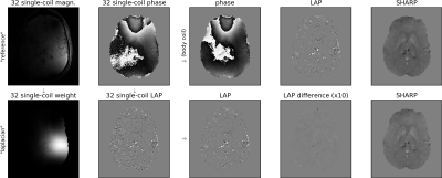
Rüdiger Stirnberg, Lino Lemmer, Tony Stöcker
We propose a novel, computationally efficient coil combination technique for multi-channel phase data based on the Laplacian of single-channel phase images. This renders explicit knowledge or estimation of the receive sensitivities unnecessary. The combined phase Laplacian can be either be transformed back to unwrapped phase domain (Laplacian-based unwrapping) or directly utilized for further analyses based on the phase Laplacian, e.g. harmonic background-field removal. At 3T we demonstrate similar-to-improved phase reconstruction compared to the vendor-provided state-of-the-art coil combination, which uses the body-coil as a uniform reference, and successfully apply the technique at 7T.
|
|
3165.
 |
91 |
Resting-state fMRI study of brain activation using low-intensity repetitive transcranial magnetic stimulation in rats.
Video Permission Withheld
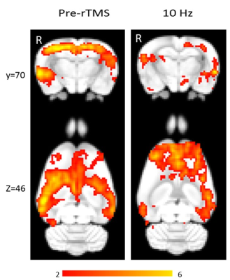
Bhedita Seewoo, Kirk Feindel, Sarah Etherington, Jennifer Rodger
Repetitive transcranial magnetic stimulation (rTMS) is a non-invasive neuromodulation technique used to treat many neuropsychiatric conditions. However, the mechanisms underlying its mode of action are still unclear. This is the first rodent study using resting-state fMRI to examine low-intensity (LI) rTMS effects, in an effort to provide a direct means of comparison between rodent and human studies. Our study shows that similar to human rTMS, 10 Hz LI- rTMS alters the resting brain activity of rats directly at the site of stimulation (e.g. cortex) as well as in remote but inter-connected brain regions (e.g. hippocampus).
|
|
3166.
 |
92 |
Imaging toxin-induced inflammation in the mouse brain with hyperpolarized 13C MRS
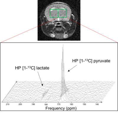
Lydia Le Page, Caroline Guglielmetti, Brice Tiret, Myriam Chaumeil
13C MRS of hyperpolarized (HP) [1-13C] pyruvate has recently shown promise in assessing neuroinflammation in mouse models of MS and TBI. Here, we expanded on previous reports and evaluated whether HP 13C MRS could detect the effect of the inflammation-inducing toxin lipopolysaccharide (LPS), using mice injected intracranially with either LPS or saline. LPS-injected mice showed significantly elevated HP [1-13C] lactate:pyruvate ratios in the LPS-injected hemisphere compared to contralateral, in line with increased microglial number. In contrast, saline-injected mice showed no such changes. Our results further confirm the potential of hyperpolarized 13C MRS for non-invasive assessment of neuroinflammation in the brain.
|
|
3167.
 |
93 |
A real-time metabolic investigation of the effect of hypothermia on hypoxic ischemia during mouse brain development using hyperpolarized 13C
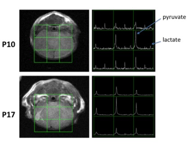
Yiran Chen, Alkisti Mikrogeorgiou, Robert Bok, Subramaniam Sukumar, R Sheldon, A Barkovich, Donna Ferriero, Duan Xu
In this study, we applied dynamic nuclear polarization (DNP) technique to investigate C1 labeled 13C pyruvate to lactate conversion to study the effect of hypothermia treatment on hypoxic ischemia (HI) injured neonatal mouse brains during development. Our results showed that lower pyruvate delivery to the injured hemisphere in comparison to the non-injured hemisphere at the day of injury (P10) for all subjects, and difference narrows as the brain matures. There were different individual responses to the lactate to pyruvate ratio between two hemispheres. With this technique, we are able to examine individual responses to treatment during brain development.
|
|
3168.
 |
94 |
19F Signal Distribution in Pre-Symptomatic Experimental Autoimmune Encephalomyelitis
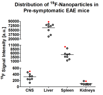
Paula Ramos Delgado, Jason M. Millward, Christian Prinz, Ludger Starke, Stefanie Münchberg, Andreas Pohlmann, Thoralf Niendorf, Sonia Waiczies
Experimental autoimmune encephalomyelitis (EAE) is an animal model used to study the pathogenesis of autoimmune neuroinflammatory diseases, such as multiple sclerosis (MS). In MS, recruitment of immune cells into the central nervous system (CNS) occurs already at early stages of the disease. Magnetic resonance imaging (MRI) can be used as a non-invasive technique suited for tracking immune cell migration, following intravenous administration of fluorine (19F)-loaded nanoparticles which can then be followed using 19F MR techniques. The present study aims to investigate the distribution of immune cells during the pre-symptomatic disease phase in the EAE mouse model using 19F MR methods.
|
|
3169.
 |
95 |
Multi-contrast Brain MRI Image Super-resolution with Gradient-guided Edge Enhancement
Hong Zheng, Kun Zeng, Di Guo, Jiaxi Ying, Yu Yang, Xi Peng, Zhong Chen, Xiaobo Qu
Since magnetic resonance imaging (MRI) can offer images of an object with different contrasts, e.g., T1-weighted or T2-weighted, the shared information between inter-contrast images can be used to benefit super-resolution. Multi-contrast images are assumed to possess the same gradient direction in a local pattern. We proposed to establish a relation model of gradient value between different contrast images, to restore a high-resolution image from its input low-resolution version. The similarity of image patches is employed to estimate intensity parameters, leading a more accurate reconstructed image. Then, iterative back-projection filter is applied to the reconstructed image to further increase image quality. The reconstructed edges are more consistent to the original high-resolution image, indicated with higher PSNR and SSIM than the compared methods.
|
|
3170.
 |
96 |
Novel Imaging Phantom for Accurate and Robust Measurement of Brain Atrophy Rates Using Clinical MRI
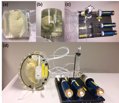
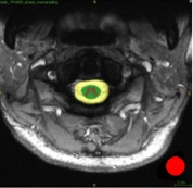
Houshang Amiri, Iman Brouwer, Joost Kuijer, Jan de Munck, Frederik Barkhof, Hugo Vrenken
MR images are widely used to measure brain atrophy in neurodegenerative diseases. However, reliable evaluation of atrophy is hampered by scanner-induced systematic variability. Here, we developed an MR-compatible phantom and analysis software for robust and reliable evaluation of the brain volume loss. The phantom was made using 3D-printing and contains three inflatable brain structures equipped with a precise volume change system. The phantom was imaged at three different clinical 3T MR scanners and images were analyzed by our developed software. This phantom can accurately and robustly provide a selected volume change to mimic a certain disease.
|
|
Neuroimaging: Pulse Sequences & Reconstruction
Electronic Poster
Neuro
Monday, 18 June 2018
| Exhibition Hall |
08:15 - 09:15 |
| |
|
Computer # |
|
3171.
 |
97 |
Brain Vessel Extraction without MRA / V using Deep Convolutional Neural Network
Hyungseob Shin, Yohan Jun, Taeseong Kim, Taejoon Eo, Sungsoo Ahn, Dosik Hwang
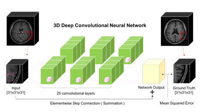
In this paper, we introduce a deep residual learning approach to extract brain vessels from contrast-enhanced(CE) magnetic resonance images. Our experiment results show that we can successfully achieve and visualize brain vessel information from CE MRI without magnetic resonance angiography and magnetic resonance venography(MRA/V) that are currently used for brain vessel extraction.
|
|
3172.
 |
98 |
High resolution atlasing of the venous brain vasculature from 7T quantitative susceptibility
Julia Huck, Yvonne Wanner, Audrey Fan, Anna-Thekla Schmidt, Sophia Grahl, Uta Schneider, Arno Villringer, Christopher Steele, Christine Tardif, Pierre-Louis Bazin, Claudine Gauthier
We present the first atlas of the venous vasculature using quantitative susceptibility maps (QSM) acquired at 7T with a 0.6mm isotropic resolution. The atlas was created from 16 datasets in young and healthy volunteers by using a three step registration method on the inflated skeletons of the venous vasculature. This cerebral vein atlas shows the average vessel location and diameter.
|
|
3173.
 |
99 |
High Resolution Black-blood T1-weighted Turbo Spin Echo with Variable Flip Angles for Visualization of Small Perforating Arteries at 3 and 7 Tesla
Samantha Ma, Lirong Yan, Kay Jann, Danny Wang
Cerebral small vessel disease frequently affects the small perforating arteries, resulting in silent strokes, which contribute to progressive cognitive impairment in elderly persons. Previous studies have demonstrated the ability of time-of-flight (TOF) MRA to non-invasively image these small arteries at 7T; In this study, we introduce and optimize a T1-weighted turbo spin echo sequence with variable flip angles (T1w-VFA-TSE) sequence for high resolution black blood MRI to delineate the lenticulostriate arteries (LSA) at 3T and 7T. Our results show T1w-VFA-TSE provides high contrast for visualizing LSAs and delineates small arteries better than TOF MRA at 7T.
|
|
3174.
 |
100 |
Systematic comparison of DTI metrics as potential biomarkers in cerebral small vessel disease
Ana Fouto, Rita Nunes, Joana Pinto, Luísa Alves, Sofia Calado, Carina Gonçalves, Pedro Vilela, Miguel Baptista, Patrícia Figueiredo
Cerebral small vessel disease (SVD) is the major cause of dementia among the elderly and sensitive biomarkers of disease progression are needed. Here, we investigated the potential of DWI to provide sensitive biomarkers of SVD, by evaluation multiple metrics extracted from DTI in terms of their predictive power of cognitive performance. We considered different white matter regions of interest to perform histogram analysis and extract multiple FA and MD metrics, and showed that specific DTI metrics were better than conventional structural MRI at explaining impairments in processing speed and execute function in a group of SVD patients.
|
|
3175.
 |
101 |
Feasibility and Evaluation of Multi-Delay Quantitative 3D GRASE pCASL MRI in Children at 3 Tesla
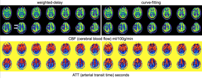
Houchun Hu, Ruiyue Peng, Xingfeng Shao, Mark Smith, Jerome Rusin, Ramkumar Krishnamurthy, Bhavani Selvaraj, Danny JJ Wang
Single post-labeling-delay (PLD) pCASL are commonly used to measure cerebral blood flow (CBF). A PLD of 1500-2000ms is commonly used in children and adults. Multi-delay pCASL has been developed as an alternative approach to better account for prolonged arterial transit times (ATT) and to improve the accuracy of CBF perfusion quantification. In this study, we evaluate the feasibility of multi-delay pCASL in children. We compare two algorithms (weighted-delay linear mapping vs. nonlinear iterative curve fitting) for estimating ATT and CBF. We further compare estimations of weighted-delay CBF derived from multi-delay pCASL data with those traditionally calculated from a single PLD measurement.
|
|
3176.
 |
102 |
Imaging Ultrashort-T2* structures in the Eye with UTE MRI
Peder Larson, Peng Cao, Kevin Chan
Ultrashort echo time (UTE) MRI has the potential to image all structures in the eye, including the sclera, cornea and lens that have relatively short T2* relaxation times. UTE MRI was used for motion-robust imaging of the eye in vivo, and multiple TE measurements were combined for 3D T2* mapping. We report measurements of in vivo T2* relaxation times in the range of several ms that suggest that UTE MRI can be used effectively to study ocular structures with short T2* in vivo.
|
|
3177.
 |
103 |
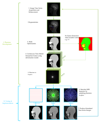
Realistic dynamic speech numerical phantom for the evaluation of real-time MRI acquisition and reconstruction methods
Joseph Martin, Redha Boubertakh, Matthieu Ruthven, Marc Miquel
Real time MRI (rtMRI) in human speech is an active field of research, with a particular clinical focus on the assessment of speech disorders. In this work, a numerical phantom is developed to allow acquisition and reconstructions schemes for rtMRI to be compared to a ‘gold standard’. Previously acquired 2D rtMRI images of speech were used to create anatomical masks of various speech organs. An interpolation method was then used to create a continuous time model of the moving structures, which forms the dynamic phantom. The model is then tested using different k-space sampling schemes (Cartesian, radial and spiral).
|
 |
3178.
 |
104 |
Improved susceptibility-weighted imaging of the thalamic nuclei at 7T with enhanced contrast and venous vessel exclusion
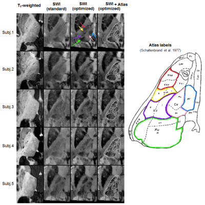
João Jorge, Elena Najdenovska, Constantin Tuleasca, José Marques, Marc Levivier, Philippe Maeder, Rolf Gruetter, Meritxell Bach Cuadra
The thalamus plays a key role in neuronal signal transmission and modulation. While most of the current non-invasive imaging techniques fail to achieve substantial contrast between the thalamic nuclei, susceptibility-weighted imaging (SWI) has recently shown promising capabilities for their visualization at 7T, despite having been originally designed primarily for venous vessel imaging. The aim of the present work was to optimize the SWI technique specifically for improved thalamic nuclei visualization, by jointly modifying the SWI combination and suppressing venous vessels. These modifications yielded substantially improved contrast and delineation of various thalamic nuclei, in good agreement with histological atlas information.
|
|
3179.
 |
105 |
$$$T_1$$$ dependency of magnetization transfer effect in human brain
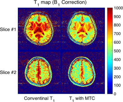
Yuki Kanazawa, Toshiaki Sasaki, Hiroaki Hayashi, Kotaro Baba, Ikuho Kosaka, Yuki Matsumoto, Mitsuharu Miyoshi, Masafumi Harada
The purpose of this study is to develop a T1 mapping method derived from the variable flip angle with an MT pulse. T1 mapping of the brain with an MT pulse was performed in five healthy subjects. The mean T1_MT values were significantly decrease than the T1 in all regions (P < 0.05). The difference of T1 and T1_MT in deep gray matter (included caudate nucleus and putamen) were more decreased than those in white matter. In conclusion, determination of T1 with MT pulse makes it possible to obtain more detailed information of the macromolecular pool and the free water pool.
|
|
3180.
 |
106 |
Utility of real-time field control in subjects with mild cognitive impairment: T2* weighted imaging at 7T.
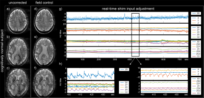
Laetitia Vionnet, Jiri van Bergen, Yolanda Duerst, Rafael Meyer, Nicole Fichtner, Sonja Kagerer, Michael Wyss, Paul Unschuld, Klaas Pruessmann
Subjects with Alzheimer disease or mild cognitive impairment are known to produce strong field perturbation by particular breathing pattern and limb motion. In this work we explore the feasibility and success of real-time field control in subjects with mild cognitive impairment and age-matched controls in the scenario of high resolution T2*-weighted imaging at 7T. Real-time field control shows to be feasible and yield greatly enhanced data quality in both type of subjects: mild cognitive impairment and cognitively normal.
|
|
3181.
 |
107 |
Multi-echo OxFlow for quantification of the cerebral metabolic rate of oxygen at 1.5T, 3T, and 7T
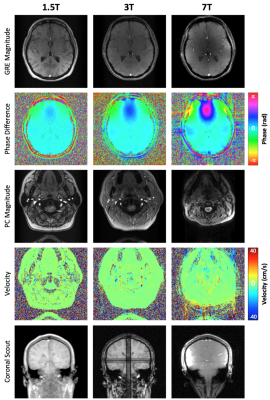
Erin Englund, Ana Rodriguez-Soto, Felix Wehrli
Fick’s principle can be used to quantify the cerebral metabolic rate of oxygen (CMRO2) as the product of oxygen extraction and blood flow. An interleaved dual-slice multi-echo GRE and phase contrast sequence, termed OxFlow, has previously been used for simultaneous measurement of SvO2 and blood flow. Here, we developed and evaluated an extended multi-echo OxFlow sequence designed for operation at multiple field strengths. The rate of phase accumulation, rather than the inter-echo phase difference, is used to compute SvO2. Results were obtained in 5 healthy subjects at 1.5T, 3T, and 7T, with agreement of the physiologic parameters between field strengths.
|
|
3182.
 |
108 |
Comparing MEGA editing techniques for in-vivo measurement of 2-hydroxyglutarate
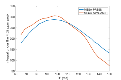
Ross Callaghan, Bhavana Solanky, Sotirios Bisdas, Hui Zhang, Enrico De Vita
MEGA-semiLASER is compared to a previously proposed MRS sequence for 2HG detection, MEGA-PRESS. The sequences are assessed using simulations of SNR with TE and the chemical shift displacement error (CSDE). Both sequences are shown to maximise 2HG SNR at TE of approximately 100ms. MEGA-semiLASER displays marginally higher SNR whilst reducing the CSDE of MEGA-PRESS. MEGA-semiLASER should offer a comparable reduction in CSDE to another proposed sequence, MEGA-LASER, whilst requiring fewer refocusing pulses and thus a lower SAR.
|
|
3183.
 |
109 |
A novel minimally invasive, image-guided neurablation technique using MRI - MINIMA
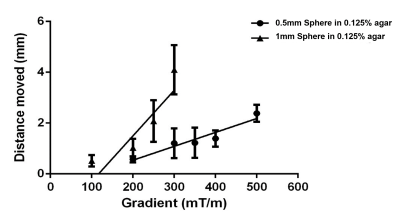
Christopher Payne, John Connell, Matin Mohseni, Stephen Patrick, Yichao Yu, Bernard Siow, Quentin Pankhurst, Mark Lythgoe
During tumour resection the goal is to remove a discrete region of cancerous tissue causing minimal damage to surrounding healthy tissue. Therefore, there is a demand for minimally invasive techniques, alongside MR imaging, for precise location of the tumour boundary. Presented here is the development of a minimally invasive, image-guided neurosurgery technique, whereby the position of an untethered surgical implant can be controlled and imaged in real time using an MRI scanner. We have demonstrated image-guided, precise movement of millimetre sized magnetic spheres inside ex vivo brain tissue by controlling the magnetic field gradients inside an MRI scanner.
|
|
3184.
 |
110 |
NODDI-DTI as proxy for Axonal Volume Fraction: Is g-ratio-weighted imaging feasible using single-shell DTI data?
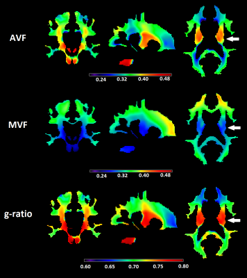
Gergely David, Maryam Seif, Patrick Freund
G-ratio-weighted imaging is an active field of research with the goal of better characterizing white matter in both health and disease. However, clinical adoption is significantly hampered by the fact that most g-ratio protocols rely on time-intensive multi-shell diffusion data which is typically not available in clinical settings. In this study, we adopted the recently introduced NODDI-DTI in combination with magnetization transfer saturation to calculate g-ratio maps based on a single diffusion shell in healthy subjects. The so-acquired g-ratio maps greatly resembled maps from the literature and had high scan-rescan repeatability, which has great implications for clinical g-ratio-weighted imaging.
|
|
3185.
 |
111 |
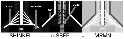
MR Myelo-Neurography: Improved Visualization of MR Neurography in the Brachial Plexus using a Combination of SHINKEI and Phase-Cycling balanced SSFP
Hitoshi Tadenuma, Kayoko Abe, Masami Yoneyama, Yasuhiro Goto, Isao Shiina, Mamoru Takeyama, Isao Tanaka, Shuji Sakai
The brachial plexus could be involved in various kinds of diseases, which may lead to serious functional disorders. MR neurography is a useful technique for evaluating the abnormal state of the peripheral nerves, however, it is still difficult to visualize the entire brachial plexus, including nerve roots, using conventional MR neurography due to its complicated anatomical structure. In this study, we evaluated a new MR neurography using a combination of SHINKEI and Phase-Cycling balanced SSFP sequence to visualize the entire brachial plexus.
|
|
3186.
 |
112 |
Nonlocal multispectral image filtering to improve determination of cerebral blood flow from pseudo-continuous arterial spin labeling imaging
Video Permission Withheld
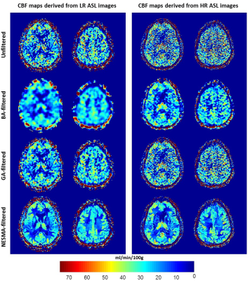
Mustapha Bouhrara, Diana Lee, Abinand Rejimon, Richard Spencer
Changes in the cerebral blood flow (CBF), measured using arterial spin labeling (ASL), are an emerging biomarker for normal aging, Alzheimer's disease, and other neurodegenerative conditions. However, ASL signal-to-noise ratio (SNR) is inherently low, diminishing the quality of CBF determination. While attempts have been made to improve SNR in ASL images using post-processing filters, performance is limited and several user-defined parameters are required adding further complexity in implementation. Here, we introduce a simple, novel filtering algorithm and demonstrate its potential to enhance the quality of CBF mapping.
|
|
3187.
 |
113 |
Rapid Myelin Measurement: Comparison Between SyMRI (Simultaneous Tissue Relaxometry), Magnetization Transfer Saturation Index, and T1w/T2w Ratio Methods
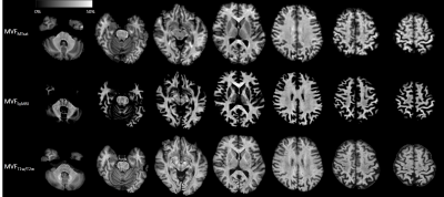
Akifumi Hagiwara, Masaaki Hori, Koji Kamagata, Misaki Nakazawa, Christina Andica, Tomoko Maekawa, Saori Koshino, Ryusuke Irie, Lydia Chougar, Osamu Abe, Shigeki Aoki
In 20 healthy adults, we examined the correlation between three rapid MR myelin measurement methods, including simultaneous tissue relaxometry of R1 and R2 relaxation rates and proton density (SyMRI), magnetization transfer saturation (MTsat) index, and the ratio of T1-weighted to T2-weighted images (T1w/T2w ratio). Even though SyMRI and MTsat showed strong correlation in the white matter, only weak to moderate correlation was found between T1w/T2w and SyMRI or MTsat. In conclusion, SyMRI and MTsat seem to be suitable for evaluating myelin in the white matter, but T1w/T2w ratio may be less optimal.
|
|
3188.
 |
114 |
Interleaved Multi-Slice Averaged Magnetization Inversion Recovery Acquisitions (imsAMIRA) for Fast Spinal Cord Imaging
Video Permission Withheld
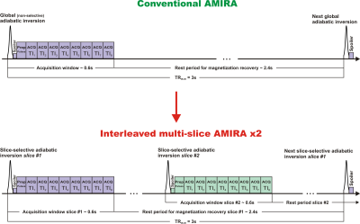
Matthias Weigel, Zarko Celicanin, Oliver Bieri
To increase the acquisition efficiency for the averaged magnetization inversion recovery acquisitions (AMIRA) approach for spinal cord imaging, an interleaved multi-slice AMIRA implementation was developed. Utilizing a slice-selective adiabatic inversion pulse with optimized slice thickness, the interleaved multi-slice AMIRA sequence provides spinal cord imaging with the same high gray matter and white matter contrast and very similar image quality like the conventional AMIRA approach, however, in considerably reduced scan times per slice.
|
|
3189.
 |
115 |
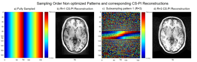
Sampling order optimization preserves contrast and improves clinical diagnostic utility of accelerated prospective 3D brain MRI: a radiological assessment study on healthy volunteers
Arnold Julian Vinoj Benjamin, Wajiha Bano, Grant Mair, Michael Davies, Ian Marshall
This study shows the importance of sampling order optimization for the contrast preservation of accelerated prospective 3D MRI leading to the improvement in clinical diagnostic utility of accelerated scans using compressed sensing and parallel imaging reconstructions.
|
|
3190.
 |
116 |
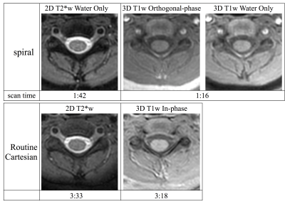
Axial Gradient Echo Spiral MRI of the Pediatric Spine
Ryan Robison, Melvyn Ooi, Amber Pokorney, Zhiqiang Li, Dinghui Wang, James Pipe, Jeffrey Miller
MRI spine examinations are an important tool in pediatric care. Long acquisition times and artifacts related to motion and flow are a regular challenge in these examinations. Spiral MRI offers substantial potential benefits with regards to both acquisition time and artifact reduction. This work studies spiral MRI for gradient echo imaging of the pediatric spine and demonstrates promising results in preliminary patient data. The results from a contrast and SNR optimization study in a volunteer are also presented.
|
|
3191.
 |
117 |
Improved brain MR imaging from a compact, lightweight 3T scanner with high performance gradients.
Norbert Campeau, Yunhong Shu, Joshua Trzasko, Erin Gray, Thomas Foo, Matt Bernstein, John Huston
A compact, low-cryogen 3T MRI scanner with high-performance gradients capable of simultaneously achieving 80 mT/m and 700 T/m/s was compared to a 60-cm, whole body 3T system (50 mT/m, 200T/m/s) for 5 routine brain MR imaging sequences in 9 clinical patients, graded by two neuroradiologists. The compact 3T system performed equally well to a standard whole-body scanner in terms of motion artifacts, and performed better in terms of signal-to-noise ratio, lesion conspicuity, gray/white contrast, susceptibility artifacts and overall exam quality.
|
|
3192.
 |
118 |
Dynamic imaging of hyperpolarized xenon-129 uptake in the human brain with spiral MRI
Madhwesha Rao, Guilhem Collier, Rolf Schulte, Graham Norquay, Jim Wild
In this study we explore the use of continuous 2D spiral k-space sampling to dynamically image the uptake of inhaled hyperpolarized 129Xe dissolved in human brain tissue. The sliding window reconstruction enables the monitoring of xenon uptake dynamics in the gray matter at high temporal resolution. Dynamic 129Xe brain MRI may be useful in pathologies related to cerebral perfusion and may provide insight in to blood brain barrier permeability.
|
|
3193.
 |
119 |
Comparison between 2D and 3D MEDIC in human cervical spinal cord at 3T
Abdullah Asiri, Franky Dimpudus, Aiman Alnajjar, Katie McMahon, Nyoman kurniawan
High-resolution MRI of the cervical spinal cord is important to provide accurate diagnosis and pathological assessment of injuries. MEDIC (Multiple Echo Data Image Combination) sequence appears promising for use in clinical imaging, however the comparison in the performance of two-dimensional (2D) and three-dimensional (3D) MEDIC sequences for spinal cord imaging has not been reported. This study aims to compare axial 2D and 3D MEDIC sequence for the visualization of the grey matter (GM) and white matter (WM) of the human cervical spinal cord.
|
|
3194.
 |
120 |
Correcting a slice distortion artifact in the multiband diffusion images by postprocessing with the known diffusion gradients
Video Permission Withheld
Jiancheng Zhuang
The diffusion weighted images acquired with the multiband sequence or the Lifespan protocols shows a type of slice distortion artifact. This artifact is caused by the eddy currents, which can be induced by the diffusion gradient associated with either the current DW image or the previous DW images. The artifact can be corrected by a correction algorithm which includes the diffusion gradients from both the current and previous DW images.
|
|
Brain Morphometry
Electronic Poster
Neuro
Monday, 18 June 2018
| Exhibition Hall |
09:15 - 10:15 |
| |
|
Computer # |
|
3243.
 |
49 |
Brain morphometry using diffusion MRI data (DTBM) reveals abnormalities in Down Syndrome that are not detected by conventional DTI analysis.
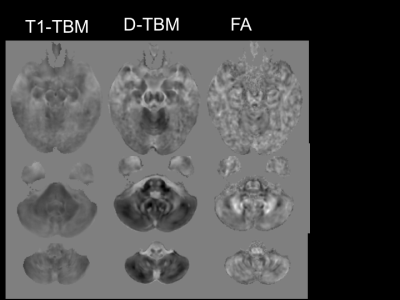
Carlo Pierpaoli, Amritha Nayak, Okan Irfanoglu, Neda Sadeghi, Nancy Raitano-Lee
We performed Tensor Brain Morphometry (TBM) as well as conventional FA analysis to compare the brains of subjects with Down Syndrome (DS) to typically developing Healthy Controls (HC). TBM deformation fields were computed from T1 weighted images (T1-TBM), as well as from diffusion data (D-TBM). D-TBM identifies differences between DS and HC brains that would have gone completely undetected by conventional TBSS analysis of FA results.
|
|
3244.
 |
50 |
An Automatic Classification of Alzheimer’s Disease Based on Structural MRI Data Compared with Voxel-Based Morphometry Method
Did Not Present
Xiangzhu Zeng, Huishu Yuan, Yan Liu, Ling Wang, Ying Liu, Zheng Wang, Lizhi Xie
In this study, a compartmental sparse feature selection method was used with feature parameter identified, and compared with classical voxel-based morphometry method (VBM) for classification of Alzheimer ’s disease (AD) from the healthy subjects. Our method had high classification accuracy for AD diagnosis and a strong linear correlation between the extracted feature parameter and volume of hippocampus obtained by VBM. The feature parameter of hippocampus had a higher linear correlation with mini-mental state examination (MMSE) score than volume of hippocampus with MMSE. Hence, compartmental sparse feature selection is an effective computer-aided diagnosis method to help clinician identify AD.
|
|
3245.
 |
51 |
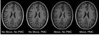
Accuracy of Morphometry Measures from MPRAGE data with Prospective Motion Correction Based on an Optical Tracking System
Joelle Sarlls, Francois Lalonde, J Andrew Derbyshire, Sean Marrett, Patrick Hucker, Maxim Zaitsev, S Lalith Talagala
Subject motion during MRI results in poor image quality and may cause bias in the morphormetric measures extracted from segmentation algorithms. Prospective motion correction (PMC) techniques can mitigate these effects by tracking brain motion and updating the scan parameters in realtime. Here, we compared the accuracy of cortical thickness and volume extracted from MPRAGE data of non-moving and intentionally moving subjects when using a PMC method based on a Moire phase tracking marker and an optical system. Data show that the PMC method used here can greatly reduce image artifacts and provide more accurate segmentation resuIts during intentional motion.
|
|
3246.
 |
52 |
Brain volumetric and fractional anisotropy differences in mice selected for high and low empathy-like traits
Diana Cash, Tobias Wood, Francesca Zoratto, Simone Macri, Camilla Simmons, Eugene Kim, Steve Williams, Jeffrey Glennon, Giovanni Laviola
High resolution ex vivo imaging of mice with high and low empathy-like behavior revealed widespread volumetric and fractional anisotropy (FA) changes. Low empathy mice had decreased volumes of the dorsal and ventral hippocampi, periaqueductal grey and the cerebellar cortex, and increased volumes of the olfactory bulb and the hypothalamus compared to high empathy mice. FA was decreased in the low empathy group, specifically in the hippocampus and in the periaqueductal grey. Functional significance can be inferred as these affected brain circuits mediate olfactory cues-based communication of pain, predatory odor fear responses and autonomic stress responses.
|
|
3247.
 |
53 |
Multicentric test-retest reproducibility of human hippocampal volumes: FreeSurfer 6.0 longitudinal stream applied to 3D T1, 3D FLAIR and high-resolution 2D T2 structural neuroimaging
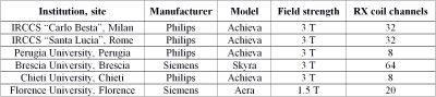
Andrea Chiappiniello, Roberto Tarducci, Cristina Muscio, Giovanni B. Frisoni, Maria Grazia Bruzzone, Marco Bozzali, Daniela Perani, Pietro Tiraboschi, Anna Nigri, Claudia Ambrosi, Massimo Caulo, Elena Chipi, Stefano Chiti, Enrico Fainardi, Stefania Ferraro, Cristina Festari, Roberto Gasparotti, Andrea Ginestroni, Giovanni Giulietti, Lorella Mascaro, Riccardo Navarra, Valentina Nicolosi, Lucilla Parnetti, Cristina Rosazza, Laura Serra, Fabrizio Tagliavini, Jorge Jovicich
This study evaluates across-session test-retest reproducibility of automatic hippocampus subfields segmentation. A customized acquisition protocol was designed to enhance segmentation reliability in FreeSurfer 6.0 longitudinal analysis stream. Images were processed performing a within-session T1 averaging, using FLAIR images for PIAL surface reconstruction and a high-resolution T2 for the hippocampal subfield segmentation. Results on 12 healthy subjects suggest high reproducibility for different hippocampal subfields and whole hippocampus, generally better than those achievable without T1 averaging and without using FLAIR and high-resolution T2 images.
|
|
3248.
 |
54 |
Regularized k-means clustering for segmentation of brain tissues using hemodynamic features in DSC-MRI
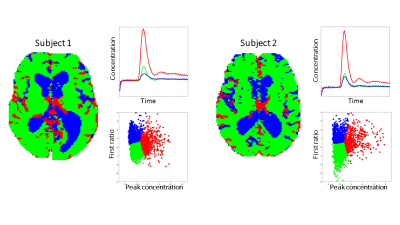
Jonathan Arvidsson, Oscar Jalnefjord, Fredrik Kahl, Magnus Båth, Göran Starck
Inclusion of voxels containing CSF and/or blood vessels can bias ROI statistics used in DSC-MRI analysis. In order to address this problem we propose an automatic method for tissue segmentation based on hemodynamic features obtained from DSC-MRI data. Application of the method in test subjects shows promising results.
|
|
3249.
 |
55 |
Brain extraction and segmentation framework for bias field rich cranial MRI scans of rats
Jacob Hansen, François Lauze, Sune Darkner, Kristian Mortensen, Simon Sanggaard, Helene Benveniste, Maiken Nedergaard
This abstract presents a framework to extract brain tissue and internal Cerebrospinal fluid in cranial magnetic resonance imaging of rats with strong bias fields. Desired segments are obtained through bias field correction and several passes of segmentation. A refinement procedure is proposed to remove brain surface CSF. Promising planar and 3D visualizations of results are presented and demonstrate the capabilities of the framework.
|
|
3250.
 |
56 |
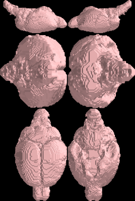
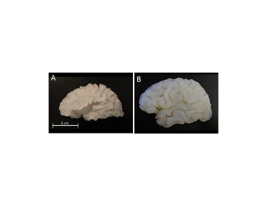
A 3D-printed anatomical multimodal phantom for brain segmentation validation
Anna Altermatt, Francesco Santini, Xeni Deligianni, Stefano Magon, Till Sprenger, Ludwig Kappos, Philippe Cattin, Jens Wuerfel, Laura Gaetano
Brain tissue segmentation algorithms applied on magnetic resonance imaging (MRI) data lack a ground truth for evaluating their performance. For this purpose, an anatomical brain phantom prototype mimicking T1 relaxation times and the complex 3D geometry of the human brain was created for use with MRI and computed tomography (CT). A scan-rescan experiment showed a low within-session variability of white matter (WM) and grey matter (GM) volumes when MRI images of the phantom were segmented with a commonly used software. Compared to the ground truth volumes derived from CT, the software overestimated the WM, while the GM was slightly underestimated.
|
|
3251.
 |
57 |
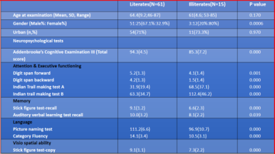
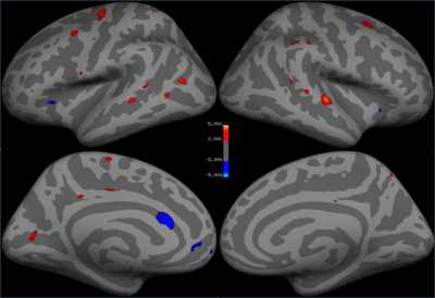
Multiparametric MRI characterization of microstructural substrate of literacy: Observations from a community cohort of literate and illiterate elderly in India
Kenchaiah Raghavendra, Alladi Suvarna , Jala Sireesha, Mekala Shailaja , Bapi Raju
Few studies have evaluated neuroanatomical differences between illiterates and literates and majority have been unimodal investigations. We explore multiparametric, neuroanatomical substrates of literacy by studying grey and white matter microstructural neuroimaging biomarker differences between 61 literate and 15 illiterate elderly subjects with normal cognition. Univariate and multivariate methods for grey matter density, diffusion tensor parameters and cortical/subcortical morphometric measures were employed. Literacy provided an advantage through increase in grey matter density, white matter integrity, cortical thickness, area and volumes in brain areas related to reading, writing, language, Visuospatial and sensorimotor processes. Enhanced white matter integrity was the most discriminating factor on the machine learning.
|
|
3252.
 |
58 |
Alterations in Cortical Thickness with Hydroxyurea Therapy in Children Treated for Sickle Cell Anemia
John Glass, Kathleen Helton, Wilburn Reddick
Sickle cell anemia is a devastating hematological disease leading to brain injury and neurocognitive deficits. Twenty-five patients (16 Hydroxyurea treated; 9 not) were imaged twice one year apart. Cortical thickness was assessed using FreeSurfer and compared between groups. Treated patients had thicker cortex in frontal and bilateral parietal lobes and in superior temporal and bankssts regions. Preservation of cortex in these regions may have implications for neurocognitive functions supported by associated networks such as central executive, attention and memory networks. These findings should be further evaluated for their impact on neurocognitive performance and Diffusion Tensor Imaging.
|
|
3253.
 |
59 |
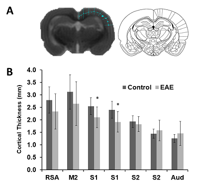
Alterations in cortical thickness and axonal density due to Experimental Autoimmune Encephalomyelitis in the Lewis Rat
J. McCreary, Brietta Gerrard, L. Truica, Gerlinde Metz
The animal model of experimental autoimmune encephalomyelitis (EAE) is characterized by in?ammatory lesions and demyelination which leads to axonal damage and subsequent neuronal death within the central nervous system. The effect is debilitating, resulting in loss of motor and sensory functions. Here, we investigated changes in cortical thickness using MRI, and axonal density in the corpus callosum using a neuronal tract tracer, biotinylated dextran amine, in Lewis rats induced with EAE. Our study found that EAE leads to a decrease in cortical thickness, particularly in the primary somatosensory trunk region, and axonal density in the corpus callosum.
|
|
3254.
 |
60 |
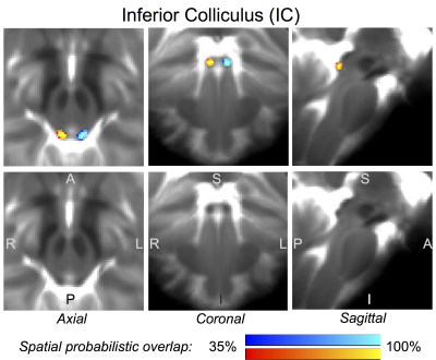
In-vivo probabilistic structural atlas of the inferior colliculus, superior colliculus, medial geniculate nucleus, and lateral geniculate nucleus based on 7 Tesla MRI
Christian Strong, Nicola Toschi, Bruce Rosen, Lawrence Wald, Marta Bianciardi
Brainstem and thalamic nuclei such as the inferior-colliculus, superior-colliculus, lateral-geniculate-nucleus, and medial-geniculate-nucleus modulate visual/oculo-motor and auditory/auditory-motor functions. Dysfunction of these nuclei is implicated in disease states such as auditory-agnosia, pure-word deafness, eye-movement and visual-field deficits, Parkinson’s hallucinations, and glaucoma. However, a stereotaxic probabilistic atlas of these nuclei in humans does not exist. We used segmentation of 1.1mm-isotropic 7Tesla T2-weighted- and diffusion-fractional-anisotropy-images to generate and validate an in-vivo probabilistic neuroimaging-based structural atlas of these nuclei in stereotaxic-MNI space. We constructed this atlas to aid the localization of these nuclei in conventional images for future research and clinical investigations of visual/auditory functions.
|
|
3255.
 |
61 |
Dual Fully Convolutional Networks for Multiscale Context based Robust MRI Skull Stripping
Pascal Ceccaldi, Benjamin Odry, Boris Mailhe, Mariappan Nadar
Brain Segmentation is a standard preprocessing step for neuroimaging applications, but can however be subject to differences in MR acquisition that can lead to added noise, bias field and / or partial volume effect. To address those protocol differences, we therefore present a generic supervised framework, using consecutively two deep learning networks, to produce a fast and accurate brain extraction aimed at being robust across MR protocol variations. While we only trained our network on Human Connectome Project 3T dataset, we can still achieve state-of-the-art results on1.5T cases from LPBA dataset.
|
|
3256.
 |
62 |
Precision of Manual vs. Automated Corpus Callosum Atrophy Measurements in Multiple Sclerosis
Michael Platten, Katarina Fink, Juha Martola, Tobias Granberg
Corpus callosum atrophy is a favorable imaging biomarker in MS. Manual measurements of the corpus callosum are considered the best current standard, but their repeatability and reproducibility are uncertain. FreeSurfer is an automatic software that can volumetrically measure the corpus callosum. Using a representative cohort of 9 MS patients, scanned twice with repositioning on 3 different MRI scanners, we compared the manual and automatic measurements of corpus callosum. We found the longitudinal FreeSurfer method to be the most precise method. Thus, we recommend that this method be used in future studies of measuring corpus callosum atrophy in patients with MS.
|
|
3257.
 |
63 |
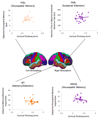
Morphological Correlates of Objective and Subjective Cognitive Control in Healthy Adults
Adam Clemente, Emma Lawrence, Phoebe Imms, Derek Jones, Karen Caeyenberghs
There is evidence showing the neural basis of cognitive control utilizing morphological measures of the brain in healthy adults. In the present study, we complement and extend on previous voxel-based morphometry based research by utilizing the more specific brain macrostructure metric of cortical thickness to investigate the differential morphological correlates of objective and subjective cognitive control. Here, we used a rigorous cognitive control test battery implemented on 25 healthy adults. Further research on morphological correlates of objective and subjective cognitive control in healthy populations is necessary to provide baseline data for future clinical populations.
|
|
3258.
 |
64 |
Hippocampal subfield analysis at 7 tesla in young adults with Down syndrome
Katherine Koenig, Sehong Oh, Melissa Stasko, Emma Lissmore, Elizabeth Roth, Anne Birnbaum, Thomas Scheidemantel, Hudson Taylor, Nancy Roizen, Stephen Ruedrich, Mark Lowe, Alberto Costa
This work uses ultra-high resolution at 7 tesla to assess hippocampal subfield volume in young adults with Down syndrome (DS). As compared to matched controls, individuals with DS show decreases in total hippocampal volume and in select regions, particularly on the left.
|
|
3259.
 |
65 |
Gray matter alterations in childhood obesity
Video Permission Withheld
Gergely Orsi, Gabor Perlaki, Gergely Darnai, Denes Molnar, Peter Bogner, Jozsef Janszky
|
|
3260.
 |
66 |
A 3D Convolutional Neural Network for Hippocampal Volume Estimation
Luca Schmidtke, Ricardo Corredor-Jerez, Jonas Richiardi, Bènèdicte Marèchal, Alexis Roche, Tobias Kober
Accurate estimation of hippocampal volume is essential for exploiting its sensitivity to pathological changes caused by Alzheimer’s disease (AD) and other forms of dementia. We built and trained a 3D convolutional neural network for fast and accurate segmentation of the hippocampus in T1-weighted structural MR images of the brain. Compared to two software packages (MorphoBox prototype and FreeSurfer), we achieved good disease classification results based on estimated hippocampal volume in a significantly shorter amount of time.
|
|
3261.
 |
67 |
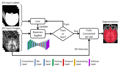
Bayesian Convolutional Neural Network Based Nonhuman Primate Brain Extraction in Fully Three-dimensional Context
Gengyan Zhao, Fang Liu, Jonathan Oler, Mary Meyerand, Ned Kalin, Rasmus Birn
Brain extraction of MR images is an essential step in neuroimaging, but current brain extraction methods are often far from satisfactory on nonhuman primates. To overcome this challenge, we propose a fully-automated brain extraction framework combining deep Bayesian convolutional neural network and fully connected three-dimensional conditional random field. It is not only able to perform accurate brain extraction in a fully three-dimensional context, but also capable of generating uncertainty on each prediction. The proposed method outperforms six popular methods on a 100-subject dataset, and a better performance was verified by different metrics and statistical tests (Bonferroni corrected p-values<10-4).
|
|
3262.
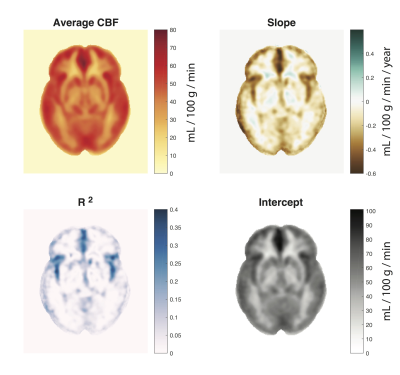 |
68 |
Cerebrovascular Brain Aging Examined with Arterial Spin Labelling and Applied to Age Prediction
M. Ethan MacDonald, Nils D. Forkert, Yuhan Ma, Rebecca J. Williams, Alexandru Hanganu, Hongfu Sun, Randall Stafford, Cheryl R. McCreary, Richard Frayne, G. Bruce Pike
Changes in both cortical thickness and cerebral blood flow are observed with age. In this work, we look at how these parameters are modulated across the lifespan. T1-weighted and arterial spin labelling data from 146 subjects were analyzed, with 68 cortical regions selected in each subject to obtain mean cortical thickness and cerebral blood flow. We calculated rates of change, correlation, and laterality for both parameters. Finally, we explored predictive modeling using cortical thickness, CBF and a model combining the two. Predictive modelling was slightly improved when both measures were included.
|
|
3263.
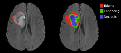 |
69 |
Evaluation of tumor shape features for overall survival prognosis in glioblastoma multiforme patients
Parita Sanghani, Ang Ti, Nicolas King, Hongliang Ren
We evaluated 13 shape features of glioblastoma multiforme (GBM) tumor for overall survival (OS) prognosis in 75 patients using univariate and multivariate Cox regression analysis. Age and Karnofsky performance scale were used as covariates for the multivariate analysis. Three shape features were found to be significant for OS prognosis in GBM patients. Kaplan-Merier survival curves were obtained for the significant features to illustrate their effectiveness. In future works, these shape features can be used along with volumetric and texture features derived from the tumor for OS prediction of GBM patients.
|
|
3264.
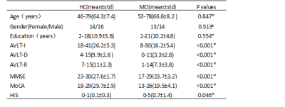 |
70 |
Investigation of Cerebral Structural Alterations of Patients with Mild Cognitive Impairment based on VBM and DBM Analysis
Did Not Present
Zhe Ma, Zhizheng Zhuo, Lijiang Wei, Bin Jing, Haiyun Li, Yingjie Mei
Voxel-based morphometry (VBM) and deformation-based morphometry (DBM) are wildly-used automated analysis pipelines for neuro-structural images. This study aims to investigate gray matter abnormalities in mild cognitive impairment (MCI) patients. Some altered brain regions could be detected with VBM and DBM method for MCI patients compared to healthy controls. Besides, the features extracted from VBM and DBM could effectively identify the MCI from healthy controls.
|
|
3265.
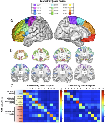 |
71 |
Rostro-caudal architecture of the frontal lobes in humans
Did Not Present
Michel Thiebaut de Schotten, Marika Urbanski, Leonardo Cerliani, Emmanuelle Volle
Functional models of the frontal lobes suggest a rostro-caudal organization that is essential for goal-directed behaviour and cognitive control, in which higher processing-level anterior regions send control signals to lower processing-level posterior regions. Here we show that tractography can divide the frontal lobes into 12 regions organized in a rostro-caudal axis showing a gradient of cortical thickness, myelination and cell body density.
|
|
3266.
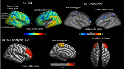 |
72 |
Volume and surface-based structural analyses in active Crohn’s Disease Patients
Gita Thapaliya, Sally Eldeghaidy, Shellie Radford, Gordon Moran, Susan Francis
Structural brain changes in Crohn’s Disease (CD) have been studied in remission, however the data is inconsistent and not assessed in the active disease state. Grey matter volume (GMV) and cortical thickness (CT) were measured using VBM and CAT12/FreeSurfer in 25 active CD patients and age-matched healthy controls (HC). CD patients showed reduced CT and GMV in frontal and motor areas. Both CT and GMV were negatively correlated with proinflammatory markers, indicating these changes could be due to chronic inflammatory response. CAT and Freesurfer measures were highly comparable.
|
|
Functional & Structural Connectivity in the Brain
Electronic Poster
Neuro
Monday, 18 June 2018
| Exhibition Hall |
09:15 - 10:15 |
| |
|
Computer # |
|
3267.
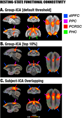 |
73 |
Investigation of the default mode network in awake marmosets
Cirong Liu, Cecil Yen, Frank Ye, David Leopold, Afonso Silva
We investigated the default mode network (DMN) in awake marmosets, by combining both resting-state and task-based fMRI, and by performing diffusion tractography and neuronal tract tracing. We found that dorsolateral (dlPFC) but NOT medial prefrontal cortex (mPFC) is a major region of the marmoset DMN. The dlPFC has direct anatomical connections and strong functional connectivity with the posterior DMN and also exists in macaques and humans, suggesting that it is more evolutionary conservative than the mPFC. However, its functions as a default mode are poorly understood and worth investigation.
|
|
3268.
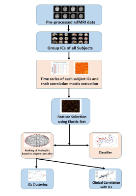 |
74 |
Resting state fMRI “Epilepsy networks”.
Rose Bharath, Rajanikant Panda, Jeetu Raj, Sanjib Sinha, Kenchaiah Raghavendra, Ravindranadh Mundlamuri, Ganne Chaitanya, Anita Mahadevan, Arivazhagan Arimappamagan, Malla Rao, Kandavel Thennarasu, Kaushik Majumdar, Parthasarathy Satishchandra, Tapan Gandhi
Resting state functional MRI (rsfMRI) research typically focuses on few well identified networks though many more networks (15-80) are often visualized, in the course of investigating functional networks. It is customary to discard these networks as they are presumed to have no functional relevance. We used machine learning methods to identify “epilepsy networks” in 45 individuals with TLE using FSL derived 88 independent components. In line with evidence from experimental models, the current results indicates that TLE is associated with disease specific “rsfMRI epilepsy networks” which can be visualised in-vivo at individual subject level.
|
|
3269.
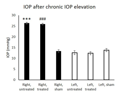 |
75 |
Effects of intraocular pressure elevation and oral citicoline treatment on resting state functional connectivity in the visual system
Yolandi van der Merwe, Matthew Murphy, Leon Ho, Xiaoling Yang, Yu Yu, Ying Chau, Christopher Leung, Kevin Chan
Glaucoma is a neurodegenerative disease that causes irreversible damage to the visual system. While elevated intraocular pressure (IOP) is a major risk factor, its neurobehavioral effects on the visual system remain unclear. Here we showed that increasing magnitude and duration of IOP elevation resulted in differential effects on the visuomotor behavior and resting-state functional connectivity between visual brain nuclei. In addition, under similar levels of chronic IOP elevation, oral citicoline treatment appeared to ameliorate visual behavioral deficits and functional connectivity decrease in some brain regions. These results suggest new potential mechanistic targets for treatment of glaucoma beyond IOP lowering.
|
|
3270.
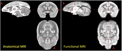 |
76 |
Construction of an MRI-Based Connectome for the Marmoset Brain: Methods and Initial Results
Cecil Yen, Cirong Liu, Afonso Silva
The common marmoset, a small New World primate, is an excellent translational animal model due to the similarity between its brain network and humans. However, marmoset’s brain networks are not fully explored because of lacking a large group of resting-state functional MRI (rfMRI) data, like human connectome project. To facilitate collecting rfMRI in awake marmosets across multiple marmoset MRI sites, an MRI protocol was developed and optimized for ultra-high filed small animal MRI. Preliminary results show high data quality and robust detection of brain networks such as default mode network. More data are being acquired and collaboration is desired for the success of marmoset connectome database.
|
|
3271.
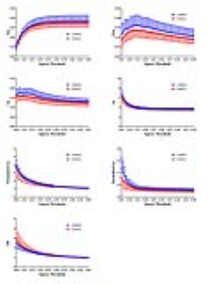 |
77 |
Disrupted Nodal Organization of brain functional Network in Children with Type I Gaucher Disease
Video Permission Withheld
Miao Zhang, Di Hu, Shengpei Wang, Huiying Kang, Yun Peng
In our study, we utilize graph-based network analysis to investigate the topological properties of resting-state brain functional connectivity in children with type I GD. Sixteen children diagnosed as type I GD and sixteen age- and sex- matched healthy controls were recruited. No alterations were found at global level. Three nodes within the executive control network showed decreased nodal degree or efficiency. Our provide new insight to further understand the neurophysiological change of these patients.
|
|
3272.
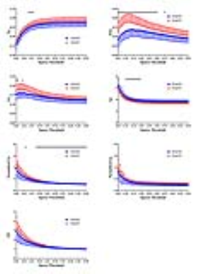 |
78 |
Altered Topological Properties of Brain Functional Networks in Type I GD Patients During a Near Five-year Follow-up
Video Permission Withheld
Di Hu, Miao Zhang, Shengpei Wang, Huiying Kang, Yun Peng
Our previous had shown altered nodal properties within the executive control network in children with type I GD. This time we further investigate the variation trend of brain topological properties in type I GD patients during a near five-year folloe-up under regular enzyme replacement. Results showed that the efficiency of functional segregation in brain network were increased and more nodes were involved and reorganized. The result may explain the improved mood and global functioning that have previous reported.
|
|
3273.
 |
79 |
Multimodal study of hyperbaric oxygenation effects on normal human brain metabolism and functional connectivity at 3 Tesla.
Andrei Manzhurtsev, Olga Vasiukova, Dmitry Kupriyanov, Victoria Sergeeva, Tolib Akhadov, Petr Menshchikov, Natalia Semenova
This study is aimed to reveal the effects of hyperbaric oxygenation (HBO) on human brain metabolites using 1H and 31P MRS, and functional connectivity using resting-state fMRI. The MRS and rs-fMRI studies were performed twice: before and after one HBO session (p=1.2 atmosphere, 100% O2). The results demonstrate the positive effect of hyperbaric oxygenation on human brain metabolism and functioning.
|
|
3274.
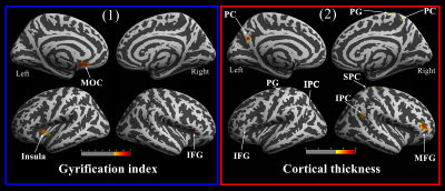 |
80 |
Brain structural plasticity associated with emotional processing in postpartum women: A longitudinal voxel-based morphometry study
Kaihua Zhang, Mengxing Wang, Xueyun Su, Jilei Zhang, Junyao Xie, Haifeng Lu, Zhong Chen*, Xiaoxia Du*
Pregnancy constitutes a significant period in women’s lives, after which they often experience numerous physiological and psychological changes. However, structural changes in the brains of postpartum women remain unclear. To investigate these phenomena, we recruited forty-seven postpartum women to participate in a longitudinal magnetic resonance imaging study. Our results first suggest that brain structures in postpartum women show adaptive plasticity, especially regarding alterations in empathy-related regions, including grey matter volume, white matter volume, and cortical thickness, that can facilitate effective adaption and behavioural and emotional adjustments towards nurturing infants.
|
|
3275.
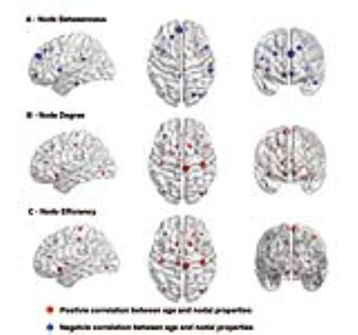 |
81 |
Age related changes in topological properties of brain functional network and structural connectivity
Chandan Shah, Jia Liu, Peilin Lv, Huaiqiang Sun, Yuan Xiao, Jieke Liu, Youjin Zhao, Wenjing Zhang, Li Yao, Qiyong Gong, Su Lui
Title of the abstract was written. The body of the abstract was within word limits. Acknowledgements were mentioned. one figure was uploaded with figure caption.
|
|
3276.
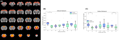 |
82 |
Resting state functional networks longitudinally correlate with working memory in a transgenic rat model of Alzheimer’s disease
Raúl Tudela, Emma Muñoz-Moreno, Xavier López-Gil, Guadalupe Soria
The TgF344-AD rats represent the most suitable and promising animal model for Alzheimer’s disease (AD) research. Resting-state functional MRI was longitudinally acquired every 3 months in a cohort of transgenic Tg344-AD and control Fisher rats between 5 and 15 months of age, together with cognitive task evaluation. Independent component analysis was applied to rs-fMRI volumes and 10 networks were anatomically identified. Spearman correlation coefficients between functional and cognitive parameters were computed. Our results show that while no differences were observed in the cognitive task between both groups, significant differences were found in the functional networks.
|
|
3277.
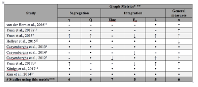 |
83 |
A meta-analysis of studies using graph theory in traumatic brain injury suggests inconclusive evidence for structural disconnectivity
Phoebe Imms, Adam Clemente, Derek Jones, Karen Caeyenberghs
The purpose of this meta-analysis is to investigate which graph metrics reliably represent changes to structural connectivity following Traumatic Brain Injury (TBI). Effect sizes were pooled for sixteen different graph metrics, from ten diffusion MRI articles that compared the structural connectome of TBI patients and healthy controls. Interestingly, no metric showed consistent differences across the studies. This analysis indicates that heterogeneity in samples are hindering progress towards clinically relevant disconnectivity biomarkers following brain trauma.
|
|
3278.
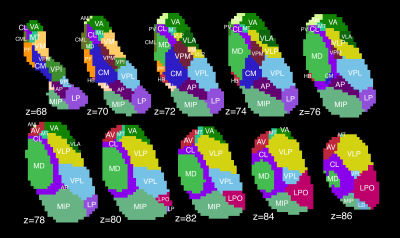 |
84 |
Structural and functional connectivity of the ipsilateral or nondecussating dentato-rubro-thalamic tract
Kalen Petersen, Jacqueline Reid, Srijata Chakravorti, Meher Juttukonda, Giulia Franco, Paula Trujillo-Diaz, Adam Stark, Benoit Dawant, Manus Donahue, Daniel Claassen
The dentato-rubro-thalamic tract (DRTT) is a cerebellar efferent pathway important to normal motor function and neurological disease. While the DRTT is described as a decussating (crossing) pathway, the existence of a nondecussating DRTT was recently demonstrated. We compared thalamic connectivity of decussating and nondecussating DRTT using both structural and functional MRI. Probabilistic tractography indicated that the two pathways contact distinct but partially overlapping sets of thalamic nuclei. These results were reinforced by significant correlations with functional connectivity. We conclude that the decussating and nondecussating DRTT exhibit different connectivity patterns, which suggests participation in divergent neural networks.
|
|
3279.
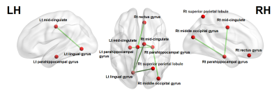 |
85 |
A Preliminary MR g-Ratio–Based Connectome Analysis in Multiple Sclerosis
Video Permission Withheld
Koji Kamagata, Andrew Zalesky, Kazumasa Yokoyama, Akifumi Hagiwara, Kouhei Kamiya, Maria Angelique Di Biase, Yuki Takenaka, Christina Andica, Asami Saito, Masaaki Hori, Keigo Shimoji, Ryusuke Irie, Akihiko Wada, Nobutaka Hattori, Shigeki Aoki
Multiple sclerosis (MS) is an inflammatory demyelinating disease of the central nervous system. We propose the use of g-ratio–based connectome for evaluating the network topology of MS since it is reported to be useful in the evaluation of demyelinating lesions in MS. Here, we evaluated the structural connectome of patients with MS, as mapped by MR g-ratio based connectome. The network-based statistic identified a subnetwork of reduced connectivity in patients with MS involving the limbic area. In conclusion, MR g-ratio–based connectome analysis can potentially detect changes in brain topology in MS with high sensitivity.
|
|
3280.
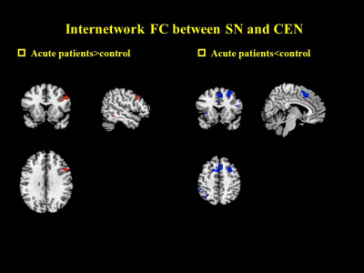 |
86 |
Temporal evolution of functional connectivity of the intrinsic networks in mild traumatic brain injury
Did Not Present
Zhuonan Wang, Lijun Bai, Qiuli Zhang, Guanghui Bai, Bo Yin, Xuan Niu, Yingxiang Sun, Ming Zhang
Mild traumatic brain injury (mTBI) often leads to disconnection of the brain intrinsic networks. Our study was to investigate how SN interacts with CEN/DMN can be viewed from the observation of how network connectivity damage affects other networks as a function of time, as well as its impact on cognition and emotion. The results suggests that intrinsic brain networks in mTBI patients showed continued damage from acute to sub-acute phase after injury. Function connectivity of SN interacts with CEN/DMN are especially susceptible in mTBI patients and the damage for inter-network functional disconnection will lead to high level cognitive dysfunction.
|
|
3281.
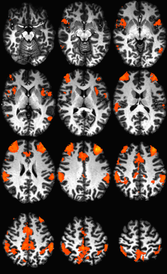 |
87 |
A preliminary report: Confirmation of the relationship of episodic memory performance and frontoparietal functional connectivity in Multiple Sclerosis
Katherine Koenig, Jian Lin, Daniel Ontaneda, Kedar Mahajan, Stephen Rao, Sanghoon Kim, Stephen Jones, Mark Lowe
This work assesses the relationship of resting state fMRI (rs-fMRI) of the dorsal lateral prefrontal cortex to episodic memory in patients with multiple sclerosis (MS). We find that rs-fMRI in the frontoparietal network is related to performance on a verbal episodic memory measure. This finding confirms previous reports and suggests this measure is appropriate to investigate as a potential predictive marker of cognitive decline.
|
|
3282.
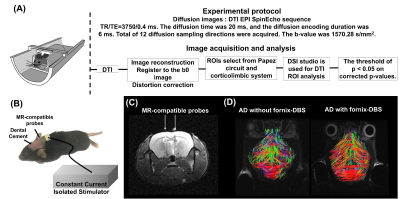 |
88 |
Diffusion MR Imaging of Structural Connectivity and Therapeutic Potential of Deep Brain Stimulation in Alzheimer’s Disease Model
Ching-Wen Chang, Yi-Chao Lee, Ssu-Ju Li, Ting-Chun Lin, Yin-Chieh Liu, You-Yin Chen, Yu-Chun Lo
Alzheimer’s disease (AD) is one of the major causes of death that currently cannot be reversed or slowed. Fornix, a major output tract of the hippocampus, has been shown to be a promising target for DBS therapy in AD patients. In this study, triple-transgenic Alzheimer’s mice were used to investigate the changes of white matter integrity and the cognitive functions after the DBS-fornix therapy. We found improvement of the cognition and increased white matter integrity after that DBS-fornix therapy in AD mice. It suggested that the DBS-fornix therapy may be a potential therapeutic intervention of AD.
|
|
3283.
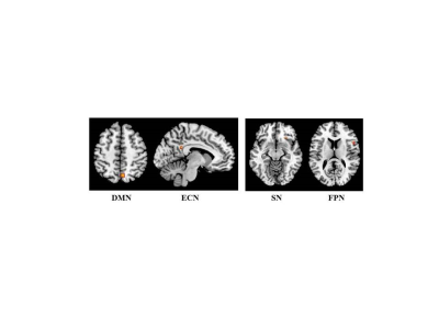 |
89 |
Modulation of cortico-subcortical functional connectivity occurs after symptomatic treatment of fatigue in patients with multiple sclerosis
Video Permission Withheld
Paola Valsasina, Bruno Colombo, Paolo Preziosa, Vittorio Martinelli, Andrea Falini, Giancarlo Comi, Massimo Filippi, Maria Rocca
In this study, 45 fatigued patients with multiple sclerosis (MS) were randomly assigned to undergo treatment with fampridine, amantadine or placebo and underwent clinical, neuropsychological and 3T resting state (RS) functional MRI at baseline and after four weeks of treatment. We found that treatment with fampridine (and, to a lesser extent, with amantadine) ameliorates fatigue in MS. Concomitant increase of RS functional connectivity (FC) in inferior frontal and parietal cortical regions, and decrease of abnormally high intra-thalamic FC were detected, suggesting an improved regulation of cortico-subcortical functional circuits.
|
|
3284.
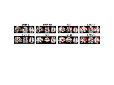 |
90 |
Relevance of functional connectivity abnormalities to cognitive impairment in neuromyelitis optica spectrum disoders
Video Permission Withheld
Paola Valsasina, Maria Rocca, Filippo Savoldi, Gianna Riccitelli, Marta Radaelli, Paolo Preziosa, Giancarlo Comi, Andrea Falini, Massimo Filippi
In this study, we explored resting state (RS) functional connectivity (FC) abnormalities of the main cognitive networks of patients with neuromyelitis optica spectrum disorders (NMOSD) and their correlation with cognitive impairment (CI). We found increased RS FC in the default mode, salience and working memory networks in cognitively preserved NMOSD, and decreased RS FC in the default mode and working memory networks in CI NMOSD. Increased RS FC seems to contribute to a better cognitive performance, probably reflecting an adaptive mechanism. Conversely, reduced RS FC is likely to be a maladaptive mechanism associated with impaired cognitive functions.
|
|
3285.
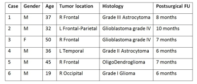 |
91 |
Longitudinal Evaluation of Post-Surgical Connectivity Changes of the Default Mode Network in Operated Glioma Patients
Domenico Zacà, Silvio Sarubbo, Monica Dalla Bona, Umberto Rozzanigo, Francesco Corsini, Giovanna Faraca, Franco Chioffi, Jorge Jovicich
In this study we evaluated the post-surgical changes in functional connectivity of the default mode network (DMN) in 6 operated glioma patients and assessed their relationship with pre-operative brain tumor size, a factor that pre-operatively has been negatively associated to cognitive performance. We found in all but one patient an increase in connectivity (average Z-score) of the DMN following brain tumor surgery. These changes were not associated with brain tumor volume, thus indicating that mechanisms other than reduction of mass effect may drive the post-surgical reorganization of the DMN.
|
|
3286.
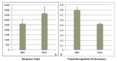 |
92 |
Short-term Visual Experience Increases Intrinsic Brain Connectivity Within Ventral Visual Pathway
Did Not Present
Bingqiang Xu, Hongmei Wang, Jiayin Tong, Xiaoyan Gao, Chenwang Jin, Ming Zhang
Medical imaging interpretation fundamentally lies in radiologists’ exceptional visual recognition skill, which enables the identification of pathological regions to render diagnosis1. Such expertise is obtained through training across review hundreds of cases2, facilitated by the plastic changes in the central visual system3. Specifically, ventral visual pathway (VTP) is responsible for visual object recognition, i.e. the fine-grained visual information processing. Visual information processing in the adult human brain is highly malleable with neural processing adapting to incoming information4. The plastic changes in the VTP in response to visual recognition tasks are well studied5. While we propose that the information embedded in the intrinsic brain activity, as revealed in the resting data, is also important6. Therefore, in the current study, we investigate how visual experience, i.e. short-term radiological training, modulates brain activity in the VTP under task-free state in the resting brain using ICA.
|
|
3287.
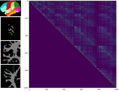 |
93 |
Individual patient-specific quantification of the network effects of lesions and diffuse disconnections with diffusion MRI
Christopher Steele, Yasser Iturria-Medina, Pierre-Louis Bazin, Bernhard Sehm, Arno Villringer
Even with its crucial role as the backbone of communication within the brain and susceptibility to damage from diseases such as stroke and multiple sclerosis, the white-matter connectional architecture of the human brain has been largely ignored. To address this, we have developed a whole-brain model for the quantification of white-matter connectional anatomy – the Tractography-based Lesion Assessment Standard (TractLAS) – which provides a complete connectional network description of the human brain. It can be used to quantify the impact of lesions on the connectivity of the brain within individuals, providing clinically useful information for individualised care.
|
|
3288.
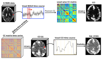 |
94 |
Normal aging differences in static and dynamic functional connectivity between men and women studied by quantitative data-driven analysis of R-fMRI data
Ninni Persson, Tie-Qiang Li
A growing number of resting-state fMRI (R-fMRI) studies have reported a significant effect of normal aging in resting-state functional connectivity (RFC) of the default mode network (DMN). One of the more intriguing findings is that gender can interact with normal aging effect to influence the RFC changes over lifespan. This adds to our understanding of gender-related cognitive differences and gender-specific nature of aging-related brain disorders. However, the precise interaction effects between sex and age on RFC remain inconclusive. The aim of the study is to quantify the normal aging induced RFC reduction difference between man and women.
|
|
3289.
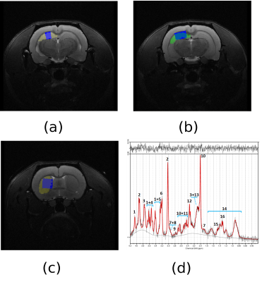 |
95 |
Brain metabolic and connectivity changes associated to the progression of Alzheimer's disease in a transgenic rat model
Emma Muñoz-Moreno, Rui Simoes, Raúl Tudela, Guadalupe Soria
TgF344-AD is a transgenic rat model of Alzheimer’s disease (AD) that shows all its pathological hallmarks in a progressive way. A cohort of transgenic rats and their control littermates were scanned at different time-points from early adulthood to aged animals, including spectroscopy and diffusion-weighted MRI. Acquisitions were processed to obtain regional structural connectivity parameters and metabolite concentrations in hippocampus and striatum. Decrease in network metrics in both regions at different ages, and increases in glutamine and decreases in glutamate, NAA, NAAt and taurine were observed in the transgenic group. Thus, multimodal MRI can improve characterization of different AD stages.
|
|
3290.
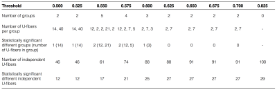 |
96 |
A novel method to detect fractional anisotropy differences in short association fibers discriminated by their functional connectivity between Multiple Sclerosis patients and healthy subjects
Cristian Montalba, Mariana Zurita, Tomas Labbe, Pamela Guevara, Claudia Carcamo, Juan Pablo Cruz, Carlos Sing-Long, Marcelo Andia, Sergio Uribe
We propose a novel method to evaluate fractional anisotropy (FA) differences in short association fibers (U-fibers) between patients with relapsing-remittent multiple sclerosis (RRMS) and healthy subjects by classifying each U-fiber in different groups according to their functional connectivity.
|
|
Neuroimaging at Ultra-High Fields
Electronic Poster
Neuro
Monday, 18 June 2018
| Exhibition Hall |
09:15 - 10:15 |
| |
|
Computer # |
|
3291.
 |
97 |
Age-related changes in cortical thickness and R1 values measured at 7T
Koji Fujimoto, Yuta Urushibata, Hideto Kuribayashi, Tobias Kober, Tadashi Isa, Tomohisa Okada
Healthy volunteers at the age between 20-30 (N=22) and over 60 (N=8) were scanned using a 7T MRI and a 1ch-Tx/32ch-Rx coil with a MP2RAGE prototype sequence at 0.7mm isotropic resolution. Cerebral cortex was segmented based on the human connectome project pipelines with some modifications to obtain cortical thickness and R1 on the gray-ordinate space. Results showed that the average cortical thickness decreased from 2.58 to 2.34 (9%) between the age groups; average R1 increased from 0.542 to 0.552 (1.9%). R1 change seems to have spatial predominance, with the preference in the frontal area.
|
|
3292.
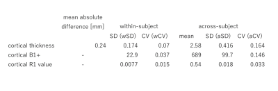 |
98 |
Test-retest reproducibility of cortical thickness, B1+, and R1 in healthy young adults measured at 7T
Koji Fujimoto, Yuta Urushibata, Hideto Kuribayashi, Tobias Kober, Tadashi Isa, Tomohisa Okada
Twenty-two healthy volunteers were scanned at 7T using MP2RAGE (R1 mapping) and Sa2RAGE (B1 mapping) sequences. Applying a modified HCP pipeline, within-subject (test-retest) reproducibility and across-subject variability of cortical thickness, B1+, and R1 were analyzed. The mean absolute difference of cortical thickness was larger at the bilateral lower temporal lobes and insula. Intra-subject variation of B1+ was larger in the lower temporal lobe, whereas across-subject variation of B1+ seems to come from reference transmitter amplitude settings. Except for the lower temporal lobes, variation in the cerebral cortex R1 was very small for both intra-subject measurement (1.5%) and across-subject measurement (3%).
|
|
3293.
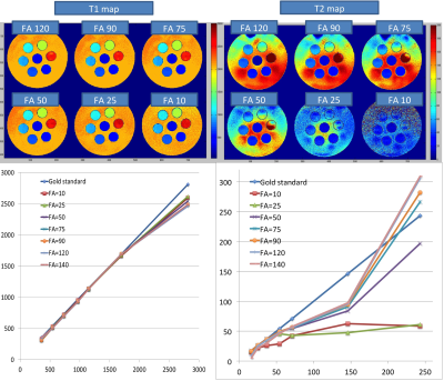 |
99 |
In-Vivo Evaluation of MR Fingerprinting at 7T
Thai Akasaka, Koji Fujimoto, Martijn Cloos, Tomohisa Okada
At 7T RF wavelength effects create a highly heterogeneous B1+ field, which can lead to contrast artifacts and signal dropouts. Instead of attempting to mitigate the B1+ inhomogeneity, the Plug-and-Play MRF (PnP-MRF) method simultaneously encodes the B1+ produced by multiple transmit channels alongside desired tissue properties (PD,T1,T2) to extract B1+ bias free multi-parametric maps. Although this method was designed for use with multiple transmit-channels, a single transmit channel may suffice if B1+ voids can be avoided. In this work, we evaluate the performance of PnP-MRF for brain imaging at 7T in a single transmit configuration.
|
|
3294.
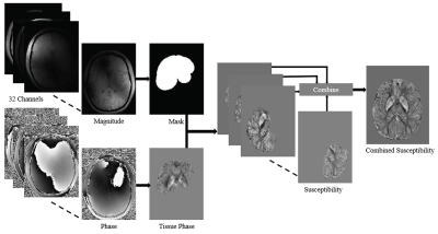 |
100 |
Echo time dependence in temporal frequency shift curves at 3T and 7T
Surabhi Sood, David Reutens, Shrinath Kadmangudi, Markus Barth, Viktor Vegh
Quantitative susceptibility mapping is an MRI tool for mapping anatomical variations. The region specific echo time dependence of frequency shift curves computed from gradient recalled echo MRI data are likely due to variations in tissue microstructure, arrangement and packing. However, the effect of field strength on frequency shift curves has not been established to date. We investigated how frequency shift curves vary with field strength (3T versus 7T) and assessed how changes in the quantitative susceptibility mapping pipeline change the result. 7T data leads to less variability in frequency shift curves and, non-linear trends are present irrespective of methodological differences.
|
|
3295.
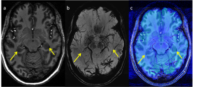 |
101 |
Quantification of venous structures in the hippocampus using segment co-registered 7T susceptibility and structural images: First application to focal temporal lobe epilepsy
Rebecca Feldman, Alexandru Rus, Lara Marcuse, John Rutland, Madeline Fields, Bradley Delman, Priti Balchandani
Susceptibility-weighted imaging offers superior contrast between veins and surrounding tissue. Our technique to isolate and quantify vein-like objects on SWI, combined with hippocampal segmentation of T1-weighted images, permits characterization of vascular density in focal temporal lobe epilepsy. We found difference in vascular density of both epilepsy patients (16%) and controls (30%), with diminished density on the left in both groups.
|
|
3296.
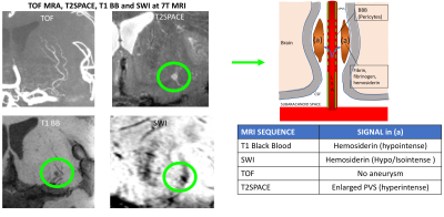 |
102 |
Imaging the perivascular spaces and lenticulostriate arteries at 3T and 7T to provide insight into the pathophysiology of neurodegenerative and neurovascular diseases
Giuseppe Barisano, Samantha Ma, Danny Wang, Yonggang Shi, Arthur Toga, Meng Law
The CSF microcirculation in the brain is not well understood and the role of the perivascular spaces in the clearance of metabolic waste products (including amyloid beta and tau) is still debated. We hypothesize that the increased permeability of the BBB and the resulting passage of blood products into the perivascular spaces may be responsible for the obstruction of CSF-ISF flow. Using a novel T1-weighted 3D Turbo spin-echo with variable flip angles at 3T and 7T, we are able to demonstrate leakage of fibrin from the small lenticulostriate arteries into the perivascular spaces.
|
|
3297.
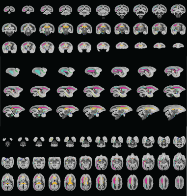 |
103 |
Dissecting white-matter fiber pathways of the marmoset brain using ultra-high-resolution diffusion MRI at 7T and 14T
Cirong Liu, John Newman, Cecil Yen, Frank Ye, David Leopold, Afonso Silva
The common marmoset has received growing interests in neuroscience. The lissencephalic brain of the marmoset grants it experimental advantages for brain mapping and stimulation, but also brings challenges for dissecting white matter tracts, which is poorly depicted in existing marmoset brain atlases. Here, we collected ultra-high-resolution diffusion MRI datasets at 7T and 14T and performed probabilistic tractography and manual segmentation. The marmoset brain demonstrates a complex multi-layer white matter structure with many regions consist of multiple different fiber pathways. With the ultra-high-resolution data, we will provide the first comprehensive map (atlas) for the white matter of the marmoset brain.
|
|
3298.
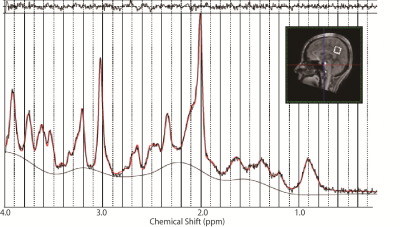 |
104 |
Age-related neurochemical changes in normal human brain: a proton MR spectroscopy study at 7T
Video Permission Withheld
Toru Ishii, Koji Fujimoto, Hideto Kuribayashi, Yuta Urushibata, Nouha Salibi, Ravi Seethamraju, Sinyeob Ahn, Tadashi Isa, Tomohisa Okada
The purpose of this study was to quantify changes of neurochemical concentrations in human brain associated with normal aging with greater sensitivity and accuracy using ultra-high field 7T-MRI. 1H magnetic resonance spectra in the posterior cingulate cortex of 54 healthy adults were measured using a stimulated echo acquisition mode (STEAM) sequence with short echo time, and analysed with LCModel. In addition to the expected result of NAA decrease with aging, both Glutamate and GABA showed significant negative correlations with age. The results may provide significant insights in understanding alterations of human brain accompanying the normal aging.
|
|
3299.
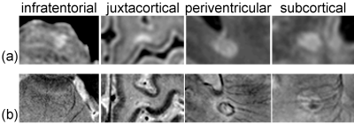 |
105 |
Validation of a Radiological Definition for Central Vessel Sign using 7T FLAIR and SWI
Zahra Hosseini, Jacob Matusinec, David Rudko, Junmin Liu, Benjamin Kwan, Fateme Salehi, Manas Sharma, Marcelo Kremenchutzky, Ravi Menon, Maria Drangova
A number of recent clinical radiology studies support the central vessel sign (CVS) as a sensitive and specific means of differentiating MS white matter lesions (WML) from non-MS WML. However, these studies have employed varying practices for imaging veins and WMLs. This has led to an inconsistent radiological definition of CVS. Recently, the North American Imaging in Multiple Sclerosis publisheda set of guidelines, which provide grounds for derivation of a more robust radiological definition of CVS. Here we employ these guidelines, together with FLAIR and SWI at high field to arrive at a sensitive and specific radiological definition of CVS.
|
|
3300.
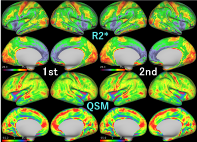 |
106 |
Cortical T2* and QSM maps at 7T: test-retest reproducibility, similarity and differences.
Tomohisa Okada, Koji Fujimoto, Yuta Urushibata, Hideto Kuribayashi, Tobias Kober, Tadashi Isa
R2* and QSM measurement of the cortex is important to investigate human brain pathology. The 7T-MRI has a large potential to provide high-resolution information; however, measurement reliability has not been much investigated. This study investigated test-retest measurement reliability of cortical R2* and QSM values in 16 healthy subjects. High-resolution 3D multi-echo GRE imaging values were mapped onto the cortical surface extracted using 3D MP2RAGE images. Average maps show similar results for both R2* and QSM with larger variance at the frontotemporal base. Except these areas, measurement of cortical R2* and QSM values were found to be reliable at 7T.
|
|
3301.
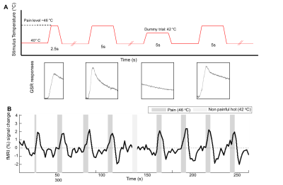 |
107 |
Localizing nociresponsive region within area 3a in human cortex using fMRI at 7T
Video Permission Withheld
Rosa Sanchez Panchuelo, Sally Eldeghaidy, Francis McGlone, Oleg Favorov, Susan Francis
A component of Brodmann area 3a has been shown to be highly responsive to thermonoxious skin stimulation in monkey studies. Here, we use BOLD fMRI at 7T to map on a fine scale the functional representation of a noxious heat stimulus in human primary somatosensory cortex (S1), and compare this to the hand representation identified from vibrotactile stimulation. Thermonoxious stimulation of both the palm and digits evoked a spatially distinct activation within S1, which extends beyond and partially overlaps with the anterior area responding to vibrotactile activation, consistent with the location of nociresponsive area 3a in monkeys.
|
|
3302.
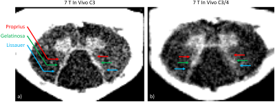 |
108 |
Spinal Cord Gray Matter Myeloarchitecture Revealed by 7T Magnetization Transfer Imaging
Alan Seifert, Joo-won Kim, Junqian Xu
The human spinal cord gray matter contains multiple nuclei and laminae, which are not usually distinguishable in MRI at conventional field strengths. The higher signal-to-noise ratio achievable at 7T provides the increased spatial resolution necessary to resolve these very small gray matter features. Using a wrap-around brainstem/cervical spinal cord RF coil at 7T and a magnetization transfer-prepared multi-echo gradient echo pulse sequence, we resolved three differentially myelinated dorsal horn gray matter structures: the dorsolateral fasciculus, substantia gelatinosa, and nucleus proprius in a single subject at 150-µm in-plane resolution.
|
|
3303.
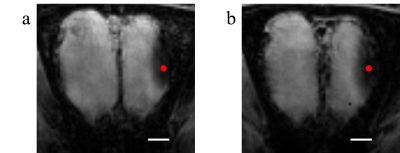 |
109 |
Probing Activated Regions in Rat’s Olfactory System by Odor Stimulations through Manganese Enhanced MRI (MEMRI) at 7T
Did Not Present
Bin Zhang, Qunchen Yuan, Zhen Qin, Liujing Zhuang, Ping Wang, Xiaotong Zhang
In vivo bioelectronic nose utilizes mammalian olfactory system as a means of odor detection and discrimination. However, electrode localization during implantation relies on the researcher’s experiences, resulting in an unreliable success rate. The goal of this research is to determine the optimal electrode implantation positions for electrophysiological recordings by using the technique of manganese enhanced MRI (MEMRI) at 7T. A small dose of manganese ion was delivered into the rat’s right naris and an odor was delivered to its nose during MRI scanning. With the MRI data, the region activated by the specific odor can be identified in the OB.
|
|
3304.
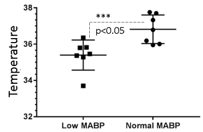 |
110 |
Concurrent changes in cerebral temperature, lactate and vasomotion in urethane anesthetized rats. Proton magnetic resonance study at 7T.
Did Not Present
Devashish Das, Aneurin Kennerley, Ben Brooks, Samuel Harris, Luke Boorman, Paolo DiCarlo, Christopher Martin, Jason Berwick
Cerebral vasomotion is frequently observed phenomenon that accompanies hypotension (mean arterial blood pressure (MABP) 45-65mmHg) in anesthetised rat models(1,2). Although unclear, this mechanism appears to play, in part, a compensatory role in replenishing oxygen reserves in the anaerobic/hypoxic brain. Although the relationship between temperature and metabolism is always interactive. Brain cell metabolism is a major determinant of brain temperature, minor changes in brain temperature can result in signi?cant changes in neural cell metabolism and therefore in brain function. During hypotension (MABP 45-65mmHg) analysis of acquired 1H-spectra revealed thalamic temperature to be ~1.5-2 °C colder than that of the core body temperature (37±0.5)°C. Further analysis of the 1H-spectra revealed dynamic pool of the lactate in the thalamus during hypotension (MABP 45-65mmHg).
|
|
3305.
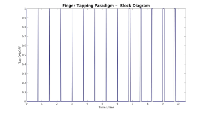 |
111 |
Ultrafast 7T EEG-fMRI for epilepsy using 3D paradigm-free models
Stephen Jones, Balu Krishnan, Anna Crawford, Wanyong Shin, Sehong Oh, Imad Najm, César Caballero-Gaudes, Mark Lowe
There is enormous benefit for non-invasive MRI techniques guiding neurosurgeons to resect tissue causing epilepsy. We extend traditional EEG-fMRI methods in 3 ways: apply 7T to increase BOLD signal; use increased temporal resolution (TR 300ms) from multiband techniques to separate primary from secondary epileptogenic zones; and use paradigm-free mapping to identify interictal spikes obtained during long scans. We test this methodology using isolated finger taps as a surrogate for epileptogenic spikes. Close correspondence between conventional event analysis and paradigm free mapping suggests epileptogenic spikes can be reliably detected if their HRF is similar to a single finger tap.
|
|
3306.
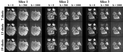 |
112 |
Evaluation of Diffusion-Weighted Imaging of the Macaque Brains Using Readout-Segmented EPI at 7T
Pinyi Wang, Jialu Zhang, Meizhen Qian, Yi Sun, Dingxin Wang, Xiaotong Zhang
Diffusion-weighted imaging (DWI) is an important tool for clinical diagnosis and neuroscience research. To evaluate DWI with higher spatial resolution and with reduced image distortion at higher field strengths, we conducted macaque brain imaging at 7T. Our results suggest that readout-segmented EPI (rsEPI) has reduced image distortion, high MR signal and image contrast. It is believed that the rsEPI can effectively benefit DWI for macaque brain researches at 7T.
|
|
3307.
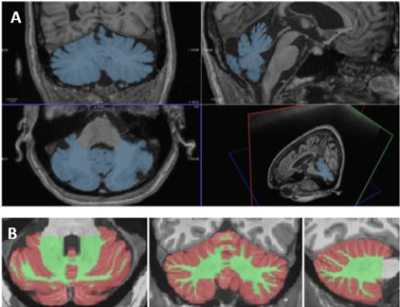 |
113 |
In vivo characterization of cerebellar microglia activation in multiple sclerosis by combined 11C-PBR28 MR-PET and 7 Tesla MRI.
Video Permission Withheld
Valeria Barletta, Elena Herranz, Constantina Treaba, Russell Ouellette, Marco Loggia, Ambica Mehndiratta, Eric Klawiter, Jacob Sloane, Caterina Mainero
We assessed microglia activation in the cerebellum and its relationship to clinical parameters in 27 subjects with multiple sclerosis (MS) and 18 healthy controls by using integrated 3 Tesla magnetic resonance-positron emission tomography imaging with 11C-PBR28. The MS cohort showed increased cerebellar microglia activation in both lesioned and normal appearing cerebellum. The highest microglia activation was found in lesions, mainly concentrated in the cerebellar white matter in relapsing remitting patients, and extensively involving the cortical grey matter in progressive patients. In MS, the tracer uptake in the cerebellar white matter correlated with neurological disability and impaired cognitive performance.
|
|
3308.
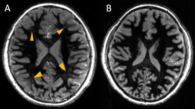 |
114 |
Comparison between 3D white-matter-suppressed MPRAGE and 3D SPACE FLAIR in the 7T MR imaging of tuberous sclerosis complex
Kaibao Sun, Jianfei Cui, Bo Wang, Zhongwei Chen, Yan Zhuo, Rong Xue, Shuli Liang, Lin Chen
Tuberous sclerosis complex (TSC) is a multisystem, autosomal dominant disorder, which was characterized by tubers at the interface of gray and white matters. The 3D SPACE FLAIR sequence has previously been applied to detect the TSC lesions at 7T. The major limitations are its insensitivity to subtle lesions and long acquisition time. 3D white matter suppressed (WMS) MPRAGE was proposed in this study. T1-weighted 3D WMS MPRAGE suppressed the normal white matter signal selectively and made the lesions stand out. In comparison with 3D SPACE FLAIR, 3D WMS MPRAGE could detect more subtle tubers and details within reasonable time.
|
|
3309.
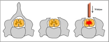 |
115 |
MR spectroscopy of mouse spinal cord injury at 9.4 Tesla
Abdullah Asiri, Gary Cowin, Marc Ruitenberg, Nyoman Kurniawan
This study aims to measure metabolic changes in the acute and chronic stages of a mouse model of spinal cord injury (SCI) using MR spectroscopy. Animal groups consisted of sham, SCI, and SCI with an intravenous immunoglobulin (IVIG) treatment. This study showed that the NAA/Cho ratio can be used as a sensitive marker for chronic injury and testing the efficacy of IVIG to promote recovery.
|
|
3310.
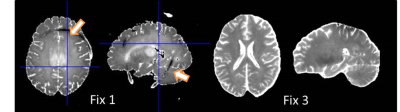 |
116 |
MRI of whole brain formalin-fixed samples at 9.4T: influence of the fixation agent and its dielectric properties on image quality
Gisela Hagberg, Thomas Shiozawa-Bayer, Christian Mirkes, Jörn Engelmann, Jonas Bause, Bernhard Hirt, Klaus Scheffler
MRI of post mortem samples in formalin is an essential tool for validation purposes and comparison with clinical histology, since fixation preserve several microstructural tissue features. We found that the dielectric properties of the fixative influences image quality attained in whole brain post mortem samples at 9.4T. The standard fixative prevented high quality MRI across the entire sample. By using a high conductivity fixative with less field focussing, a more homogeneous excitation was achieved without any drop outs and T1 mapping could be performed using rapid inversion recovery techniques.
|
|
3311.
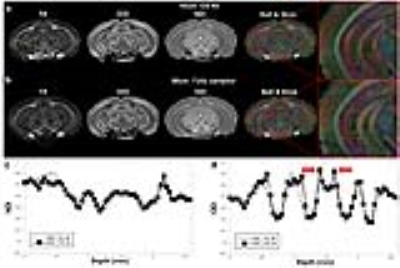 |
117 |
High-resolution neurite orientation dispersion and density imaging of mouse brain using compressed sensing at 9.4T
Nian Wang, Jieying Zhang, Gary Cofer, Robert J. Anderson, Yi Qi, G. Allan Johnson
To evaluate the potentials of compressed sensing (CS) in quantification of neurite orientation dispersion and density imaging (NODDI) index of ex vivo mouse brain at high resolution (45 µm3 isotropic). We were able to achieve compression factors of 4X through judicious choice of k-space sampling pattern. The strong heterogeneous microstructure of corpus callosum (CC) regions were illustrated using different diffusion metrics and NODDI index.
|
|
3312.
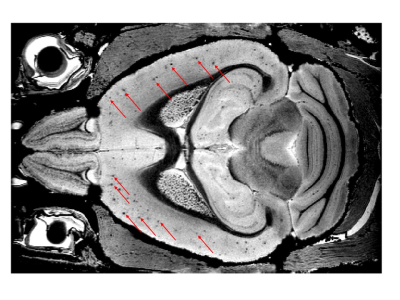 |
118 |
Comparison of high resolution ex vivo multi gradient echo sequences at 7T and 11.7T for amyloid load quantification in an Alzheimer's disease mouse model
Matthieu Gerstenmayer, Francoise Geffroy, Sébastien Mériaux, Benoit Larrat
The gold standard to quantify amyloid load in animal models of Alzheimer’s disease is histology. Despite being really reliable, this technique is destructive and still mainly 2D. Here we optimized a full 3D ex vivo protocol to image whole brain of Alzheimer’s disease mouse model. A high resolution Multi Gradient Echo sequence evidences the shortening of T2* caused by the iron content of amyloid plaques. Plaque detection capabilities were compared between 7T and 11.7T acquisitions for similar scan duration. Unlike the 7T protocol, the 11.7T setup clearly allows amyloid plaque detection and quantification.
|
|
3313.
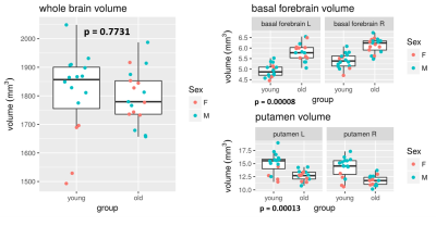 |
119 |
Resting state, gluCEST and anatomical MRI approaches at 11.7T for brain aging studies in a non-human primate
Clément Garin, Nachiket Abhay Nadkarni, Salma Bougacha, Jeremy Pepin, Julien Flament, Jean-Luc Picq, Marc Dhenain
The gray mouse lemur (Microcebus murinus) is a small non-human primate with rapid maturity. This study focuses on the development of non-invasive MRI tools applied to neurodegenerative processes. We performed three different types of analysis: anatomical volumetric measures, neuronal network assessment with resting-state fMRI and brain glutamate distribution with gluCEST imaging. We found anatomical atrophy and functional deficiency mostly in cortical regions. To our knowledge, this study is the first to characterize the functional and anatomical brain aging process in a non-human primate. Furthermore, the mouse lemur functional and gluCEST maps have never been described before.
|
|
3314.
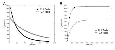 |
120 |
Fluorine-19 Magnetic Resonance at 21.1 Tesla to Detect Brain Inflammation
Sonia Waiczies, Jens Rosenberg, Christian Prinz, Ludger Starke, Jason Millward, Paula Ramos Delgado, Andreas Pohlmann, Andre Kuehne, Helmar Waiczies, Thoralf Niendorf
Detection of brain inflammation by fluorine-19 (19F) MRI was studied for the first time at 21.1 T (NHMFL, Tallahassee, FL). Studies on phantoms showed an SNR gain of 2.1 at 21.1 T, when compared to 9.4 T. A dependency between 19F-MR relaxation and magnetic field strength (B0) was demonstrated. A T1 reduction and higher SNR per unit time counterbalanced the T2 shortening-induced loss in 19F MR signal when performing high spatially-resolved MRI of post mortem experimental autoimmune encephalomyelitis (EAE) mouse samples at 21.1 T. 19F-signals, not seen at 9.4 T, were revealed in both mouse brain and draining lymph nodes.
|
|
| Back |
| The International Society for Magnetic Resonance in Medicine is accredited by the Accreditation Council for Continuing Medical Education to provide continuing medical education for physicians. |
