|
Electronic Poster Session
Diffusion |
Monday, 18 June 2018
Electronic PosterDiffusion
3075 -3098 Diffusion: In Vivo & Ex Vivo Applications: CNS
3099 -3122 Microstructure: Experiments & Applications
3195 -3218 Diffusion: In Vivo & Ex Vivo Applications: Body
3219 -3242 Diffusion MRI: Validation |
| |
Diffusion: In Vivo & Ex Vivo Applications: CNS
Electronic Poster
Diffusion
Monday, 18 June 2018
| Exhibition Hall |
08:15 - 09:15 |
| |
|
Computer # |
|
3075.
 |
1 |
 2-year-old human brain DTI atlas with comprehensive gray and white matter labels 2-year-old human brain DTI atlas with comprehensive gray and white matter labels
Limei Song, Yun Peng, Qinmu Peng, Lei Feng, Minhui Ouyang, Huiying Kang, Shuwei Liu, Hao Huang
2-year-old, marking the end of infancy, is critical for understanding not only precisely organized normal brain development but also serving as clinical anatomical references for neurodevelopmental disorders such as autism. The 2-year-old brain labels transformed from adult atlases lead to relatively large offsets due to dramatic and nonlinear neuroanatomical differences of the brains between these two populations. With DTI data from nineteen healthy 2-year-old subjects, we created a 2-year-old brain DTI atlas with comprehensive labels of 124 gray and white matter structures. The test results suggested the established atlas can be applied to label 2-year-old brain images automatically and accurately.
|
|
3076.
 |
2 |
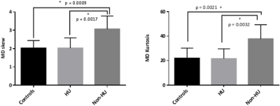
Children with Sickle Cell Disease treated with Hydroxyurea show increased CVR and White Matter Integrity: a quantitative MRI study
Daniel Kapustin, Jackie Leung, Isaac Odame, Suzan Williams, Andrea Kassner
Sickle cell disease (SCD) is a devastating genetic blood disorder leading to chronic anemia and cerebral infarctions. We sought to assess microstructural properties in the WM using diffusion tensor MRI and compare them to measures of cerebrovascular reactivity (CVR). Specifically, we investigated the effect of hydroxyurea (HU) treatment in SCD. Our results show that non-HU patients had increased skew and kurtosis of mean diffusivity in the WM compared to HU patients and healthy controls, and these parameters were correlated to WM CVR in this group. This suggests HU may have beneficial effects on WM microstructural integrity in patients with SCD.
|
|
3077.
 |
3 |
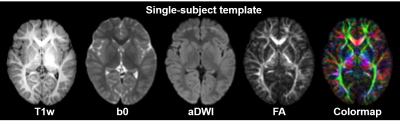
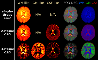
Feasibility and benefits of 3-tissue constrained spherical deconvolution for studying the brains of babies
Thijs Dhollander, Julien Zanin, Bryony Nayagam, Gary Rance, Alan Connelly
When studying white matter in baby brains with diffusion-weighted imaging, we face a range of challenges, including larger water-content, lower anisotropy, differentiated maturation and a (relatively) larger proportion of the brain being comprised of grey matter. We attempt to apply single-tissue, 2-tissue and 3-tissue constrained spherical deconvolution (CSD) to single-shell data of two 5 month old babies. 3-tissue CSD still worked successfully. The nature of benefits was in line with those obtained previously in adults, but they were greater in the babies, mostly due to a much larger presence of GM-like tissue.
|
|
3078.
 |
4 |
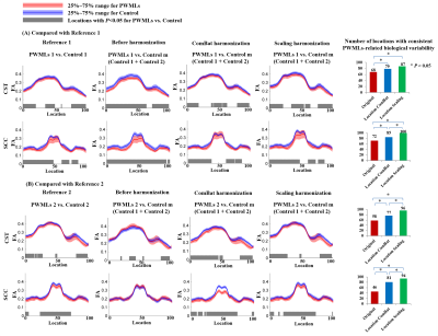
Location-wise Harmonization along White Matter Tracts on Neonatal Brain from Multiple Acquisition Protocols
Simin Liu, Xianjun Li, Miaomiao Wang, Chao Jin, Hua Guo, Jian Yang
In this work, we proposed white matter location-wise harmonization of DTI measurement on neonatal brain based on tract quantification from multiple acquisition protocols. A total of 120 term neonates examined by two DTI acquisition protocols, including 38 neonates with punctate white matter lesions and 82 without MRI abnormalities, were enrolled. Two methods, Scaling and ComBat, were adopted, each carried out also on global-wise and ROI-wise in addition to location-wise. We also proposed an evaluation framework to systematically compare the adopted harmonization approaches, which suggests the way to select the utmost harmonization level and the specific method.
|
|
3079.
 |
5 |
DTI Assessment of Regional White Matter Changes in the Cervical and Thoracic Spinal Cord in Pediatric Subjects
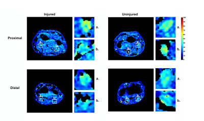
Sona Saksena, Devon Middleton, Laura Krisa, Mahdi Alizadeh, Chris Conklin, Adam Flanders, MJ Mulcahey, Feroze Mohamed, Scott Faro
Synopsis: Prior adult studies have shown that DTI allows for noninvasive assessment of the severity of spinal cord injury (SCI). The aim of this study was to determine whether DTI at sites cephalad and caudal to the injury provides measures of injury severity in pediatric subjects with chronic SCI and compared these data with normative DTI data of typically developing subjects. ROIs were drawn on whole cord and spinal cord white matter (WM) areas: ventral, dorsal, and both right and left lateral regions along the entire cervical and thoracic SC. For each SCI subject, DTI parameters for each WM region were measured at the levels cephalad and caudal relative to MR injury. We demonstrated changes in FA and AD in WM regions at levels both cephalad and caudal to the injury site. This suggests that FA and AD has the potential to be sensitive marker of true extent of cord injury and might be useful in detecting remote injuries.
|
|
3080.
 |
6 |
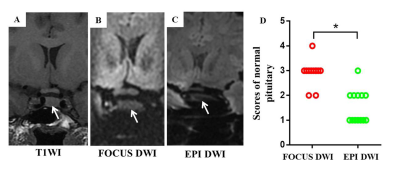
Application of Reduced Field-of-View Diffusion-Weighted Imaging in Evaluation of Normal Pituitary Glands and Pituitary Macroadenomas
Miaomiao Wang, Heng Liu, Congcong Liu, Ting Liang, Xianghui Zhang, Xianjun Li, Chao Jin, Jian Yang
Field-of-view optimized and constrained undistorted single-shot (FOCUS) imaging provides relatively high resolution images with few artifacts. However, application of this technique for evaluation of normal pituitary glands and pituitary macroadenomas has not been reported to date. The study aims to evaluate the image quality and value of FOCUS DWI in evaluation of normal pituitary glands and pituitary macroadenomas. Our results suggest that FOCUS DWI exhibited obviously superior image quality both in normal pituitary glands and macroadenomas in a clinically feasible scan time. Moreover, it might be helpful for evaluating the consistency of pituitary macroadenomas.
|
|
3081.
 |
7 |
Apparent diffusion coefficient for molecular subtyping of non-Gadolinium-enhancing WHO grade II/III glioma
Laura Mancini, Sara Hassanein, Sotirios Bisdas, Jeremy Rees, Harpreet Hyare, John Maynard, Sebastian Brandner, Carmen Tur, H Jager, Tarek Yousry, Steffi Thust
A proportion of non-enhancing intrinsic presumed low-grade-gliomas(LGG), rapidly progresses. Hypothesis: ADC can predict glioma molecular subtypes of the revised 2016 World_Health_Organization brain tumours classification. Methods..44 non-Gadolinium-enhancing LGG divided in three molecular subgroups. 2D and 3D T2-derived tumour and normal-appearing-white-matter (NAWM) masks co-registered to ADC_maps(b=1000s/mm2). Linear-regression, ROC-analysis and logistic-regression compared ADC_values with tumour type. Results..ADCmean and ADCratio(tumour/NAWM) were lowest (p<0.001) in the most malignant tumour type (IDHwt). An ADCmean(ADCratio) threshold of 1201*10-6mm2/s(1.65) identified IDHwt with sensitivity=83%(80%) and specificity=86%(92%) (AUC=0.9-0.94). Between-observers (2D-versus-3D) intraclass-correlation-coefficient=0.98(0.92). Conclusions..ADC measurements can support the distinction of non-enhancing glioma subtypes. 3D and 2D measurements were both accurate.
|
|
3082.
 |
8 |
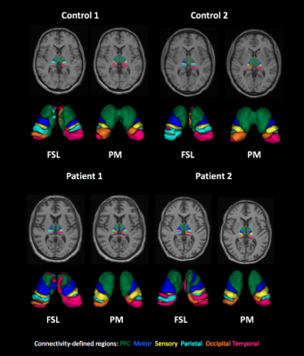
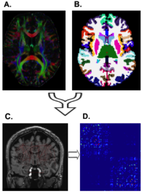
Thalamic nuclei segmentation on dementia using tractography and population-specific priors
Carla Semedo, M. Jorge Cardoso, S. B. Vos, Carole H. Sudre, Martina Bocchetta, Annemie Ribbens, Dirk Smeets, Jonathan Rohrer, Sebastien Ourselin
Thalamic changes have been reported in several neurological disorders, such as Alzheimer's disease and frontotemporal dementia (FTD). As pathologies affect different cortical and subcortical brain regions disproportionally, accurate segmentation of thalamic nuclei can provide relevant insights about brain function and neurological disorders mechanisms. Here, we used a previously developed thalamus parcellation strategy that relies on tractography and population-specific to infer any connectivity changes in presence of FTD. The obtained results were compared against to the ones derived with the commonly used probabilistic tractography pipeline available in FSL.
|
|
3083.
 |
9 |
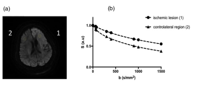
Kurtosis and IVIM measurements applied to ischemic stroke diagnosis: an initial experience.
Aude Pavilla, Alessandro Arrigo, Giulio Gambarota, Mehdi Mejdoubi, Régis Duvauferrier, Hervé Saint-Jalmes
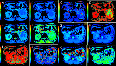
Diffusional kurtosis imaging (DKI) enables the characterization of non-Gaussian diffusion providing an additional diffusion parameter, the kurtosis (K), that may reflect microstructure heterogeneity. The DKI-IVIM model that incorporates DKI into the IVIM model has been investigated here to assess the feasibility and the potential utility of the DKI-IVIM model for both enhanced diffusion characterization and perfusion measurements in ischemic stroke. Five stroke patients were enrolled. DKI-IVIM imaging was performed using 8 b-values from 0 to 1500 s/mm2 with a 4 minutes scan duration. IVIM pseudo-diffusion coefficient D*, perfusion fraction f, blood flow-related parameter fD* in addition to the diffusion parameters D (diffusion coefficient) and K were determined in the ischemic lesion and contralateral normal tissue for the stroke patients. Diffusion and perfusion parametric maps were reconstructed.
A significant decrease for D (p<0.0001) and increase for K (p=0.0002) in the lesion was observed. The perfusion fraction exhibited a significant decrease in the ischemic regions (p=0.005).
DKI-IVIM model enables for simultaneous cerebral perfusion and enhanced diffusion characterization in an acceptable clinically acquisition time that might improve ischemic stroke diagnosis.
|
|
3084.
 |
10 |
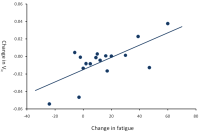
Interferon-alpha induced changes in NODDI predispose to the development of fatigue
Nicholas Dowell, Samira Bouyagoub, Mara Cercignani, Neil Harrison
Here we use NODDI modeling of multi-shell diffusion MRI to investigate whether changes in orientation-dispersion index (ODI) or intracellular volume fraction (Vic) can predict the later emergence of IFN-α-induced fatigue. Eighteen patients initiating IFN-α based treatment for hepatitis-C underwent diffusion MRI and blood sampling at baseline and 4 hours after their first IFN-α injection. They were then followed up with regular psychological assessments for 12 weeks of treatment. IFN-α injection stimulated an acute inflammatory cytokine response and evoked acute fatigue that peaked between 4 and 12 weeks of treatment. Within the brain, IFN-α induced an acute increase in intracellular volume fraction in patients that experienced a simultaneous increase in IFN-α induced fatigue but not patients that did not. Acute changes in striatal microstructure additionally predicted the continued development of fatigue but not mood symptoms 4 and 8 weeks later into treatment. Our findings highlight the value of NODDI as a potential in vivo biomarker of the central effects of peripheral inflammation. We highlight the exquisite sensitivity of the striatum to IFN-α and further implicate striatal perturbation in IFN-α-induced fatigue.
|
|
3085.
 |
11 |
Hippocampal Subfield-specific Tractography in Epilepsy Patients at 7 Tesla
John Rutland, Rebecca Feldman, Lara Marcuse, Madeline Fields, Bradley Delman, Prantik Kundu , Patrick Hof, Priti Balchandani
This is the first investigation to use diffusion tensor imaging at 7 Tesla to quantify changes in the structural connectivity of individual hippocampal subfields in epilepsy patients. Diffusion imaging and automated hippocampal subfield segmentation were performed on 19 epilepsy patients and 10 healthy controls. We found that hippocampal volumes are reduced bilaterally in epilepsy patients compared with controls. Connectivity in the left fimbria and right hippocampal-amygdaloid transition area is significantly reduced in epilepsy patients compared with controls. These findings suggest that connectivity of hippocampal subfields are independently affected in epilepsy patients.
|
|
3086.
 |
12 |
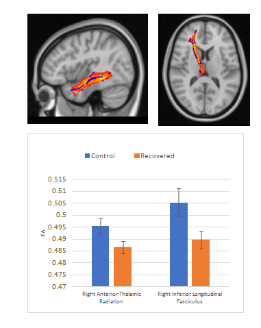
Positive DTI Findings After Recovery Following Youth Sport-Related Concussion May Be of Little Clinical Relevance
Video Permission Withheld
Najratun Nayem Pinky, Carolyn Emery, Chantel Debert, Bradley Goodyear
Mild traumatic brain injury (mTBI), including sport-related concussion, is a major health issue. Diffusion Tensor Imaging (DTI) can be useful for identifying alterations in white matter tracts following sport-related concussion. We found that fractional anisotropy (FA) significantly differed between recovered and control youth within the right anterior thalamic radiation and the right inferior longitudinal fasciculus; however, the effect size (< 0.02 change in FA) suggests that the difference may be of little or no clinical relevance, given the recovered group exhibited no symptoms. Large cohort studies are needed before statistically significant MRI findings should be used to inform return-to-play policy
|
|
3087.
 |
13 |
Regional Brain Changes in Autonomic, Mood, and Cognitive Control Areas in Adolescents with Single Ventricle Heart Disease
Sadhana Singh, Bhaswati Roy, Nancy Halnon, Alan Lewis, Mary Woo, Nancy Pike, Rajesh Kumar
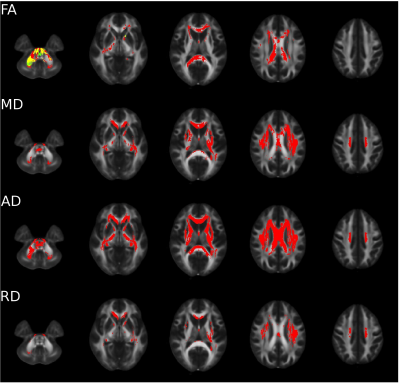
Adolescents with single ventricle heart disease (SVHD) show both white and gray matter injury in multiple brain areas that control autonomic, mood, and cognitive functions that are deficient in the condition. However, the nature and extent of brain injury in SVHD are unclear. Using diffusion tensor imaging based MD procedures, we showed wide-spread chronic tissue changes in SVHD subjects in areas involved in autonomic, mood, and cognitive regulatory functions. These findings may have resulted from hypoxia/ischemia- or developmental-induced processes accompanying the condition.
|
|
3088.
 |
14 |
Brain White Matter Microstructure Changes in Alzheimer disease with Type 2 diabetes:a DKI study
Junyi Dong, Liang Han, Xiaoxin Li, Yanwei Miao
In this study, the groups were respectively used as the people with Alzheimer disease with type 2 diabetes, Alzheimer disease without type 2 diabetes and healthy person, the effect of high blood glucose on the microstructure in patients with type 2 diabetes mellitus, and the changes on microstructure in patients with AD patients were studied used DKI study, and it was concluded that the high blood glucose level may have certain damage to the microstructure of the white matter. In conclusion, DKI study can evaluate secondary brain microstructure changes from hyperglycemia in T2DM patients.
|
|
3089.
 |
15 |
Brain Microstructure Changes of gray matter detected by DKI in Alzheimer disease with Type 2 diabetes patients
Junyi Dong, Xiaoxin Li, Liang Han, Yanwei Miao
In this paper, the experimental group and the control group were respectively used as the people with Alzheimer disease with type 2 diabetes and Alzheimer disease without type 2 diabetes, the effect of high blood glucose on the microstructure in patients with type 2 diabetes mellitus was studied used DKI study, and it is concluded that the high blood glucose level may have certain damage to the microstructure of the gray matter. In conclusion, DKI study can evaluate secondary brain microstructure changes from hyperglycemia in T2DM patients.
|
|
3090.
 |
16 |
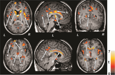
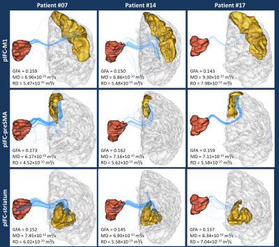
Looking at the structural connectivity of the frontal inferior cortex to better decipher the inhibitory control mechanisms in Obsessive Compulsive Disorder
Ivy Uszynski, Cyril Poupon, Cédric Pichat, Pauline Favre, Benjamin Fredembach, Hervé Mathieu, Laurent Lamalle, Alexandre Krainik, Olivier David, Emmanuel Barbier, Mircea Polosan
Obsessive-compulsive disorder (OCD) is a neuropsychiatric disease affecting 2.5-3% of the population and characterized by repetitive compulsive behaviours with severe complications such as depression, suicide and addiction. Diffusion MRI is a powerful non-invasive technique that evaluates the integrity of the white matter pathways such as those implicated in the impulse control, likely to be impaired in OCD. Here, we investigate the connectivity of the right posterior inferior frontal cortex and in particular to the presupplementary motor area (also involved in inhibition), to the striatum (involved in proactive and/or selective control) and to the primary motor cortex in the contralateral hemisphere.
|
|
3091.
 |
17 |
Predicting treatment outcome of schizophrenia based on white matter tract integrity using a support vector classifier
Wen-Bin Luo, Jing-Ying Huang, Yung-Chin Hsu, Wen-Yih Isaac Tseng
Although white matter tract microstructure has been implicated in treatment outcome of schizophrenia, its predictive capability on first-episode patients remains unknown. In the study, diffusion spectrum imaging (DSI) data were acquired from both chronic and first-episode patients, reconstructed by mean apparent propagator (MAP) MRI and analyzed with tract-based automatic analysis (TBAA). Stepwise statistical analysis was then performed to identify specific segments of white matter tracts that were significantly different between remitted and non-remitted chronic patients. We built a support vector classifier on the preprocessed data matrix. The resulting model yielded fair validation and test accuracy on chronic and first-episode patients, respectively.
|
|
3092.
 |
18 |
Diffusion MRI as an imaging marker of depression from a large and homogenous population study
Julie Coloigner, Jean-Marie Batail, Isabelle Corouge, Jean-Christophe Ferré, Dominique Drapier, Christian Barillot
Despite the extensive therapy options available for depression, up to 80% of patients will suffer from a relapse. Consequently, understanding the neural correlates underlying the depression will optimize the diagnosis and treatment of individual depressed patients. The purpose of our study was to investigate alterations of white matter integrity in a large cohort of patients suffering from depression using diffusion tensor imaging. Our findings provide robust evidence that the reduction of white-matter integrity in the interhemispheric connections and fronto-limbic neuronal circuits may play an important role in depression pathogenesis.
|
|
3093.
 |
19 |
Wired for music? – a diffusion MRI based study of normative music perception skills
Archith Rajan, Jacob Alappatt, Apurva Shah, Megha Sharda, Jeffrey Valla, Madhura Ingalhalikar, Nandini Singh
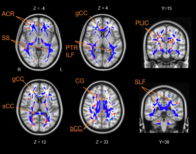
White matter micro-structural correlates of music perception skills have only been studied in expert musicians, although skills can be independent of musical training. We assessed normative variation of music perception skills in adult population by the PROMS-S musicality test. A tract based spatial statistic on high angular diffusion data revealed negative associations between Mode of Anisotropy and d’ measures of total scores, sub-scores of Accent, Embedded Rhythms and Tempo in the Corpus Callosum extending to Corona Radiata. Partial volumes of secondary fiber population also correlated positively to these scores, suggesting the recruitment of inter-hemispheric connections necessary for enhanced music perception.
|
|
3094.
 |
20 |
The neurosurgical implication of scanner, gradient performance and acquisition protocol on Meyer’s loop reconstruction
Maxime Chamberland, Chantal Tax, William Gray, Derek Jones
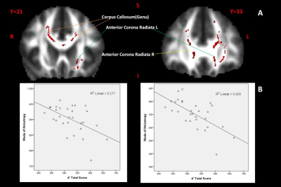
The optic radiation (OR) is a key brain fiber bundle of the visual system which must be spared as much as possible during resection of the temporal lobe in epilepsy surgery to prevent visual field defects. Therefore, it is of utmost importance to avoid underestimating its anterior location (Meyer’s loop) with diffusion MRI tractography. For this reason, it is critical that this part of the OR is reconstructed as accurately as possible. In this abstract, we demonstrate that standard diffusion MRI acquisitions potentially underestimate the true location of Meyer’s loop, when compared to state-of-the-art protocols.
|
|
3095.
 |
21 |
Retrospective Reduction of Systematic Differences Across Scanner Changes by Accounting for Noise Floor Effects in Diffusion Tensor Imaging
Ken Sakaie, Xiaopeng Zhou, Jian Lin, Josef Debbins, Mark Lowe, Robert Fox
Scanner upgrades are a persistent but important problem when conducting MRI research studies. Systematic differences introduced by a scanner upgrade can have undesirable effects on the conclusions of a study. Quantitative tissue microstructure measurements by diffuson tensor imaging (DTI) can be affected by systematic differences in noise floor effects. Noise floor effects are due to rectification of signal by magnitude reconstruction than can, in turn, bias microstructure measurements. A retrospective correction that accounts for noise statistics is proposed to limit systematic differences in DTI measurements across scanner upgrades. A practical measure, signal to noise floor ratio (SNFR) is proposed to determine the conditions under which the retrospective correction works effectively.
|
|
3096.
 |
22 |
Age-effects on cortical tissue diffusivity
Jordan Chad, David Salat, J. Chen
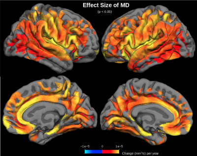
Mean diffusivity (MD) is known to increase with age in the white matter (WM), serving as a measure of age-related WM degeneration. However, age-effects on MD with age have scarcely been studied in the gray matter. Here we examine age-effects on MD across the cerebral cortex. MD is shown to correlate more strongly with age than cortical thickness measurements derived from anatomical MRI. Cortical regions showing the largest MD age-effects include the insula and anterior cingulate, suggesting greatest neurodegeneration in these regions. This work suggests that MD may be used as a sensitive measure of aging in the cerebral cortex.
|
|
3097.
 |
23 |
Investigating the performance of Diffusional Kurtosis Imaging for group-wise analyses: A study from the Human Connectome Project
Hamed Y. Mesri, Szabolcs David, Max Viergever, Alexander Leemans
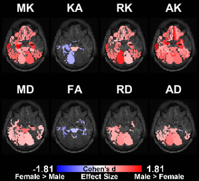
Diffusional Kurtosis Imaging (DKI) is an extension to Diffusion Tensor Imaging (DTI), which allows the quantification of non-Gaussian water diffusion and the quantification of parameters related to microstructural changes. In this work, we used high-quality datasets from the Human Connectome Project and non-parametric statistical inference to evaluate the performance of the DKI measures for group-wise studies. To this end, we used the gender information to group the subjects and study the differences. Our results demonstrated that DKI metrics could reveal the differences more accurately compared to DTI metrics.
|
|
3098.
 |
24 |
IVIM values in healthy brain
Steren Chabert, Jorge Verdu, Gamaliel Huerta, Cristian Montalba, Pablo Cox, Rodrigo Riveros, Sergio Uribe, Rodrigo Salas, Alejandro Veloz
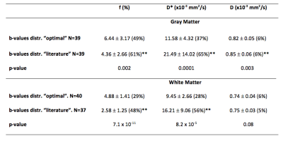
Even though there is much interest in brain IVIM imaging, it is difficult to get a clear view from literature on which values to expect. Our purpose is to obtain healthy brain D, D* and f, to add findings and get closer to reference values. Two distributions of 16 b-values were used to acquire data on 10 volunteers, at 1.5T: one commonly found in literature and the other considered as optimal. Values obtained from the “optimal distribution” were significantly different in all cases but D in white matter. This study emphasizes the dependence of IVIM results on the acquisition scheme applied.
|
|
Microstructure: Experiments & Applications
Electronic Poster
Diffusion
Monday, 18 June 2018
| Exhibition Hall |
08:15 - 09:15 |
| |
|
Computer # |
|
3099.
 |
25 |
Biomimetic numerical phantoms for white matter tissues characterization using a reduced number of design parameters
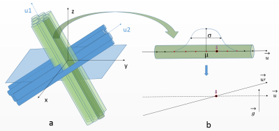
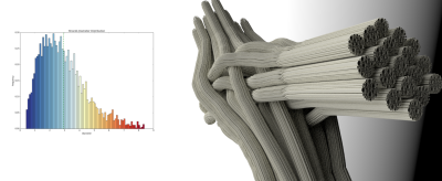
Kevin GINSBURGER, Fabrice POUPON, Felix MATUSCHKE, Jean-François MANGIN, Markus AXER, Cyril POUPON
We propose to extend the functionalities of the Diffusion Microscopist Simulator to design more realistic white matter phantoms without any input mesh and with few parameters. The biomimetic phantoms can represent crossing configurations with an arbitrary number of fiber populations, include a myelin sheath and Ranvier nodes and account for beading, tortuosity and angular dispersion of fibers.
|
|
3100.
 |
26 |
Analysis of the T2-Relaxation-Diffusion Correlation MRI in Glioblastoma
Video Permission Withheld
Yuan Li, Michelle Kim, Theodore Lawrence, Parmar Hemant, Yue Cao
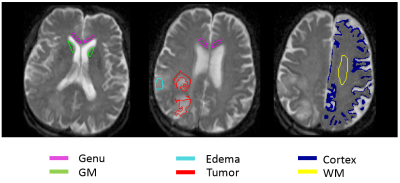
Analysis of the T2-relaxation-diffusion correlation is an emerging approach. It has the potential to reveal the biophysical behavior of tissue and tumor, which cannot be done by the analysis of T2-relaxation and diffusion MRI separately. This study applied this approach to glioblastoma (GBM) and revealed the different correlations between T2 and diffusion in tumor, normal tissue and edema.
|
|
3101.
 |
27 |
Bound Model for Extracting Small Airway Scales in Pediatric Asthma
Annie Malkus, Robert Cadman, Sean Fain
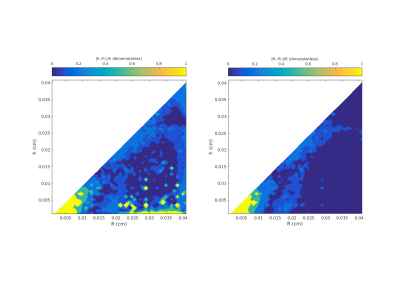
We evaluate a morphometric approach for measuring acinar airway and alveolar scales with diffusion weighted MRI of hyperpolarized helium in pediatric asthma subjects.
|
|
3102.
 |
28 |
In Vivo 3D Axonal Diameter Estimation in the Human Brain with 300 mT/m Gradient MRI
Alfred Anwander, Thomas Knösche, Thomas Witzel, Assaf Horowitz, Yaniv Assaf
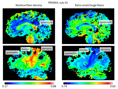
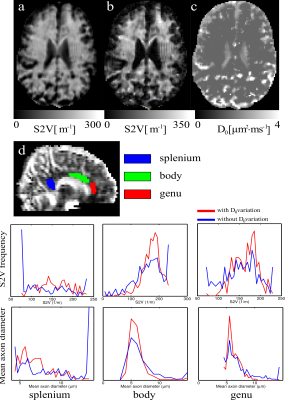
The estimation of neural micro-structure in general and axon diameter in particular became feasible using advanced diffusion imaging frameworks such as CHARMED and AxCaliber. Recently, the AxCaliber model was extended to 3D enabling to capture the axonal properties of any fiber system in the brain. In this work we challenged the utility of using the CONNECTOM MRI, that provides a gradient strength of up to 300 mT/m, for axon diameter estimation. We found that the sensitivity of the model towards small diameter axons increases dramatically with the use of the strong gradient system increasing the validity and accuracy of AxCaliber3D.
|
|
3103.
 |
29 |
Estimation of Extracellular Matrix Diffusion Properties in Decellularized Porcine Myocardium from DTI
Noel Naughton, Nicholas Gallo, Marcella Viacik, Aaron Anderson, Bradley Sutton, John Georgiadis
We present a method to estimate microstructural parameters of a decellularized pig myocardium using a two-compartment exchange model. We also show that the estimated parameters are in good agreement with other values found in the literature.
|
|
3104.
 |
30 |
MRI approaches to map focal cortical dysplasia in focal epilepsy using anomalous diffusion and magnetic susceptibility
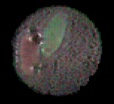
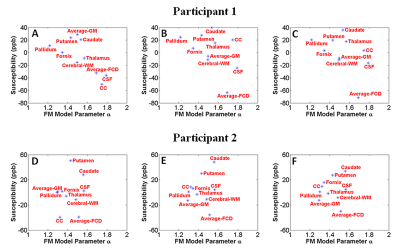
Qiang Yu, David Reutens, Javier Urriola, Surabhi Sood, Viktor Vegh
Focal cortical dysplasias (FCD) are developmental malformations of the cerebral cortex that are often highly epileptogenic. When medications fail to control seizures, surgical removal of dysplastic epileptogenic tissue may be curative. But, in 20-40% of patients current MRI scans cannot identify FCD affected brain regions. Building on the anomalous nature of diffusion and magnetic susceptibility of tissue, we aimed to improve in vivo identification of FCD in the brain. We found the combination of anomalous diffusion model parameters and tissue magnetic susceptibility can be used to differentiate FCD from healthy tissue in the brain.
|
|
3105.
 |
31 |
A two-perfusion compartment model for human placenta.
Michele Guerreri, Amanda Antonelli, Silvia Bernardo, Carlo Catalano, Lucia Manganaro, Silvia Capuani
This work proposes the use of a two perfusion comparmtent model to fit diffusion MRI data of human placenta. The aim of the work is to characterize the parameters values and compare them with results obtained in animal models.
|
|
3106.
 |
32 |
Does the g-ratio influence resting-state functional connectivity? A group-level analysis
Matteo Mancini, Charlotte Clarke, Nick Dowell, Neil Harrison, Mara Cercignani
Recent findings have shown specific relationships between the cortical myeloarchitecture of the brain and resting-state functional connectivity patterns, while little is known about the white matter myelin distribution. The aim of this work is to preliminary characterize how the g-ratio (i.e., the ratio of the inner and the outer diameters of myelinated axons) and functional connectivity are interrelated. We characterized at group level connectivity patterns using structural connectivity, functional connectivity and g-ratio. We then assessed potential differences between specific functional modules. We observed different distributions when comparing structure and function in terms of g-ratio, and reported significant differences.
|
|
3107.
 |
33 |
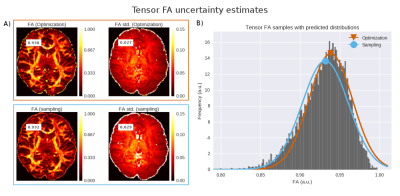
More certainty about your uncertainty in diffusion MRI microstructure estimates
Robbert Harms, Alard Roebroeck
Diffusion MRI microstructure approaches use point estimates ignoring the uncertainty in these estimates. In this work, we evaluate two general methods to quantify uncertainty and generate uncertainty maps for any microstructure model. We find that the Fisher Information Matrix method based in nonlinear optimization is fast and accurate for models with few parameters. The Markov Chain Monte Carlo (MCMC) based method takes more time, but provides robust uncertainty estimates even for sophisticated models with more parameters. Uncertainty estimates of microstructure measures can help power evaluations for group/population studies and assist in data quality control and analysis of microstructure model fit.
|
|
3108.
 |
34 |
Disentangling the Effects of Anisotropy and Orientation Dispersion Using Diffusion Spherical Mean Spectrum Imaging
Tiantian Xu, Geng Chen, Haiyong Wu, Weili Lin, Dinggang Shen, Pew-Thian Yap
Diffusion fractional anisotropy (FA) measures voxel-level anisotropy, which mingles the effects of neurite microscopic-level anisotropy and orientation dispersion. We introduce a technique, called spherical mean spectrum imaging (SMSI), that can disentangle these two effects. We applied SMSI on baby brain diffusion MRI data collected during the first year of life and show that SMSI can extract microstructural information that is elusive to diffusion tensor imaging (DTI).
|
|
3109.
 |
35 |
Quantification of white matter pathologies during multiple sclerosis disease development
Video Permission Withheld
Chunyu Song, Peng Sun, Anne Cross, Sheng-Kwei Song
A new diffusion MRI histology (D-Histo) is proposed to model both intra and extra axonal diffusion, in addition to isotropic diffusion. It not only resolves crossing fibers but also quantitatively assess axonal injury, axon loss, demyelination, edema and inflammation. Through the multiple-tensor modelling of diffusion-weighted MRI signals, D-Histo has shown promise to monitor evolving pathologies in normal appearing corpus callosum in multiple sclerosis patient brain.
|
|
3110.
 |
36 |
Determining how varying the number of gradient strengths and frequencies affects fitted mean axon diameters in the corpus callosum using oscillating spin echo gradients
Morgan Mercredi, Sheryl Herrera, Richard Buist, Kant Matsuda, Melanie Martin
There is an increasing drive to use diffusion spectroscopy to infer the sizes of structures in samples. We present here the first use of the sine OGSE to infer the effective mean axon diameters in the human corpus callosum and study the effect on accuracy of reducing the number of images used in the inference. Aiming to reduce imaging times, this study examines how the number of frequencies or number of gradients affects accuracy and precision. We found that collecting OGSE data with two gradients gives a difference in results of less than 5% compared to six gradients.
|
|
3111.
 |
37 |
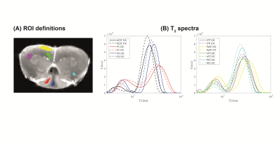
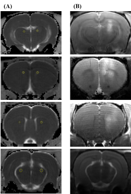
Exchange arising from myelin water revealed from temperature-dependent multiexponential T2 mapping
Noam Shemesh, Mark Does
The importance of myelin water exchange in relaxation and diffusion metrics extracted from tissues is still an open question. In particular, to what extent myelin water fraction (MWF) values derived from multiexponential T2 are due to exchange between myelin and intra/extra axonal spaces remains unclear. Here, fixed rat spinal cords were subject to temperature-dependent multiple spin echo experiments, aiming to probe how exchange modulates quantitative T2 maps in rat spinal cords. We find signatures for exchange from T2 shift patterns, which are tightly linked to the axon diameters in the spinal cord.
|
|
3112.
 |
38 |
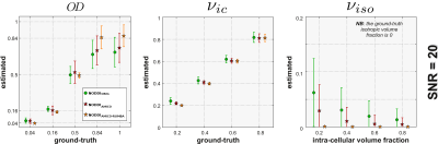
Fast and robust estimation of NODDI parameters using non-Gaussian noise models and spatial regularization
Erick Canales-Rodríguez, Jean-Philippe Thiran, Alessandro Daducci
In this study we developed a robust inversion algorithm to estimate the Neurite Orientation Dispersion and Density Imaging (NODDI) model. It is based on the Accelerated Microstructure Imaging via Convex Optimization (AMICO) framework. However, in contrast to AMICO, the proposed method relies on realistic MRI noise models. Moreover, it allows to take into account the underlying spatial continuity of the brain image by including a total variation regularization term. In simulated data the new method was effective in reducing the outliers, producing results more close to the ground-truth and with lower variability. The method was also evaluated on real data.
|
|
3113.
 |
39 |
Diffusional changes of perivascular space in focused ultrasound induced Blood-Brain Barrier disruption in rat brain
Heajung Choi, Huijin Song, Kyung Eun Jang, Hyunsil Cha, Eunji Kim, Mujin Yang, Jiung Yang, Hoesu Jung, Mun Han, Taekwan Lee, Juyoung Park, Yongmin Chang
Focused ultrasound (FUS) induces microbubble oscillation, which loosens the tight junction of the endothelial cells in the brain and opens Blood-Brain Barrier (BBB). In this study, we opened the BBB of rat brain using the MRI guided FUS to investigate the changes of diffusivity in perivascular space using diffusion-weighted imaging (DWI). Our results showed that ADC of the perivascular space increased after sonication suggesting the increased water diffusion in perivascular space after BBB opening. Therefore, our study suggests that ADC change can be a possible imaging marker for opening of BBB.
|
|
3114.
 |
40 |
Feasibility of VERDICT-MRI for non-invasive characterisation of rectal cancer microstructure
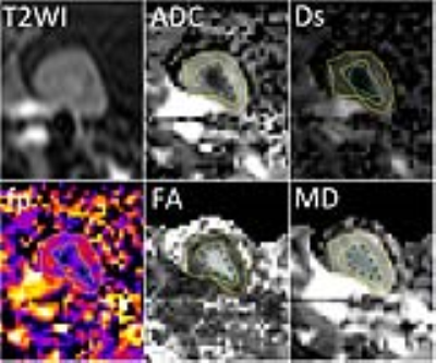
Maira Tariq, Christopher Liao, Elisenda Bonet-Carne, Andrew Plumb, Manuel Rodriguez-Justo, Daniel Alexander, Manish Chand, David Atkinson, Eleftheria Panagiotaki
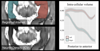
This work evaluates the feasibility of in-vivo microstructure imaging for rectal cancer using the VERDICT MRI framework. We perform a model comparison to find the form of VERDICT that can describe the rich DW-MRI data. Preliminary results from two subjects show promise for non-invasive clinical rectal cancer characterisation. We find that a multi-compartment VERDICT model that explicitly models restriction explains the signal in the rectal tissue better than the conventional cancer models and shows plausible estimates of microstructure in the rectal cancer tissue. Future work will compare these findings with corresponding histology.
|
|
3115.
 |
41 |
Towards the assessment of myelination using time-dependent diffusion MRI indices.
Abib Alimi, Alexandra Petiet, Mathieu Santin, Anne-Charlotte Philippe, Stephane Lehericy, Rachid Deriche, Demian Wassermann
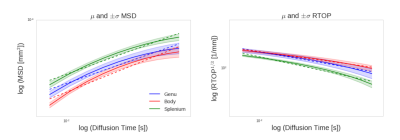
We study the sensitivity of time-dependent diffusion MRI indices or qτ-indices to demyelination in the mouse brain. For this, we acquire in vivo four-dimentional diffusion-weighted images -varying over gradient strength, direction and diffusion time- and estimate the qτ-indices from the corpus callosum. First order Taylor approximation of each index gives fitting coefficients α and β whose variance we investigate. Results indicate that, cuprizone intoxication affects mainly index coefficient β by introducing inequality of variances between the two mice groups, most significantly in the splenium and that MSD increases and RTOP decreases over diffusion time τ.
|
|
3116.
 |
42 |
Towards Non-Invasive Characterization of Intravoxel Tumor Heterogeneity: Correlation between Non-Gaussian Diffusion MRI and Histology Using Machine Learning
Muge Karaman, Lingdao Sha, Tingqi Shi, Weiguo Li, Dan Schonfeld, Tibor Valyi-Nagy, Xiaohong Zhou
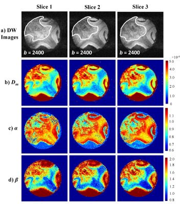
Tissue heterogeneity is an important consideration for diagnosing many diseases. Recently, a novel non-Gaussian diffusion model – continuous-time random-walk model (CTRW) – provided promising evidence indicating a possible link between voxel-level spatiotemporal diffusion heterogeneity and microscopic intravoxel tissue heterogeneity. Establishing a correlation between imaging-based and histology-based measurements, however, has been challenging because of the lack of efficient and subjective evaluation of tissue heterogeneity histologically. In this study, we applied a machine-learning algorithm to quantitatively determine microscopic tissue heterogeneity, enabling a correlation between intravoxel diffusion heterogeneity based on CTRW parameters and structural heterogeneity from histopathology.
|
|
3117.
 |
43 |
Stromal collagen content correlates with fast diffusivity signal fraction in breast lesions
Video Permission Withheld
Liv Egnell, Igor Vidic, Dennis Adams, Jr., Neil Jerome, Torill Sjøbakk, Agnes Østlie, Hans Fjøsne, Rebecca Rakow-Penner, Anders Dale, Anna Bofin, Tone Bathen, Pål Goa
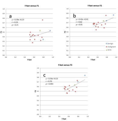
The deviation from a monoexponential of the DW-signal decay towards higher b-values (>1000s/mm2) reflects the complex tissue microstructure. The biexponential decay model assumes that the signal is composed of two components with different diffusivity, possibly originating from two physically separated tissue components. In this study, we estimate the collagenous and non-collagenous extracellular contents in sixteen breast lesions using hematoxylin-eosin-saffron stained histological specimens and compare with pre-surgical in vivo DW-MRI data. Our results show that the signal fraction of the faster diffusivity component correlated significantly with collagen content, suggesting that collagen contributes to the DWI signal decay
|
|
3118.
 |
44 |
Multimodal microstructure imaging: joint T2-relaxometry and diffusometry to estimate myelin, intracellular, extracellular, and cerebrospinal fluid properties
Marco Pizzolato, Erick Canales-Rodríguez, Alessandro Daducci, Jean-Philippe Thiran
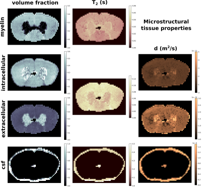
We propose a multimodal joint estimation that aims at exploiting the complementary information of diffusion and multi-echo spin echo data to disentangle the contributions and properties of the main tissue microstructure compartments. We recovered T2, diffusion coefficient, and volume fractions values of myelin, intracellular, extracellular, and cerebrospinal fluid compartments within an ex vivo spinal cord sample by means of diffusometry and relaxometry. A g-ratio map was also calculated.
|
|
3119.
 |
45 |
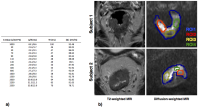
Prostate Cancer Classification Using Stretched Exponential Model Parameters of Diffusion Signal Decay
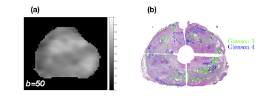
Meltem Uyanik, Michael Abern, Brandon Caldwell, Muge Karaman, Winnie Mar, Virgilia Macias, Xiaohong Joe Zhou, Richard Magin
Prostate cancer is a common malignancy among men. Using MRI to discriminating high-grade disease from benign and indolent cancer in the prostate is highly desirable for treatment planning. Single and multi- exponential models of diffusion signal decay in the prostate has proven useful for determining prostate cancer tissue structure. However, classification of cancer grade remains illusive. In this study, we investigate the stretched exponential signal decay model using histology and ROC analysis to determine if it will more accurately characterize aggressive prostate cancer.
|
|
3120.
 |
46 |
Pre-surgical high-resolution microstructural imaging in mesial temporal lobe epilepsy
Farshid Sepehrband, Ryan Cabeen, Meng Law, Kristi Clark
The gold standard for the treatment of medically refractory temporal lobe epilepsy continues to be surgical resection. This technique is not significantly different from when it was first popularized by Wilder Penfield in 1952. Significant advances in treatment are limited by our understanding of the structural abnormalities within the hippocampus prior to resection. In addition, pre-surgical planning for minimized resection demand accurate localization of hippocampal sclerosis (HS), which is limited by the achievable neuroimaging resolution. With advances in structural and diffusion MRI, microstructural imaging of brain tissue in high resolution is made possible, which can aid pre-surgical planning.
|
|
3121.
 |
47 |
Isotropic diffusion weighted 3D oscillating gradients at 7T
Ivan Maximov, Sebastian Vellmer, Rüdiger Stirnberg, Tony Stöcker
The diffusion MRI represents a signal obtained from the relatively large voxel size consisting of complex tissue microstructure. Modern diffusion MRI strategies typically work with one parametric dimension associated with either b-value or diffusion time. In turn, spatial anisotropy of biological tissue demands to take into account a high angular resolution. In order to simplify the interpretation of the diffusion signal, we introduce isotropic diffusion weightings. Essentially, we sample the diffusion signal by 3D oscillating gradient method. Novel biomarkers such as surface-to-volume ratio and mean neurite diameter are presented.
|
|
3122.
 |
48 |
Diffusion MRI in muscles at high b-values: towards a quantification of microscopic organelles
Nicolas Moutal, Denis Grebenkov, Sylvie Clerjon, Guilhem Pages, Jean-Marie Bonny
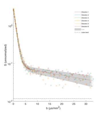
We present an application of diffusion MRI at high b-values to a non-invasive quantification of micron-sized organelles such as mitochondria. The experiments were conducted ex vivo on pork muscle and analyzed with a bi-exponential tensorial model, which allows us to estimate the mitochondria content in the muscle. Even though a more systematic comparison between mesoscale diffusion and microscale histology is deserved, this work is a proof of concept and a prerequisite for developing in vivo methods for quantifying the content of various organelles in muscles, e.g. for studying mitochondrial dysfunction in aging.
|
|
Diffusion: In Vivo & Ex Vivo Applications: Body
Electronic Poster
Diffusion
Monday, 18 June 2018
| Exhibition Hall |
09:15 - 10:15 |
| |
|
Computer # |
|
3195.
 |
1 |
Atypical Imaging Features of Renal Pelvic Urothelial Carcinoma That Mimics Central Renal Cell Carcinoma:Utility of monoexponential, biexponential, and stretched exponential Diffusion-weighted imaging models
Video Permission Withheld
Haojie Li
Multi-b values DWI are feasible and useful in the noninvasive tissue characterization of renal tumors. DDC and f may provide additional information and could lead to improved differentiation with better sensitivity and specificity between central renal cell carcinoma (RCC) from renal pelvic urothelial carcinoma compared with conventional diffusion parameters.
|
|
3196.
 |
2 |
A pilot study of the effect of high pressure renal pelvic perfusion on the renal microstructure and microcirculation using multiparametric magnetic resonance imaging (mpMRI)
Qiong Ye, Zhixian Yu, Honghui Zhu, Zhao Zhang, Jiance Li
Multiparametric MRI is widely used for tissue characterization. High pressure perfusion is commonly used in endoscopic surgery. In this study we compared the quantitative change of renal microstructure and microcirculation using DTI and simplified intravoxel incoherent motion imaging (sIVIM) in an operation simulating high pressure renal pelvic perfusion in the process of endoscopic surgery. Additionally, we compared the cortical and medullar difference. The results of this pilot study showed the feasibility of mpMRI to characterize renal physiology and investigate its quantitative change, with the potential value in early detection of renal function. But further study with larger sample size is required to draw a clear conclusion.
|
|
3197.
 |
3 |
IVIM DWI in the assessment of renal diffusion and perfusion alternations in ischemic acute kidney injury (AKI) animals
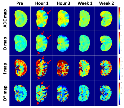
Chengyan Wang, Hanjing Kong, Fei Gao, Li Jiang, Jue Zhang, Xiaoying Wang
Intravoxel incoherent motion (IVIM) DWI is able to simultaneously detect diffusion and perfusion characteristics of renal tissue, which provides more sensitive measurement of renal function than serum creatinine. This study investigates the feasibility of using IVIM DWI to evaluate renal diffusion and perfusion changes in ischemic AKI animals. IVIM DWI was performed on rabbits prior to (24 hours before surgery) and after the surgery (1 hour, 3 hours, 1 week and 2 weeks after surgery). After injection of 0.8 mg microspheres, a noticeable change of renal diffusion and perfusion can be seen in the cortex immediately after the surgery. Pathological results also confirmed the renal injury with findings of ischemic and wrinkled features with dilated change of Bowman’s capsule.
|
|
3198.
 |
4 |
Diffusion kurtosis imaging in the characterization of rectal cancer: Evaluation of segmentation strategies and repeatability
Did Not Present
Yiqun Sun, Qin Xiao, Feixiang Hu, Chao Xin, Huixun Jia, Sanjun Cai, Robert Grimm, Caixia Fu, Xu Yan, Weijun Peng, Tong Tong, Yajia Gu
The aim of this study was to evaluate the influence of different segmentation strategies on diffusion parameters and the performance of diffusion kurtosis imaging in predicting rectal cancer histopathological characteristics before a treatment decision is made. The results show that the whole-tumor-volume segmentation strategy could achieve the best inter- and intra-observer repeatability among the six different strategies, and DKI with this segmentation strategy performed accurately for differentiating between well-differentiated and poorly to moderately differentiated patients.
|
|
3199.
 |
5 |
Evaluation of cervical cancer staging using readout segmentation of long variable echo-trains and single-shot diffusion weighted echo-planar imaging: a comparison study
Did Not Present
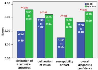
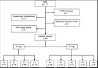
WeiLiang Qian, Qian Chen, Zhongshuai Zhang, Hong Wang, Jibin Zhang, Jianming Xu
Diffusion weighted imaging (DWI) sequence based on readout segmentation of long variable echo-trains (RESOLVE) is superior to that based on single-shot echo-planar imaging (SS-EPI) with respect to improved the image quality, which makes the images more valuable for clinical needs. In this study, we propose an idea to evaluate whether such differences in these images have an impact on staging of cervical cancer. The results show that RESOLVE can improve the accuracy of staging of cervical cancer because of the reductions of image artifacts and geometric deformation.
|
|
3200.
 |
6 |
Predictive and prognostic value of intravoxel incoherent motion (IVIM) MR imaging in patients with advanced cervical cancers undergoing concurrent chemo-radiotherapy
Zhengyang Zhou, Weibo Chen
Pelvic IVIM MR imaging were performed on 30 women with advanced cervical cancers at three time points. The performance of tumour size and IVIM-derived parameters in predicting long-term prognosis was evaluated. After a median follow-up of 24 months, 83.33% patients were alive, 70.00% remained free of disease. A shrinkage rate of maximum diameter≥ 58.31% was useful in predicting a good long-term prognosis. The IVIM-derived ADCIVIM value at time point 2 and the ADCIVIM and f values at time point 3 also performed well in predicting a good prognosis. IVIM has great potential in predicting long-term prognosis in patients with advanced cervical.
|
|
3201.
 |
7 |
Whole-lesion apparent diffusion coefficient histogram analysis: significance in T and N staging of gastric cancers
Zhengyang Zhou, Weibo Chen
Eighty patients with pathologically confirmed gastric carcinomas underwent DWI-MR imaging before surgery prospectively. Whole-lesion ADC histogram analysis was performed. The differences of ADC histogram parameters among different T and N stages were compared with independent-samples Kruskal-Wallis test. ROC analysis was performed to evaluate the performance of ADC histogram parameters in differentiating particular T or N stages of gastric cancers.There were significant differences of all the ADC histogram parameters at different T (except ADCmin and ADCmax) and N (except ADCmax) stages. Whole-volume ADC histogram parameters held great potential in differentiating different T and N stages of gastric cancers preoperatively.
|
|
3202.
 |
8 |
Diffusional kurtosis imaging of parotid glands in Sjögren's syndrome: Initial findings
Zhengyang Zhou, Weibo Chen
A total of 40 patients with SS and 40 healthy volunteers underwent DKI-MR imaging, which generated ADC, D, and K values. The MR nodular grade was determined on the basis of MR morphological findings. The parotid ADC, D, and K values in patients with SS were significantly higher than those of healthy volunteers. The K values correlated positively with the MR nodular grade significantly in patients with SS.All parotid DKI parameters differed significantly among patients with SS at different MR nodular grades. Parotid DKI parameters hold great potential in diagnosing SS, especially in early-stage SS without MR morphological changes.
|
|
3203.
 |
9 |
Assessment of Liver Fibrosis: Comparison of Diffusion Kurtosis Imaging, Conventional DWI, Aspartate Aminotransferase-to-Platelet Ratio Index and Fibrosis-4
Video Permission Withheld
Li Yang, Mengsu Zeng, Shengxiang Rao, Caizhong Chen, Robert Grimm, Caixia Fu, Xu Yan
Diffusion kurtosis imaging (DKI) is a recently developed diffusion model that measures the non-Gaussian diffusion of water molecules in biological tissue. Few studies reported the potential of DKI on assessing hepatic fibrosis. Aspartate aminotransferase-to-platelet ratio index (APRI) and fibrosis-4 (FIB-4) are widely used non-invasive serum tests that estimate liver fibrosis. We compared the diagnostic performance of DKI, conventional DWI, APRI, and FIB-4 for evaluating the severity of liver fibrosis. Our results showed that diffusion-based measurements offer a similar diagnostic performance to the serum fibrosis biomarkers APRI and FIB-4 index for predicting liver fibrosis in patients with chronic liver disease.
|
|
3204.
 |
10 |
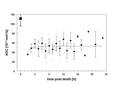
Postmortem MR diffusion-weighted imaging of the liver: Time behavior of the hepatic apparent diffusion coefficient in the early death interval
Did Not Present
Jin Yamamura, Tony Schmidt, Anne Kim, Roland Fischer, Gerhard Adam, Sarah Keller
Postmortem liver ADC values shows a characteristic change over time in the first 16 hours postmortem, which seemed to be influenced by the core body temperature. The postmortem time behavior of liver DWI values could be of interest for postmortem MRI in virtual autopsy.
|
|
3205.
 |
11 |
Towards a definition of the biophysical bases of transient Anomalous Diffusion (tAD) parameters. Evaluation of tAD, DKI and DTI in normal and cancer prostate tissue with Magnetic Resonance micro-imaging at 9.4 Tesla
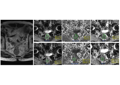
Maria Giovanna Di Trani, Alessandra Caporale, Marco Nezzo, Silvia Capuani
Since DKI and transient anomalous diffusion imaging (tADI) are based on statistical models, they can be performed without the need of a-priori hypothesis on tissue micro-structures. However, the relation between tissue micro-structure DKI and tADI derived parameters have not been clearly established yet. In this work, we evaluated DKI, tAD and DTI diffusion parameters in normal and high-grade cancer prostate, by MR microimaging at 9.4T with a 70μmx70μm in plane resolution. As prostate tissue is a complex tissue, composed by several micro-compartments that exhibit different diffusion behaviors, it is an ideal tissue to investigate the biophysical features of diffusion parameters.
|
|
3206.
 |
12 |
Prostate Cancer: Influence of the Diffusion Time on Diffusion Kurtosis Imaging
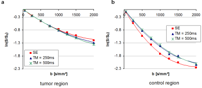
Tristan Kuder, Frederik Laun, David Bonekamp, Matthias Röthke
Diffusion MRI is routinely used in prostate cancer diagnosis. Diffusion kurtosis imaging allows measuring the kurtosis Kapp, related to deviations from free diffusion, additionally to the diffusion coefficient Dapp. Varying the diffusion time may yield additional information about the investigated tissue by probing the diffusion barriers at different length scales. Here, Dapp and Kapp were measured at three diffusion times in 27 patients with histologically confirmed prostate cancer. A reduction of Kapp was observed in tumor and normal control regions with increasing diffusion time, while a Dapp reduction was mostly seen in control regions.
|
|
3207.
 |
13 |
METastasis Reporting and Data System for Prostate Cancer (MET-RADS-P) for castration-resistant prostate cancer: prediction of clinical course, and identification of oligo-progressive lesions as targets for loco-regional ablative therapy.
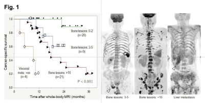
Soichiro Yoshida, Taro Takahara, Chikako Ishii, Thomas Kwee, Keiko Nakagawa, Kazuma Toda, Yuki Arita, Toshiki Kijima, Minato Yokoyama, Junichiro Ishioka, Yoh Matsuoka, Kazutaka Saito, Ryoichi Yoshimura, Kazunori Kihara, Yasuhisa Fujii
Whole-body diffusion-weighted MRI is a new-generation imaging tool for detecting prostate cancer. The extent of bone metastasis and the presence of visceral metastasis on whole-body diffusion-weighted MRI according to METastasis Reporting and Data System for Prostate Cancer (MET-RADS-P) were associated with a lower cancer-specific survival in castration-resistant prostate cancer. Furthermore, whole-body diffusion-weighted MRI facilitates identification of oligo-progressive lesions, which can be targets for loco-regional radiotherapy. MET-RADS-P score of whole-body diffusion-weighted MRI can be an imaging biomarker for castration-resistant prostate cancer in predicting clinical course, and identifying oligo-progressive lesions as targets for loco-regional ablative therapy.
|
|
3208.
 |
14 |
Acute Ankle Sprain: Demonstration of Reliability and Reproducibility of DTI in Imaging Peripheral Nerves
Natalia I Lopez, Nadia Barakat, Andrew M Youssef, Katie E Silva, Jürgen Finsterbusch, Laura Simons, David Borsook
We investigated potential structural changes in peripheral nerves following ankle sprain injuries. Specifically, we assessed the integrity of the sciatic nerve and its major divisions: the tibial and peroneal nerves. Reduced field-of-view DTI was used, and the reproducibility of the DTI measures was examined. Our results revealed excellent reliability of DTI measures in injured versus control nerves across each parameter (FA, AD, RD, MD). A comparison of injured versus control nerves in the associated nerve branch indicated significant difference in AD. Given these results, DTI may have potential as a powerful tool to determine disease profile.
|
|
3209.
 |
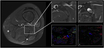
15 |
Evaluation of two collagen conduits and autograft in sciatic nerve regeneration in a rabbit nerve gap model with DTI, histology and electrophysiology
Tina Jeon, Emil Vutescu, Eliana Saltzman, Jordan Villa, Scott Wolfe, Steve Lee, Joseph Feinberg, Sarah Pownder, Jonathan Dyke, Darryl Sneag
DTI has been used primarily to evaluate white matter tracks in the brain. More recent studies have applied DTI techniques to peripheral nerves, due to their anisotropic architecture. In this investigation, we evaluated peripheral nerve regeneration in a rabbit sciatic nerve gap model comparing two collagen conduits with nerve autograft using DTI and comparison with functional/physiologic testing and histology. We hypothesized that this study would allow us to reliably compare outcomes of nerve regeneration between collagen-based conduits and autograft nerve reconstructions and provide validation for the use of DTI techniques to non-invasively monitor nerve regeneration in-vivo.
|
|
3210.
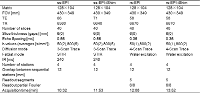 |
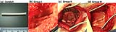
16 |
Improvements in Whole Body Diffusion Weighted Imaging: Combination of Integrated Slice-Specific Dynamic Shimming and Readout-Segmented EPI
Wei Liu, Alto Stemmer, Elisabeth Weiland, Kun Zhou
Single-shot echo planar imaging (ss-EPI) is most frequently used for whole body diffusion weighted imaging (WB-DWI) because of short acquisition time and motion insensitivity. However, ss-EPI is vulnerable to the effects of the static field inhomogeneity and poses a challenge to perform ss-EPI based WB-DWI at 3 Tesla. Integrated slice-specific dynamic shimming (iShim) combined with ss-EPI has shown a remarkable improvement on the susceptibility related artifacts in WB-DWI. In this study, we demonstrate the application of rs-EPI using iShim to WB-DWI, which can provide higher quality WB-DWI, specifically less spatial distortions.
|
|
3211.
 |
17 |
The clinical value of DWIBS in the diagnosis of bone marrow involvement in lymphoma and hyperplastic hematopoietic bone marrow
Mengtian Sun, Jingliang Cheng, Yong Zhang, Zhizheng Zhuo
This study aimed to investigate the clinical value of diffusion weighted imaging with background signal suppression(DWIBS) in differentiating bone marrow involvement(BMI) in lymphoma from hyperplastic hematopoietic bone marrow(HHBM). Eleven BMI patients, 19 HHBM patients and 20 normal controls underwent DWIBS before the bone marrow biopsy. The ADC value of the bone marrow within the biopsy regions in BMI group was lower than that of HHBM group and higher than that of normal controls. ADC values added relevant information in the potential clinical value of differentiating BMI and HHBM patients.
|
|
3212.
 |
18 |
Development of Diffusion Tensor Imaging to Assess Traumatic Peripheral Nerve Injury and Recovery
Michael Pridmore, Wesley Thayer, Mark Does, Richard Dortch
Current clinical management following traumatic peripheral nerve injuries (TPNI) and repair require physicians to rely on qualitative measures from patient history/physical exam that can cause delay in patient care. Such delays can a negative impact on outcomes because the healing of nerves must occur in a timely fashion to avoid permanent loss of sensory and/or motor function. The current study aims to test the feasibility of performing DTI measurements in TPNI patients to better improve clinical outcomes.
|
|
3213.
 |
19 |
A Local Coregistration for Quantitative Evaluation of Distortion of Parotid Gland Tumors in Echo-Planar DWI and PROPELLER DWI
Video Permission Withheld
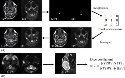
Kai-Min Chien, Yi-Jui Liu, Yi-Hsiung Lee , Hing-Chiu Chang , Hui-Chu Chiu , Ta-Wei Chiu , Kang Hsu , Hsian-He Hsu , Chun-Jung Juan, Yu-Shu Fu, CHENG-YI JUAN
The study is to quantitatively compare the morphology distortion in distinguishing parotid pleomorphic adenomas (PMA) between PROPELLER-DWI and EP-DWI. This retrospective study enrolled 14 PMAs. All participants underwent 1.5-T fat-saturated diffusion-weighted imaging with PROPELLER-DWI and EP-DWI. A local coregistration method to quantitatively evaluate the distortion of parotid gland tumors between single shot EP-DWI and PROPELLER DWI. Imaging distortion represented by Dice coefficient was quantitatively analyzed. Our results showed that PROPELLER-DWI allows distinguishing PMAs with less distortion than EP-DWI.
|
|
3214.
 |
20 |
Is ADC Affected by Distortion of EP-DWI for Distinguishing Parotid Gland Tumor- To Compare ADC of Parotid Gland in Echo-Planar DWI and PROPELLER DWI
Video Permission Withheld
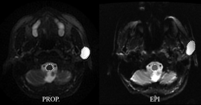
Kai-Min Chien, Yi-Jui Liu, Yi-Hsiung Lee , Hing-Chiu Chang , Hui-Chu Chiu , Ta-Wei Chiu , Kang Hsu , Hsian-He Hsu , Chun-Jung Juan, Yu-Shu Fu, CHENG-YI JUAN
The study is to investigate if the perceptible geometric distortions could bias for parotid pleomorphic adenomas (PMA) by ADC measurements by comparing PROPELLER-DWI with EP-DWI. This retrospective study enrolled 14 PMAs. All participants underwent 1.5-T fat-saturated diffusion-weighted imaging with PROPELLER-DWI and EP-DWI. ADCs were measured on normal parotid gland and PMA for PROPELLER-DWI and EP-DWI. Our results showed that PMAs had significantly higher ADC than normal parotid glands no matter on PROPELLER-DWI or EP-DWI. The ADC measured by PROPELLER-DWI was significantly higher than by EP-DWI, but they were proportional to each other. EP-DWI allows distinguishing PMAs even under image distortion.
|
|
3215.
 |
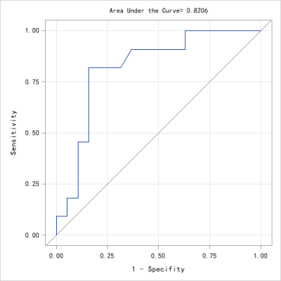
21 |
The value of DWI with Readout Segmentation of Long Variable Echo-trains in Differentiating Benign and Malignant Lesions of the Tongue
Baohong Wen, Jingliang Cheng
The purpose of this study was to assess the role of readout segmentation of long variable echo-trains(RESOLVE) DWI in differentiating benign and malignant lesions of the tongue. 120 patients with lingual lesions underwent preoperative RESOLVE DWI. The results demonstrated that the mean ADC values of benign lesions of the tongue were significantly higher than malignant lesions. Using an ADC value of 1.25×10⁻³ mm2/s as the threshold value, the sensitivity and specificity were 94.64%, 93.75% respectively. RESOLVE can offer high quality DWI of tongue. The ADC value can be applied in differential diagnosis between benign and malignant lesions of the tongue
|
|
3216.
 |
22 |
Diffusion kurtosis imaging in evaluating non-Hodgkin’s lymphoma: associations with tumor aggressiveness and proliferation
Jing Zhong, Yunbin Chen, Ying Chen, Cuifang Chen, Weibo Chen
This study tries to evaluate the usefulness of diffusion kurtosis imaging (DKI) parameters in segregating the pathological subtypes of NHL lymphoma, and to explore its associations with aggressiveness and proliferative index. Significant differences were found among the WHO classified subgroups. Meanwhile, the stronger tumor aggressiveness (higher Ki67 percentage) was accompanied with more restricted water diffusivity (lower ADC and D values) and more complex cell micro-structural environment (higher K value). The DKI technique may help estimation of tumor proliferation in NHL lymphoma and DKI parameters can be used as imaging biomarker of the biological aggressiveness of the tumor.
|
|
3217.
 |
23 |
Assessment of the Specificity and Sensitivity of DTI Metrics for Evaluation and Diagnosis in Degenerative Cervical Myelopathy
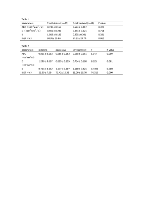
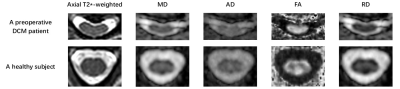
Guangqi Li, Xiaodong Ma, JinChao Wang, Donghang Li, Xiao Han, Wen Jiang, Xiaoguang Cheng, Hua Guo
Diffusion Tensor Imaging (DTI) can detect diffusion information of water molecules, and is used to diagnose the severity of degenerative cervical myelopathy (DCM). However, the diagnostic capability of DTI metrics is not fully investigated. In this study, DTI metrics are employed to evaluate the spinal cord function in preoperative DCM patients and healthy volunteers. Nonparametric t-test results show that MD, FA and RD have significant differences between patients and healthy volunteers. In addition, ROC results indicate that FA has higher sensitivity, RD has higher specificity for evaluation and diagnosis in DCM.
|
|
3218.
 |
24 |
L-spine Bone Marrow on Female: A Intravoxel Incoherent Motion MR Imaging Study
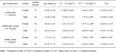
Teng Zhao, Yunsong Zheng, Hui Xu, Yuanyuan Chen, Yanbing Guo, Dong Han, Nan Yu
To our knowledge, no studies have employed IVIM diffusion-weighted MRI to explore the variation trend of bone marrow in female. Whether gender difference exists in marrow structure is not well investigated. Therefore, we explored the diagnostic utility of IVIM diffusion-weighted MRI parameters in this context. We found that D, D* and f value showed a decreased trend with age, and the D, D* value of bone marrow in female was significantly higher than that in male except the f value. IVIM diffusion-weighted MRI was useful in the evaluation of bone marrow.
|
|
Diffusion MRI: Validation
Electronic Poster
Diffusion
Monday, 18 June 2018
| Exhibition Hall |
09:15 - 10:15 |
| |
|
Computer # |
|
3219.
 |
25 |
How do current diffusion-based MR methods reflect hypomyelination – comparison of diffusion tensor, neurite orientation dispersion and density, and diffusion kurtosis imaging
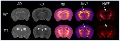
Rakshit Dadarwal, Amir Moussavi, Wiebke Möbius, Susann Boretius
Diffusion-based MRI comprises an exciting toolset to analyze tissue microstructures under normal and pathological conditions. Among numerous diffusion-based methods compared, all reflected the differences in myelination in a mouse model expressing only reduced levels of the myelin basic protein. However, diffusion tensor was more robust than diffusion kurtosis imaging. Intra-neurite volumes, as revealed by neurite orientation dispersion and density imaging or by the spherical mean technique, were not specific for the numbers of axons but were also affected by difference in myelination.
|
|
3220.
 |
26 |
Realistic 3D Fiber Crossing Phantom Models for Monte Carlo Diffusion Simulations
Jonathan Rafael-Patino, Gabriel Girard, David Romascano, Muhamed Barakovic, Gaëtan Rensonnet, Jean-Philippe Thiran, Alessandro Daducci
Monte-Carlo Diffusion Simulations has proved to be a powerful approach to study Diffusion-Weighted MRI; from generating ground-truth data, to study the diffusion process in complex media. However, a major problem with the current approaches is that they oversimplify the geometrical properties of the diffusion media. In this work we present a framework to create 3D meshes for realistic configurations that can be used for MCDS. The synthesized signals from this models can be used to study microstructure and tractography methods, which is of vital importance since novel methods require better ground-truth that mimics real tissue properties to avoid oversimplifications.
|
|
3221.
 |
27 |
Critical Choices in ROI Analysis of Diffusion MRI Data
Mohammad Alipoor, Stephan Maier
Diffusion parameters such as diffusivity, compartment fractions and diffusion signal itself are informative bio-markers in understanding and analyzing pathological changes in biological tissue. One frequently needs to compute a representative value of a diffusion parameter in a homogeneous ROI. Here we compare and contrast 3 common choices that researcher would make in the course ROI analysis. Though ROI analysis is deemed to be a common practice, the critical choices of computational methods (depending on noise distribution and underlying model) can considerably affect its findings and conclusions.
|
|
3222.
 |
28 |
Transient anomalous diffusion micro-MRI parameters reflect white matter morphology: comparison with histology of the mouse spinal cord.
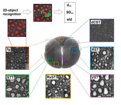
Alessandra Caporale, Giovanni Bonomo, Giulio Tani, AdaMaria Tata, Bice Avallone, Felix Wehrli, Silvia Capuani
Transient-anomalous diffusion (tAD) has previously been used for tumor delineation and human brain tissue characterization, however, comparison with histology is largely missing. This work aims to compare α and γ tAD parameters, DTI and q-space-imaging parameters obtained at 9.4T with micro-MRI, with the morphologic characteristics provided by optical microscopy of mouse spinal cord white matter (MSC-wm). We found that γ- and q-space-imaging are sensitive to axon diameter and effective local axon density, while α-imaging is sensitive to the heterogeneity or degree of disorder of the wm tracts. These techniques outperform DTI as a means to probe MSC-wm morphology.
|
|
3223.
 |
29 |
Developing 3D perfusion bioreactor for MRI and optical imaging
Video Permission Withheld
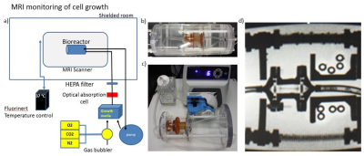
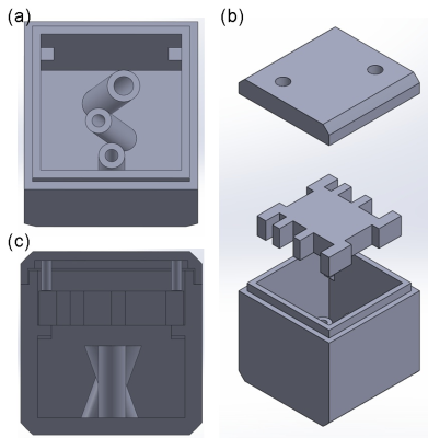
Slavka Carnicka, Jeanne Barthold, Kathryn Keenan, Karl Stupic, Corey Neu, Stephen Russek
Whole-body medical imaging (such as MRI) can map many physical tissue parameters; however, there are currently many questions in the field regarding how changes in MRI are representative of changes in the underlying cells. To better understand these processes, we need to correlate MRI measurements with changes in microstructure. We created a living phantom for evaluation of techniques such as diffusion tensor imaging (DTI) that can be monitored and validated by optical techniques. Our future plan is to use MRI to study cell growth and monitor response to chemical and mechanical stimuli.
|
|
3224.
 |
30 |
Test-retest reliability of graph theoretic metrics in adolescent brains
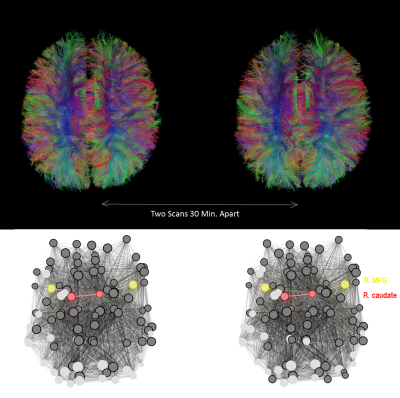
Justin Yuan, Eva Henje Blom, Trevor Flynn, Yiran Chen, Tiffany Ho, Colm Connolly, Rebecca Dumont Walter, Tony Yang, Duan Xu, Olga Tymofiyeva
Graph theory analysis of structural brain networks derived from diffusion tensor imaging (DTI) has been utilized to study neurological and psychiatric disorders but its reliability remains understudied, especially in the still-developing brain. Repeated DTI scans of adolescents were acquired to assess the test-retest reliability of different weighting schemes of brain networks: fractional anisotropy (FA), streamline count (SC), and binary (B). The test-retest scans were performed at two time intervals: 12 weeks apart and within the same scan session, approximately 30 minutes apart. Results suggest that FA-weighting outperforms the other schemes.
|
|
3225.
 |
31 |
Design of multi-purpose and 3D-printed fibre phantoms for investigating complex tissue microstructures
Husan-Han Chiang, Kuan-Hung Cho, Ezequiel Farrher, Johannes Lindemeyer, Richard Buschbeck, Ming-Jye Chen, Farida Grinberg, Nadim Shah, Chang-Hoon Choi, Li-Wei Kuo
Investigating complex tissue microstructures has become of great interest during the past decade. One of the most promising MRI methods to map tissue microstructures is diffusion MRI. However, the validation of its accuracy in mapping fibre orientation and microstructural characteristics is still challenging. In this work, we have successfully designed and prototyped a fibre phantom using 3D printing and micro-scale fused silica capillaries. Our results show that susceptibility of the capillaries and/or the coating material is different from that of distilled water and suggest that our phantom design could provide detectable microstructures for further studies.
|
|
3226.
 |
32 |
Multisite Reproducibility of Radiomics and ADC Measurements for temperature-controlled phantom: Preliminary Results.
Video Permission Withheld
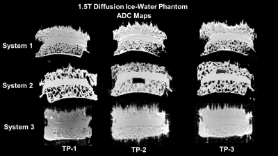
Michael A. Jacobs, Dariya I. Malyarenko, David C. Newitt, Vishwa S. Parekh, Nola M. Hylton, Thomas L. Chenevert
Radiomics is an emerging field which deals with high throughput extraction of quantitative features from radiological images. Radiomic features correspond to textural information that is otherwise not visually perceivable. To establish confidence intervals for extracted features the longitudinal reproducibility of the radiomic metrics should be assessed with the standard. This work has demonstrated radiomic reproducibility for ADC mapping acquired for the ice-water phantom over several years on three independent systems at two different field strengths (1.5 and 3T).
|
|
3227.
 |
33 |
MR Characterization and Temperature Dependence of Aqueous Polyvinylpyrolidone (PVP) Solutions for use as MR Phantoms
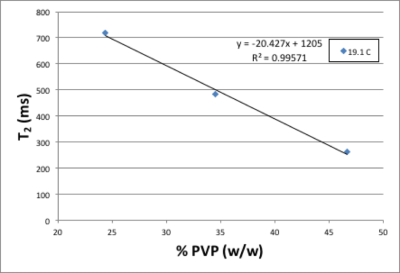
Joelle Sarlls, Michal Komlosh, Ferenc Horkay, Uri Nevo, Peter Basser, Carlo Pierpaoli
Diffusion-weighted MRI methods often contain variability and bias in diffusion parameters that are measured between sites, scanners, and vendors. There is a clear need for a calibrated diffusion phantom to help identify and mitigate these differences. Here we utilize a 7T spectrometer to characterize MR parameters of T1, T2, and the water self-diffusion coefficient in aqueous Polyvinylpyrolidone (PVP) solutions and their dependence on temperature and PVP concentration, without imaging confounds. An empirical formula is presented for use wtih PVP aqueous solutions as a calibrated diffusion phantom. Data show that aqueous PVP solutions are well suited as a MR phantom material.
|
|
3228.
 |
34 |
Does neurite density as measured by diffusion-weighted MR imaging relate to neuronal density as measured by MR spectroscopy?
Hamied Haroon, Ben Dickie, Faezeh Nezhad, Martyn McFarquhar, Stephen Williams, Geoff Parker, Laura Parkes
Capturing the earliest signs of dementia with MR imaging relies on techniques that are sensitive to the subtle loss or disconnection of neurons before atrophy occurs. Models of multi-shell HARDI such as NODDI claim to quantify neurite density in vivo and non-invasively, but the specificity of these HARDI-based metrics remain unvalidated. This study aims to determine the sensitivity of NODDI’s neurite density and orientation dispersion index to regional variation of MRS markers of neuronal and glial cell density. We find that caution must be exercised when interpreting NODDI’s neurite density as related to neuronal density. Orientation dispersion instead appears to be a closer marker of neuronal density and may be a more sensitive marker of disease-related change.
|
|
3229.
 |
35 |
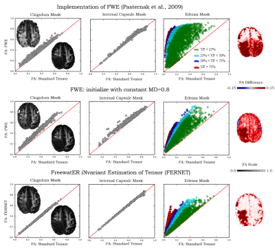
Free watER iNvariant Estimation of Tensor (FERNET): Addressing the Issue of Edema in Clinically Feasible Acquisitions
Abdol Aziz Ould Ismail, Drew Parker, Simon Alexander, Emmanuel Caruyer, Ofer Pasternak, Ragini Verma
Despite the growing research in free water elimination (FWE) methods with advanced diffusion acquisition protocols, the need for robust single-shell based FWE remains, as this is the standard acquisition protocol in the clinic. This is especially important in the characterization of peritumoral regions with infiltration. However, single-shell FWE is an ill-posed problem, dependent on parameter initialization, solutions to which often fail to obtain a balanced correction between healthy and abnormal tissue. We introduce FERNET, a robust FWE protocol for single-shell data with a comprehensive investigation of initialization parameters based on a software simulated phantom where the ground truth is known.
|
|
3230.
 |
36 |
Multi-shell multi-tissue fODF tractography improves V1-V2 macaque connectivity mapping
Guillaume Theaud, Maxime Descoteaux, Rémi Cossette-Roberge, Jean-Christophe Houde, Chuyang Ye, Nathalie Richard, Yujie Hou, Loïc Magrou, Kenneth Knoblauch, Henry Kennedy, Bassem Hiba
We show that multi-shell (multi b-value), multi-directional, and high spatial resolution (300 microns isotropic) diffusion MRI combined with multi-tissue fiber orientation distribution function (fODF) tractography increases by 6% the number of true positive connections and uniformly increases the cortical coverage by 3%, while preserving the same percentage of false positive connections, with respect to a more standard single-tissue single-shell tractography. As a result, it is possible to find all 5 ground truth V1-V2 bundles (true positives), while reconstructing only 4 invalid bundles (false positives) corresponding to 4 pairs of spatially neighboring regions.
|
|
3231.
 |
37 |
Apparent exchange rate mapping: relation to membrane permeability
Dominik Ludwig, Frederik Laun, Peter Bachert, Tristan Kuder
Apparent exchange rate (AXR) mapping might provide an insight into the exchange of water between intra- and extracellular space by using a double-diffusion encoded sequence with varying mixing time between the two gradient pairs. To investigate the connection between AXR and membrane permeability and to test the assumptions of the underlying theory, Monte Carlo simulations using simplified tissue models were performed. Simulations covered a broad range of membrane permeabilities to determine limits of the applicability of this technique. For the considered simplified tissue model, AXR-values could be reliably related to membrane permeabilities typically occurring in vivo.
|
|
3232.
 |
38 |
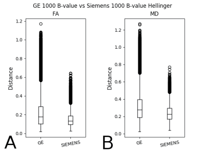
Characterization of Diffusion Metric Map Similarity in MRI Data from a Clinical PACS using the Histogram Distance
Graham Warner, Karl Helmer
As data reuse becomes more popular, it is critical to develop methods that characterize the similarity of data. Methods have been developed that characterize raw image files, but users often only have access to calculated parameter maps. Here we describe a histogram-distance-based method applied to diffusion metric maps generated from MRI data extracted from a clinical data repository. We find that metric maps from GE scanners are less similar than that from Siemens scanners. We also find within vendor differences at any selection of the acquisition parameters considered here (field strength, number of gradient directions, b-value and vendor).
|
|
3233.
 |
39 |
Pre-treatment intra- and inter- voxel magnetic resonance diffusion heterogeneity correlates with chemoradiotherapy treatment outcome of patients with head and neck squamous cell carcinoma
Marianthi-Vasiliki Papoutsaki, Harbir Sidhu, Nikolaos Dikaios, David Atkinson, Timothy Beale, Simon Morley, Martin Forster, Dawn Carnell, Ruheena Mendes, Shonit Punwani
Heterogeneity assessment and diffusion weighted magnetic resonance imaging (DW-MRI) have been considered powerful diagnostic tools in predicting chemoradiotherapy treatment outcome in patients with cancer. In this study, pre-treatment microstructural heterogeneity derived by intra- and inter-voxel MR diffusion rates was assessed in patients with neck squamous cell carcinoma (HNSCC). A correlation was presented between the pre-treatment MR diffusion heterogeneity and the chemoradiotherapy treatment outcome of patients with HNSCC. Future work, to ascertain the mechanisms of these correlations would open the opportunity to tailor therapies to individuals in clinical practice.
|
|
3234.
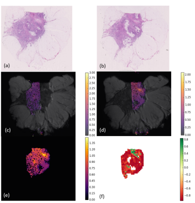 |
40 |
Histological breast cancer stroma patterns correlate with diffusion MR signal
Sara Reis, Colleen Bailey, Thomy Mertzanidou, Bernard Siow, Eleftheria Panagiotaki, John Hipwell, Julie Owen, Patrycja Gazinska, Sarah Pinder, Daniel Alexander, David Hawkes
We report on the histological analysis of the stroma compartment of two formalin-fixed breast cancer ex-vivo samples that were scanned under a wide range of PGSE acquisitions. Our histological analysis of stroma approach shows that mature regions present higher ADC values compared to immature regions. Mature regions are mainly composed by highly organised fibres, normally following the same direction, which may facilitate directional movement of molecules. This increases the effective tortuosity in the tumour and its interstitial fluid pressure and osmotic pressure, which inhibits water diffusion and subsequently results in a lower ADC value.
|
|
3235.
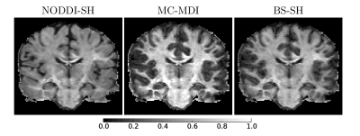 |
41 |
Investigating Diffusion-MRI based neurite density estimation model dependency: an in-vivo study on the HCP dataset
Mauro Zucchelli, Maxime Descoteaux, Gloria Menegaz
Diffusion MRI can be used to estimate the brain tissue neurite density from Multi-Compartment models. This index corresponds to the “stick” compartment volume fraction estimated in every voxel. In this work, we provide evidence that the distribution of stick volume fraction is characteristic of the brain tissue and is highly reproducible between subjects but strongly depends on the underlying multi-compartment model definition. In particular, in-vivo results on 10 subjects of the Human Connectome Project show that the neurite density distribution depends on both the stick parallel diffusivity and the extra-axonal compartment model.
|
|
3236.
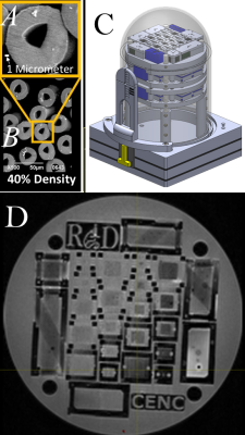 |
42 |
Time-dependent diffusion modeling using a hollow textile-based Phantom
Sudhir Pathak, Vinod Kumar, Catherine Fissell , Anthony Zuccolotto, Walter Schneider
A Textile Anisotropic Brain Imaging Phantom incorporating textile water filled hollow fibers (taxons with inner/outer diameter 12/34 micron) is used to examine time-dependent diffusion. In this study, impermeable hollow tubes (taxons) with 12-micron diameter are used to test the relationship between axial and radial diffusivities with diffusion time (Δ) for a given taxon packing density. An inverse relationship of radial diffusivity with diffusion time (Δ) is established. A constant relationship of axial diffusivity with diffusion time is established. The dependence of these relationships on packing density is then tested and the radial diffusivity relationship is shown to vary with packing density.
|
|
3237.
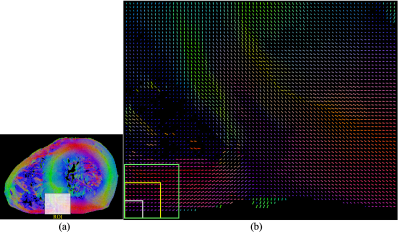 |
43 |
Investigation of Myocardial Fiber Crossings in the Human Heart Using Realistic HARDI Simulation Based on PLI
Lihui WANG, Feng YANG, Gabrielle Michalowicz, Yves Usson, Pierre-Simon Jouk, Rongpin Wang, Yuemin Zhu
We investigate fiber crossings in the myocardium of the human heart using realistic HARDI simulation based on polarized light imaging (PLI). The whole human heart was first imaged using PLI. Based on the fiber orientations measured by PLI, cardiac fiber structures were then modeled, and finally diffusion-weighted images were simulated at different scales using Monte Carlo method and the corresponding ODFs were calculated. The results show that fiber crossings clearly appeared in the myocardium with the increase of scale and that the accuracy of estimating the number of fiber crossings degraded with variable false positive and/or false negative errors.
|
|
3238.
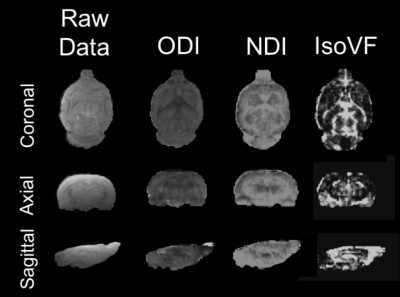 |
44 |
Reliability of Neurite Orientation Dispersion and Density Imaging (NODDI) at 9.4 Tesla
Video Permission Withheld
Patrick McCunn, Alex Li, Peter Zeman, Kyle Gilbert, Ali Khan, Robert Bartha
Neurite Orientation Dispersion and Density Imaging (NODDI) is a rapidly emerging diffusion MRI (dMRI) technique used to characterize microstructural complexity through the compartmental modelling of neural water fractions into Intra-neurite, Extra-neurite and CSF volume fractions. This project aimed to further the ability of preclinical diffusion imaging through the application of NODDI to a rodent model with the objective to determine its utility, precision and reliability at 9.4 Tesla.
|
|
3239.
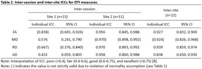 |
45 |
Test-Retest and Between-Site Reliability in a Multisite Diffusion Tensor Imaging Study
Ikbeom Jang, Sumra Bari, Yukai Zou, Nicole Vike, Pratik Kashyap, Thomas Talavage
Diffusion tensor imaging (DTI) has been frequently employed in the identification of brain biomarkers for neurodevelopmental and neurodegenerative disorders due to its ability to measure spatial organization of brain tissue. Due to the need for larger sample size to address substantive questions of interest, many studies try to merge data from several scanners, and ideally, a reliability study should come first. In this study, we assess reliability of DTI measures across two systems using the intraclass correlation coefficient, such that we may pool data in future multi-site DTI studies.
|
|
3240.
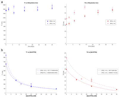 |
46 |
Optimal tissue preparation for ex vivo preclinical imaging
Rachel Barrett, Diana Cash, Camilla Simmons, Tobias Wood, Anthony Vernon, Marco Catani, Flavio Dell'Acqua
Ex vivo imaging is beneficial for studying rodent brain microstructure in healthy and pathological tissue at high resolution. There are challenges however associated with changes in tissue properties resulting from fixation. We present a tissue preparation protocol optimised for diffusion MRI in the rat brain by varying fixative concentration, gadolinium concentration and rehydration time. By altering T1 and T2 relaxivity, we show how these factors can be combined to maximise SNR efficiency. Improving SNR efficiency in ex vivo diffusion MRI will allow higher spatial and angular resolution for studying tissue microstructure in the rodent brain.
|
|
3241.
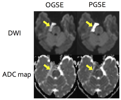 |
47 |
Oscillating gradient spin echo (OGSE) diffusion weighted imaging of the epidermoid cysts: simulation application
Saori Koshino, Akiyuki Sakakibara, Antoine Choppin, Christina Andica, Akifumi Hagiwara, Tomoko Maekawa, Mihiro Takemori, Atsushi Arakawa, Lydia Chougar, Masaaki Hori, Shigeki Aoki
Diffusion weighted imaging with shorter diffusion time using oscillating gradient spin echo (OGSE) may reveal microstructural features among brain disorders. Here we observed apparent diffusion coefficient (ADC) values in three patients with intracranial epidermoid cysts. ADC values measured by OGSE were higher than those measured by pulsed gradient spin echo (PGSE; used in conventional MRI), indicating restricted diffusion due to spatial restriction. The results of our diffusion simulation based on the pathological feature of epidermoid cysts suggest that spatial restriction of “Rugby Ball” regions formed by keratin layers should mainly affect the extent of restricted diffusion.
|
|
3242.
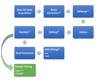 |
48 |
Correlation of baseline DSI based metrics with clinical motor outcomes at 6 weeks after acute ischemic stroke
Kyler Hodgson, Ganesh Adluru, Lorie Richards, Jennifer Majersik, Nagesh Adluru, Edward DiBella
The difference between the baseline ipsilesional and contralesional mean values in the internal capsule from Orientation Dispersion Index and Generalized Fractional Anisotropy correlate strongly with upper extremity clinical outcomes at 6 weeks. These models account for regions of crossing fibers and demonstrate improvements over DTI in using brain microstructure to make clinical judgments.
|
|
| Back |
| The International Society for Magnetic Resonance in Medicine is accredited by the Accreditation Council for Continuing Medical Education to provide continuing medical education for physicians. |
