|
Electronic Poster Session
Cardiovascular |
Monday, 18 June 2018
Electronic PosterCardiovascular
3315 -3338 Cardiac Function & Myocardial Perfusion
3339 -3361 Vascular
3433 -3456 Velocity & Flow
3457 -3480 Atherosclerosis & Vessel-Wall Imaging |
| |
Cardiac Function & Myocardial Perfusion
Electronic Poster
Cardiovascular
Monday, 18 June 2018
| Exhibition Hall |
13:45 - 14:45 |
| |
|
Computer # |
|
3315.
 |
1 |
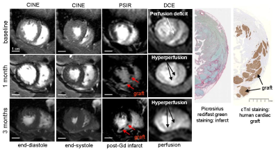
 Temporal changes in cardiac contractility, perfusion and infarct size after human cardiomyocyte transplantation to the infarcted heart of non-human primates. Temporal changes in cardiac contractility, perfusion and infarct size after human cardiomyocyte transplantation to the infarcted heart of non-human primates.
Anna Naumova, William Kerwin, Yen-Wen Liu, Billy Chen, Xiulan Yang, Hiroshi Tsuchida , R. Scott Thies, Charles Murry
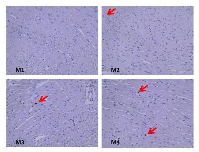
This study demonstrates structural and functional benefits of human embryonic stem cell derived cardiomyocyte engraftment to the infarcted heart of nonhuman primates. Those benefits were manifested in improved global and regional left ventricle contractile function, improved myocardial perfusion, decrease in infarct size and partial regeneration of the scarred myocardium. The benefit from human cardiomyocyte therapy was durable with the potential for further improvement in function between 1 and 3 months. The functional recovery was larger than were observed previously on small animal models of myocardial infarction, which might be due to the greater physiological match between human and macaque species.
|
|
3316.
 |
2 |
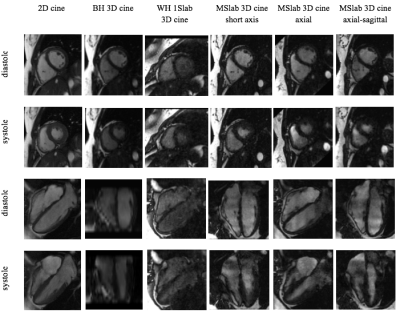
Feasibility of Multi-Thin-Slab Whole-Heart 3D Cine with Isotropic Resolution and High Contrast in Free Breathing
Peng Lai, Haonan Wang, Anja Brau, Martin Janich
Conventional 2D cine is hindered by its needs of repeated breathhold and imaging at each individual view, while breathheld 3D cine suffers from limited slice coverage and resolution. This work developed a new 3D cine sequence with free-breathing capability for whole-heart coverage and isotropic resolution and multi-thin-slab acquisition for high blood-to-myocardium contrast.
|
|
3317.
 |
3 |
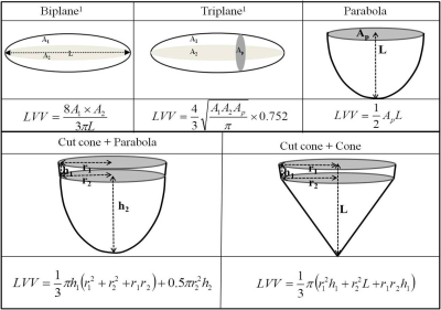
Evaluation of geometric models for estimating left ventricular (LV) mass in 980 children using cardiac cine magnetic resonance imaging
Jiming Zhang, Carlo Uribe, Benjamin Cheong, Paolo Angelini, Raja Muthupillai
LV mass computed from commonly used bi-plane and tri-plane ellipsoidal models can deviate significantly when compared to LV mass estimated from a stack of short axis balanced SSFP cine MR images. The results from the study show that by using different geometric assumptions for the shape of the endocardium (Cut-cone+cone) and epicardium (Cut-cone+parabola), it is possible to estimate LV mass with just two projections that is comparable to that obtained from a full stack of short axis slices.
|
|
3318.
 |
4 |
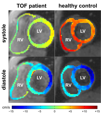
Tissue Phase Mapping for Assessment of Biventricular Myocardial Motion in Pediatric Patients with Repaired Tetralogy of Fallot
Alexander Ruh, Arleen Li, Michael Rose, Haben Berhane, Joshua Robinson, Michael Markl, Cynthia Rigsby
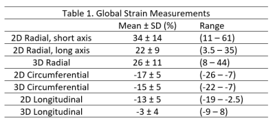
The purpose of this study was to assess regional biventricular myocardial function in pediatric patients with repaired tetralogy of Fallot (TOF) by tissue phase mapping (TPM). Segmental left (LV) and right ventricular (RV) peak velocities were calculated in systole and diastole based on an extended AHA 16+10-segment model. Inter-ventricular dyssynchrony was quantified from the correlation between global LV and RV radial, long-axis and circumferential velocity time courses. Compared to age-matched healthy controls, TOF patients exhibited reduced long-axis peak velocities in systole and diastole as well as increased inter-ventricular dyssynchrony for radial and circumferential myocardial motion.
|
|
3319.
 |
5 |
Volumetric Measurements with Real-Time Imaging for Cardiac Stress MRI
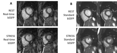
Shams Rashid, Yang Cheng, William Schapiro, Kathleen Gliganic, Ann-Marie Yamashita, Marie Grgas, Michelle Maragh, Jie Cao, Yu Li
Cardiac stress MRI is a valuable diagnostic tool for heart disease. However, volumetric measurements are challenging because stress introduces rapid heartbeats and body movements. Real-time MRI is advantageous for this application due to its robustness to motion and ability for ungated imaging during free breathing. Here, we demonstrate an undersampled radial balanced steady state free precession (bSSFP) sequence to acquire real-time cine images at high spatial (1.7 mm) and temporal (40 ms) resolutions, applied in cardiac stress MRI. We show that, compared to standard breath-held bSSFP cine MRI, our technique provides more robust and reliable volumetric measurements.
|
|
3320.
 |
6 |
Global left ventricular myocardial deformation measures by CMR tissue tracking in isolated diastolic dysfunction (DD) spontaneous T2DM rhesus monkeys: comparison with tagging
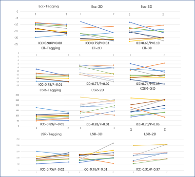
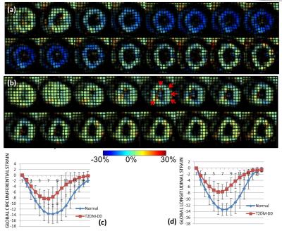
Tong Zhu, Li Gong, Yushu Chen, Yu Zhang, Wen Zeng, Jie Zheng, Fabao Gao
In this study, the myocardial deformation characteristics of early diabetic cardiomyopathy were evaluated by using CMR strain imaging in a spontaneous non-human primate T2DM disease model. We found the early cardiac dysfunction characteristics of diabetic cardiomyopathy, and verified the effectiveness of CMR-tissue tracking in its early diagnosis.
|
|
3321.
 |
7 |
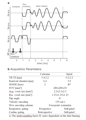
Whole heart First-pass spiral perfusion imaging with 1.25mm resolution at 3T
Yang Yang, Christopher Kramer, Michael Salerno
First-pass contrast-enhanced myocardial perfusion imaging is a useful noninvasive tool to evaluate patients with coronary artery disease, but current techniques are still limited in spatial-temporal resolution, and ventricular coverage which reduces the sensitivity to detect perfusion differences between the endocardium and epicardium and quantify ischemic burden. Outer-volume suppression (OVS) can achieve good signal suppression around the heart, but may have SAR limitations at 3T. In this study, we designed a spiral pulse sequence with slice-interleaved or simultaneous multi-slice (SMS) acquisition without OVS to achieve comparable high quality ultra-high 1.25mm resolution perfusion imaging. The sequences were tested in heathy volunteers and demonstrated high image quality.
|
|
3322.
 |
8 |
Impact of native T1 on pixel-wise myocardial blood flow quantification
Corina Kräuter, Ursula Reiter, Clemens Reiter, Albrecht Schmidt, Michael Fuchsjäger, Rudolf Stollberger, Gert Reiter
Native myocardial T1 varies between subjects and between segments, yet its impact on pixel-wise quantification of myocardial blood flow (MBF) has not been studied. 15 patients with coronary heart disease underwent 3T cardiac magnetic resonance native myocardial T1 mapping and perfusion imaging at rest. Nonlinearity correction for MBF calculation was performed employing literature native T1 values and patient-specific global as well as local native T1, respectively. Since reference T1 revealed substantial individual MBF errors and application of patient-specific global T1 overestimated MBF in perfusion deficit regions compared to local T1, patient-specific local native T1 should be employed for MBF quantification.
|
|
3323.
 |
9 |
Accelerated Cardiac Perfusion MRI with Radial k-space Sampling, Compressed Sensing, and KWIC filtering to Enable Qualitative and Quantitative Analyses of Perfusion.
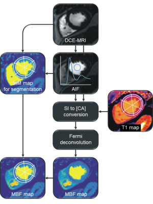
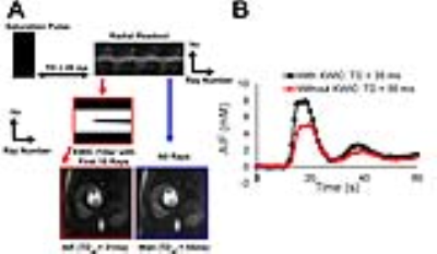
Nivedita Naresh, Hassan Haji-Valizadeh, Ali Serhal, Pascale Aouad, Daniel Lee, Daniel Kim
First-pass cardiac perfusion MRI is widely used as an important diagnostic tool for cardiovascular disease and extensive efforts are focused on improving spatial coverage, minimizing dark rim artifacts and quantifying absolute myocardial blood flow. In this study, we used a combination of radial k-space sampling, compressed sensing, and KWIC filtering to address these issues. Compared to the conventional perfusion technique, the accelerated method improved spatial coverage, minimized dark rim artifact and enabled quantification of myocardial blood flow.
|
|
3324.
 |
10 |
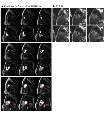
Low-Rank plus Sparse Matrix Decomposition for Accelerated Radial MS-CAIPIRINHA in First-Pass Myocardial Perfusion Imaging
Tobias Wech, Julius Heidenreich, Daniel Gensler, Tim Salinger, Peter Nordbeck, Daniel Stäb, Peter Speier, Thorsten Bley, Herbert Köstler
The anatomical coverage in first-pass myocardial perfusion imaging was extended by applying undersampled radial MS-CAIPIRINHA and a model-based reconstruction exploiting low-rank plus sparse matrix decomposition. The technique was tested in a patient with acute left ventricular myocardial infarction, yielding six short-axis slices from base to apex with a temporal resolution of one heartbeat. The reconstructed images exhibited a quality which is comparable to the conventional approach of acquiring only three slices per RR interval.
|
|
3325.
 |
11 |
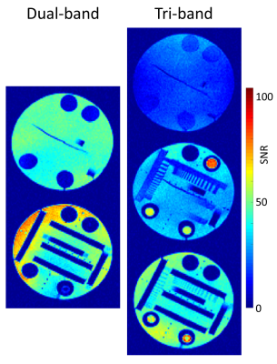
Comparison of Dual- and Tri-band Excitation for MS-CAIPIRINHA-accelerated First-Pass Myocardial Perfusion Imaging
Tobias Wech, Tina Urbanek, Andreas Weng, Daniel Stäb, Peter Speier, Thorsten Bley, Herbert Köstler
MS-CAIPIRINHA is a valuable technique to extend the anatomical coverage in myocardial first-pass perfusion imaging. In our previous studies, dual-band excitation was applied in conjunction with three SR-prepared acquisition blocks per RR interval to obtain six 2D-slices with a temporal resolution of one heartbeat. Especially in obese patients, however, the SNR occasionally turned out to be marginal, ultimately complicating the assessment of perfusion defects. In this work, an according approach using tri-band RF excitation was tested with respect to potential SNR benefits.
|
|
3326.
 |
12 |
Radial-CAIPI myocardial first-pass perfusion for high spatiotemporal resolution “iso-phase” multi-slice imaging at multiple cardiac phases per cycle
Merlin Fair, Peter Gatehouse, Ricardo Wage, Aleksandra Radjenovic, David Firmin
A high-resolution myocardial perfusion sequence is implemented with radial-CAIPI to allow simultaneous acquisition of the conventional three myocardial slices, all at identical cardiac phase, which can be repeated for multiple phases of the cardiac cycle and throughout the first-pass. The synchronised cardiac phases and repeat acquisitions aim to enable improved specificity, through better artifact identification.
|
|
3327.
 |
13 |
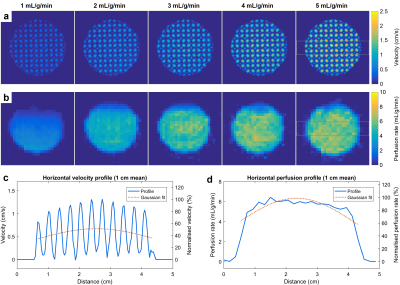
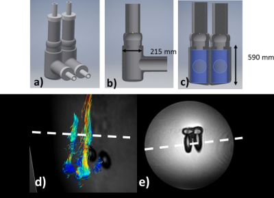
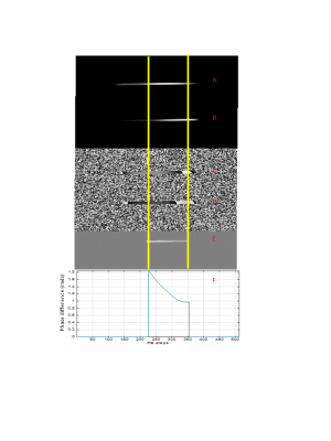
Dynamic contrast-enhanced and phase contrast MRI of a novel 3D printed cardiac phantom mimicking transmural myocardial perfusion gradients
Xenios Milidonis, Muhummad Nazir, Myles Capstick, Sita Drost, Gertjan Kok, Nikola Pelevic, Christian Poelma, Tobias Schaeffter, Amedeo Chiribiri
In recognition of the lack of a physical standard for the assessment and validation of myocardial perfusion imaging methodologies, a phantom simulating first-pass perfusion has recently been developed. This study builds on this work by introducing a novel 3D printed myocardial compartment with a radial variation in flow that mimics physiological transmural perfusion gradients. Velocity and perfusion rate estimates using phase contrast and dynamic contrast-enhanced MRI of the myocardium, respectively, were found to be repeatable. The myocardium shows potential in multi-modality evaluation and validation of perfusion pulse sequences and quantification algorithms before their introduction into routine clinical use.
|
|
3328.
 |
14 |
MRI-derived myocardial strain in patients with mild cognitive impairment (MCI)
Heng Ma, Jun Yang, Haizhu Xie, Fang Wang, Xiao Xu, Wei Bai, Jing Liu, James Carr, Kai Lin
Our data show that patients with mild cognitive impairment (MCI) have a lower regional peak myocardial strain and peak systolic strain rate at left ventricle (LV) as compared with healthy controls. Patients with MCI seem to have a heavier burden of subclinical cardiovascular diseases (CVDs).
|
|
3329.
 |
15 |
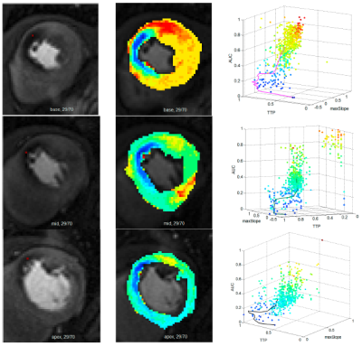
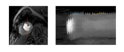
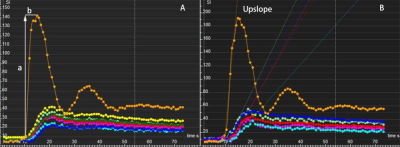
Fully automated spatio-temporal segmentation approach for myocardium ischemic lesions detection and tissue classification
Clément Daviller, Thomas Grenier, Shivraman Giri, Pierre Croisille, Magalie Viallon
CMR Perfusion Imaging proved its role in patient triage, identifying visually ischemia and its capability in quantifying heart perfusion1,2, but failed to transfer this technology to clinical routine and to show how this worth information could be used to improve tissue lesions comprehension. Deconvolution techniques are sensitive to noise present on time intensity curves S(t), when observation scale decreases. Automated segmentation prior modelling would be a powerful adjunct. Indeed, prior tissue classification would optimize perfusion quantification accuracy since enabling advanced modelling leading to additional markers while reducing processing time. Such automated method is proposed here.
|
|
3330.
 |
16 |
Rapid Motion Compensation Reconstruction for Dynamic MRI using Pixel Tracking Temporal Total Variation Constraint
Ye Tian, Apoorva Pedgaonkar, Jason Mendes, Mark Ibrahim, Brent Wilson, Edward DiBella, Ganesh Adluru
We present a novel motion compensation reconstruction method based on spatiotemporal constrained reconstruction (STCR) by tracking the movements of every pixel in each time frame, and constrain the temporal total variation along the pixel tracks. The proposed method can handle both respiratory and cardiac motion, and has comparable reconstruction speed but offers better image quality compared with STCR.
|
|
3331.
 |
17 |
Myocardial microvascular dysfunction in patients with end-stage renal disease and the risk factors for the heart damage in hemodialysis
Rong Xu, Yingkun Guo, Zhigang Yang, Huayan Xu
Cardiovascular disease is the major cause of death in patients with chronic kidney disease, this study prospectively enrolled 67 patients with ESRD to quantify evaluate the difference in left ventricular (LV) regional myocardial microvascular function using cardiac magnetic resonance (CMR), and to discuss the factors that may affect myocardial damage in the clinical treatment. The results confirmed that the first-pass perfusion CMR can early defect the myocardial deformation and dysfunction in ESRD patients, and the treatment time may be a risk factor for the cardiovascular disease in the patients with CKD.
|
|
3332.
 |
18 |
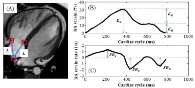
Importance of Right Atrial Strain on Right Ventricular Dilation in Pediatric and Adult Patients with Repaired Tetralogy of Fallot: Fast CMR Feature Tracking Study
Shuang Leng, Liwei Hu, Xiaodan Zhao, Ju Le Tan, Wen Ruan, Ru San Tan, Yumin Zhong, Liang Zhong
Right ventricular (RV) volume overload is common in patients after initial repair of tetralogy of Fallot (rTOF) and is associated with adverse long-term outcomes. We aimed to determine the effect of right atrial (RA) strain derived from feature tracking cardiovascular magnetic resonance (CMR) on the RV volume in both pediatric and adult rTOF patients. Results revealed that RA strain and strain rates were impaired in rTOF and RA reservoir strain impairment was significantly associated with RV dilatation. Hence, unloading of the RA and augmentation of RA function may be important future therapeutic targets in rTOF.
|
|
3333.
 |
19 |
MR-compatible Echocardiography and Predictive Slice Tracking Towards Ultrasound-driven Cardiovascular MRI
Lindsey Crowe, Francesco Santini, Laura Gui, Pauline Guillemin, Orane Lorton, Pamina Bernou, Myriam Roth, Gibran Manasseh, Oliver Bieri, Jean-Paul Vallée, Rares Salomir
MR imaging of cardiac valves is challenging as out-of-plane motion limits the use of 2D cine acquisitions. Ultrasound is well accepted as the clinical tool for observing valve motion. Here we suggest a hybrid imaging technology using in-bore ultrasound for direct observation of the motion of interest and subsequent on-the-fly adaptation of the MR slice position. MR-compatibility and workflow of echocardiography in-situ was demonstrated on a volunteer. Dynamic correction of motion in MR images was quantified with a moving phantom. Future-predicting algorithms yielded a reduction of apparent motion amplitude by a factor of twenty and dramatically improved “cine” image sharpness versus uncorrected data.
|
|
3334.
 |
20 |
Systolic and Diastolic Strain Abnormalities are Observed in Spontaneously Diabetic Non-Human Primates with Diastolic Dysfunction and Preserved Ejection Fraction.
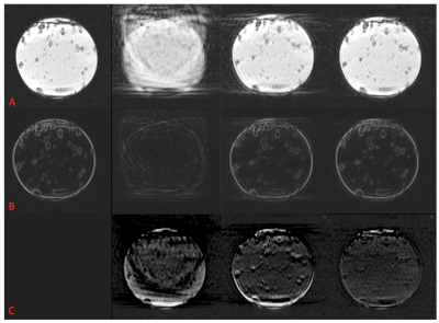
Smita Sampath, Li Gong, Yushu Chen, Chin Han Cheng, Zhigang Liang, Stephanie Seah, Jie Zheng, Sarayu Parimal, Zhu Tong, Zhang Yu, Wei Chen, Asad Abu Bakar Ali, Jeffrey Evelhoch, Wen Zeng, Chih-Liang Chin, Fabao Gao
Non-human primate (NHP) models of cardiovascular disease or metabolic disorder offer a unique framework to evaluate novel therapeutics. Herein, we perform cardiac strain MRI on a 3T MRI scanner to characterize systolic and diastolic function in naïve and spontaneously diabetic NHPs with diastolic dysfunction (T2DM-DD) and preserved ejection fraction (which may represent an early phenotype of HFpEF). In addition, the naïve animals were imaged twice to perform test-retest analysis of the strain-based biomarkers. Peak strains and peak diastolic strain-rates (both circumferential and longitudinal) are significantly impaired in the T2DM-DD monkeys compared to the naïve monkeys indicating impaired systolic and passive diastolic function. In addition, peak untwist rate is also decreased depicting impairment in active diastolic function as well. The test-retest results in the naïve animals show that all biomarkers, with the exception of peak longitudinal strain, are reproducible.
|
|
3335.
 |
21 |
Cardiac MRI detects functional deterioration prior to apoptosis in a mouse model of doxorubicin-induced cardiotoxicity using tissue phase mapping
Bradley Allen, Nivedita Naresh, Alexander Ruh, Sol Misener, Zhuoli Zhang, Daniele Procissi, James Carr
Doxorubicin-induced cardiotoxicity is an important limiting factor preventing dose-effective cancer treatment, and the associated cardiac sequelae appears to be mediated through cardiac myocyte apoptosis. Multiple studies have suggested there is potential for cardiovascular MRI (CMR) multiparametric analysis to detect doxorubicin-induced cardiotoxicity. In the current study, we performed multiparametric CMR including DENSE strain assessment and tissue phase mapping (TPM) for myocardial velocity measurement in a mouse model of doxorubicin-induced cardiotoxicity. Our results suggest that advanced CMR functional assessment shows promise in identifying treatment-related decrease in myocardial longitudinal systolic and diastolic velocity prior to the onset of cardiac myocyte apoptosis.
|
|
3336.
 |
22 |
The assessment of global and regional strain in patients with preserved ejection fraction after Fontan operation using feature tracking technique as compared with healthy children
Video Permission Withheld
Li-wei Hu, Rong-zhen Ouyang, Yu-min Zhong
The quantification of myocardial deformation may allow detection of early abnormalities and provide independent prognostic information, as demonstrated in echocardiographic studies.Meanwhile, some studies have suggested that CMR-FT may be evaluated earlier than ejection fraction to detect early abnormalities of the ventricular myocardium in postoperative follow-up of CHD. But, there is still limited experience with cardiac magnetic resonance feature tracking strain analysis in child patients. To the best of our knowledge, the significance of quantifying ventricular myocardial deformation in post-Fontan patients with pEF using CMR-FT has not been investigated. Therefore, the aim of this study was to evaluate the myocardial strain in children with pEF after the Fontan operation using feature tracking technique compared to healthy children.
|
|
3337.
 |
23 |
Hypertrophic Cardiomyopathy: The Potential Value of Tissue-Tracking Strain Analysis
Lindsay Griffin, Emily Ferris, Scott Nagle, Christopher Francois
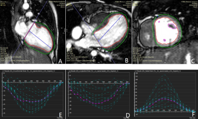
In hypertrophic cardiomyopathy (HCM), myocardium is disorganized, causing contraction abnormalities, perhaps before wall motion abnormalities are visually apparent. Tissue-tracking, a post-processing technique using routinely-acquired cine images, can assess strain, a multidimensional measure of contraction. We assess strain in 19 HCM cases. Global circumferential strain (GCS) and radial strain (GRS) correlated well (r > -0.85, p < 0.0001) and were worse in those with late gadolinium enhancement (p < 0.05). GCS modestly correlated with segment thickness (r = 0.46, p < 0.05). These data suggest strain may add value as a diagnostic/prognostic tool in assessment of HCM, available without additional imaging time.
|
|
3338.
 |
24 |
Subtle differences in left ventricular cardiotoxicity remodeling between risk groups of cancer survivors based on strain analysis from cine-DENSE MRI
Delphine Perie-Curnier, Denis Corbin, Frederik Epstein, Daniel Auger, Tarik Hafyane, Daniel Curnier
The aim of this study was to evaluate a reliable clinical tool to assess subtle differences in left ventricular cardiotoxicity remodeling in acute lymphoblastic leukemia survivors. Cine-DENSE MRI provided accurate evaluation of heart’s functionality of young cancer survivors in the short-axis view. Significant strain differences between groups were mostly observed in basal septal and apical septal segments while most of the other segments did not show significant differences. The next step of this study will be to include a control group of healthy volunteers.
|
|
Vascular
Electronic Poster
Cardiovascular
Monday, 18 June 2018
| Exhibition Hall |
13:45 - 14:45 |
| |
|
Computer # |
|
3339.
 |
25 |
Three-dimensional MRA Demonstrates Eccentric Enlargement of the Non-Conjoined Cusp-Sinus in Bicuspid Aortic Valve Patients
Pascale Aouad, Hector Michelina, Ian Murphy, James Carr, Jeremy Collins, Alex Barker
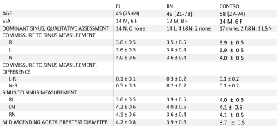
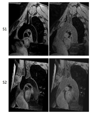
Bicuspid aortic valve is associated with ascending aortic dilatation, including the aortic root. The etiology of this aortopathy is controversial, with contributions proposed to come from both genetic and hemodynamic origins. While the pattern of aortic dilatation has been loosely associated with cusp fusion patterns and valve function, no study has investigated if eccentric sinus dilation is present and whether it varies in relation to the BAV phenotype. Thus, this study uses b-SSFP cine imaging and gated CE-MRA or 3D IR SSFP to assess the structure of the bicuspid aortic valve and identify the presence of a dominant sinus.
|
|
3340.
 |
26 |
MR Venography with Ferumoxytol in Central Venous Occlusion
Puja Shahrouki, John Moriarty, Biraj Bista, Sarah Khan, Stephen Kee, Brian DeRubertis, Takegawa Yoshida, Kim-Lien Nguyen, J. Paul Finn
Treatment of central venous occlusion is guided largely by anatomic considerations determined by pre-procedural imaging. Current approaches to imaging such as ultrasound and conventional cross-sectional imaging of central veins face many technical challenges and may be contraindicated in patients with renal impairment. We demonstrated that ferumoxytol-enhanced MR venography (FE-MRV) is a safe and highly accurate diagnostic tool that can be used as a reliable pre-interventional vascular map.
|
|
3341.
 |
27 |
Whole Body Vascular MR Imaging in Five Minutes for Patients With Claustrophobia
Puja Shahrouki, John Moriarty, Biraj Bista, Sarah Khan, Stephen Kee, Brian DeRubertis, Takegawa Yoshida, Kim-Lien Nguyen, J. Paul Finn
Patients with claustrophobia represent a significant proportion of patients who would otherwise be suitable candidates for MR angiography. With conventional acquisition techniques, examination times for MRA typically exceed 30 minutes and claustrophobic patients are often unwilling or unable to undergo the study. We implemented a new approach to minimize time in the magnet bore for patients with claustrophobia, acquiring comprehensive vascular evaluation of the thorax, abdomen and pelvis in as little as 5 minutes.
|
|
3342.
 |
28 |
Feasibility and Optimization of Ultra-Short Echo Time MRI for Improved Imaging of IVC Filters
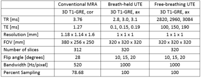
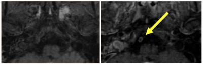
Gesine Knobloch, Scott Nagle, Timothy Colgan, Tilman Schubert, Kevin Johnson, Peter Bannas, Nathan Artz, Christopher François, Mark Schiebler, James Holmes, Scott Reeder
Monitoring of inferior vena cava (IVC) filters for complications is commonly performed using CTA. It would be desirable to evaluate IVC-filters using MRA to avoid the need for ionizing radiation and to exploit the superior soft tissue contrast of MRA. Unfortunately, conventional contrast enhanced MRA (cMRA) techniques are limited by distortion and signal voids arising from metal in the IVC-filter. In this pilot study, we evaluated the feasibility of ultra-short echo time (UTE) MRA at 3.0T in nine patients with IVC-filters. Results demonstrate feasibility of free-breathing UTE-MRA for the assessment of IVC-filters with comparable IVC-depiction compared to cMRA.
|
|
3343.
 |
29 |
MR time optimization: 2-year institutional experience in clinically applied high-resolution intracranial vessel wall imaging
Laura Eisenmenger, Lizhen Cao, Chengcheng Zhu, Christopher Hess, David Saloner
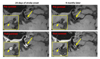
High-resolution intracranial vessel wall imaging (VWI) can provide valuable information not only regarding vascular morphology but also about the presence or absence of vessel wall enhancement; however, VWI acquisition times are often long, limiting routine use in clinical practice. We sought to investigate the use of the VWI in routine clinical practice to evaluate its application within our institution as well as optimize the imaging protocol to meet clinical needs. Our study found post-contrast VWI better demonstrated the vascular pathology compared to pre-contrast VWI. We also found only one case that may have benefited from the addition of pre-contrast VWI; however, the addition of pre-contrast VWI in this case would not have changed clinical management. Our findings suggest that the routine use pre-contrast VWI may not be needed to obtain the imaging necessary for clinical diagnosis and patient management.
|
|
3344.
 |
30 |
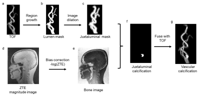
MRI and Non-Contrast CT Image Fusion to Guide Vascular Intervention: Feasibility Using Ferumoxytol in Patients with Renal Failure
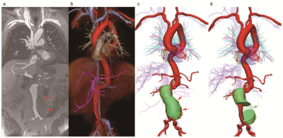
Takegawa Yoshida, Puja Shahrouki, Kim-Lien Nguyen, John Moriarty, Stephen Kee, J Paul Finn
Accurate pre-procedural vascular mapping may be crucial to guide successful intervention. Whereas MRA provides excellent definition of the perfused vascular lumen, it is insensitive to vascular calcification and may fail to image indwelling devices. CTA can address the latter limitations, but may be contraindicated in patients with renal impairment, as is the case for gadolinium based contrast agents (GBCA). Our early results suggest that, in patients with renal failure, 3D fusion of non-contrast CT and ferumoxytol-enhanced MR images leverages the complementary strengths of both modalities while avoiding both iodinated contrast agents and GBCA.
|
|
3345.
 |
31 |
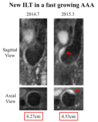
Monitoring intraluminal thrombus (ILT) progression in abdominal aortic aneurysm (AAA) using 3D black blood MRI: a longitudinal analysis
Chengcheng Zhu, Lizhen Cao, Zhaoying Wen, David Saloner, Michael Hope
The composition of intraluminal thrombus (ILT) is uniquely identified by MRI and has been suggested as a marker of abdominal aortic aneurysm (AAA) growth. However, the natural history of ILT progression is still unknown. This study followed 25 AAA patients over 19±9 months using repeated high resolution black-blood MRI. We found baseline ILT types did not predict AAA growth, however, AAAs with new ILT formation or fresher ILT during follow-up grew 3 times faster than AAAs without ILT change or older ILT (4.0±2.3mm/year vs. 1.3±2.5 mm/year, p=0.009). Monitoring ILT change provides new insights into the AAA risk assessment.
|
|
3346.
 |
32 |
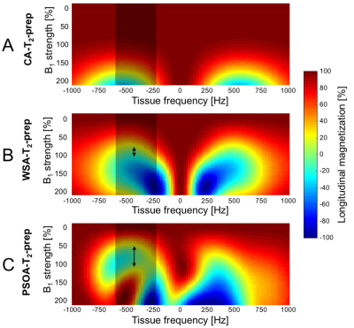
Particle swarm optimization (PSO) and comparison of a water selective T2 preparation module for simultaneous robust fat suppression and tissue contrast enhancement at 3T
Lionel Arn, Ruud van Heeswijk, Andrew Coristine, Matthias Stuber, Jessica Bastiaansen
Gradient echo based pulse sequences at 3T may lack the required contrast to distinguish blood from muscle. To overcome this, T2 preparation (T2-prep) modules are used in cardiac imaging to distinguish the blood pool from the myocardium. To suppress unwanted fat signals, we exploited the additional degrees of freedom that offer the multiple radiofrequency pulses of an adiabatic T2-prep and we used particle swarm optimization (PSO) to develop a T2-prep with robust fat suppression capabilities that works in the presence of flow. Its robustness against B1 and B0 inhomogeneities were predicted by the Bloch simulations for a range of fatty tissue frequencies, and could be confirmed experimentally both in phantoms and in volunteers.
|
|
3347.
 |
33 |
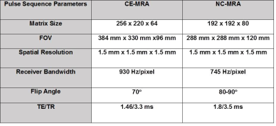
Rapid, non-contrast thoracic MRA using a combination of stack-of-star k-space sampling, compressed sensing, and self-navigation of respiratory motion
Hassan Haji-valizadeh, Nivedita Naresh, Jeremy Collins, Joshua Robinson, Pascale Aouad, Ali Serhal, James Carr, Cynthia Rigsby, Daniel Kim
We sought to highly accelerate high resolution (1.5 mm x 1.5 mm x 1.5 mm) non-contrast thoracic MRA using a combination of compressed sensing, stack-of-stars k-space sampling with variable density, and self-navigation , and we compared its performance against clinical contrast-enhanced MRA in patients with suspected aortic disease.
|
|
3348.
 |
34 |
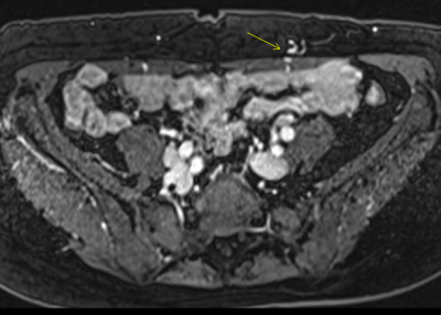
Application of non-contrast-enhanced MR angiography in hepatic arteriography
Did Not Present
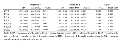
Xianlun Zou, Di Zhu, Hao Tang, Yaqi Shen, Zhen Li, Daoyu Hu
Accurate evaluation of hepatic arterial anatomy and variants is essential for preoperative planning of hepatic resection, transarterial chemoembolization and liver transplantation. In the present study, we try to explore the value of non-contrast-enhanced MR angiography using spatial labeling with multiple inversion pulses (SLEEK-MRA) in hepatic arteriography, and to compare the results with CT angiography (CTA). Although SLEEK-MRA was inferior to CTA in depicting small branches, it was comparable to CTA for depiction of the common hepatic artery, proper hepatic artery, left hepatic artery and right hepatic artery. As a noninvasive angiography method, SLEEK-MRA is valuable in hepatic arteriography.
|
|
3349.
 |
35 |
Simultaneous acquisition of motion-corrected coronary MRA and respiratory-resolved attenuation maps for whole-heart PET-MR imaging
Camila Munoz, Radhouene Neji, Gastao Cruz, René Botnar, Claudia Prieto
Motion-compensated attenuation correction is fundamental for accurate quantification in cardiac PET imaging. Here we propose a dual-echo water/fat coronary MR angiography acquisition with a motion-corrected reconstruction framework that simultaneously allows visualisation of the coronary anatomy and produces respiratory-resolved high-resolution attenuation maps. Results from healthy subjects show that the motion correction approach improves vessel contrast and sharpness compared to uncorrected water/fat images. Additionally, respiratory-resolved attenuation maps were obtained from motion fields and water/fat images with good tissue contrast. The proposed scheme can potentially be used for accurate and highly efficient whole-heart motion-corrected cardiac PET-MR imaging ensuring alignment between emission PET, attenuation maps and diagnostic MR data.
|
|
3350.
 |
36 |
Combined Assessment of Peripheral Artery Disease by MRI-based Vascular Calcification Visualization and Quiescent Interval Single-Shot (QISS) MRA
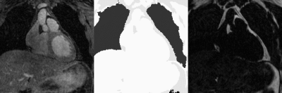
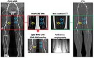
Akos Varga-Szemes, Taylor Duguay, Thomas Todoran, Megha Penmetsa, Stephen Fuller, Carlo De Cecco, Pal Suranyi, Robert Edelman, Ioannis Koktzoglou, U. Joseph Schoepf
The diagnostic accuracy of quiescent-interval single-shot (QISS) MRA to detect peripheral artery disease (PAD) has been shown to be similar to that of CTA. Unlike CTA, standard MR techniques are limited in the detection of vascular calcification. However, proton density-weighted, in-phase 3D stack-of-stars gradient-echo (PDIP-GRE) prototype pulse sequence has been shown to accurately depict calcifications in PAD. In our study, PDIP-GRE MRI provided comparable assessment to CTA. The MRI visualization of lower extremity vascular calcification improved readers’ confidence and the diagnostic accuracy of QISS-MRA in detecting significant vascular stenoses. Quantification of vascular calcium with MRI showed good agreement with CTA.
|
|
3351.
 |
37 |
Improved Golden Ratio Radial Arterial Spin Labeling Angiography Reconstruction using k-t Sparsity Constraints
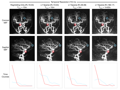
Mark Chiew, Thomas Okell
Dynamic arterial spin labeling angiography enables non-invasive visualization of arterial flow patterns, but is often time-consuming to perform. Undersampled radial trajectories help reduce acquisition time, but can result in noise-like aliasing artefacts and reduced spatial fidelity, particularly for a combined angiographic and perfusion golden ratio imaging technique, CAPRIA. An image reconstruction framework leveraging coil information and sparsity in the spatial and temporal frequency domains is presented which reduces aliasing and improves image sharpness in both 2D and 3D data. In addition, scan time reductions up to 10x are shown to be feasible whilst maintaining spatial and temporal information.
|
|
3352.
 |
38 |
Accelerated Non-Rigid Respiratory Motion Corrected Simultaneous Bright- and Black-Blood 3D Whole-Heart Coronary MR Angiography
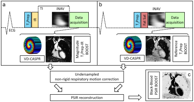
Giulia Ginami, Aurelien Bustin, Gastao Cruz, Radhouene Neji, René Botnar, Claudia Prieto
A novel 3D whole-heart sequence for simultaneous bright- and black-blood coronary angiography (named BOOST) was recently introduced. BOOST alternates the acquisition of two differently magnetization-prepared bright-blood volumes for coronary lumen visualization and from which respiratory motion information can be independently extracted. These datasets are subsequently combined in a PSIR-like reconstruction to obtain a complementary co-registered black-blood volume for thrombus/haemorrhage visualization. BOOST acquisitions, however, require prolonged acquisition times. Here, we accelerate BOOST acquisition by exploiting a variable density Cartesian trajectory that generates incoherent undersampling artefacts. Furthermore, non-rigid respiratory motion correction incorporated in the undersampled reconstruction is exploited for improved sharpness.
|
|
3353.
 |
39 |
Contrast-enhanced magnetic resonance angiography (MRA) in pre-surgical planning of deep inferior epigastric artery perforator flaps: comparison with surgical outcomes
Sze Yiun Teo, Christopher Au, Evan Woo
Deep inferior epigastric perforator (DIEP) flap reconstruction is an excellent choice because only the subcutaneous fat is used. DIEP flap reconstruction requires selection of a suitable perforator vessel, which can be highly variable in size and location. Pre-operative imaging can identify these vessels. Doppler sonography is the standard imaging modality, but has mixed results. CT angiography is accurate, but involves ionising radiation. MR angiography is less commonly used, but obviates any radiation exposure. This study shows that MR angiography is an accurate imaging modality to detect the size and location of suitable perforator vessels. Pre-operative knowledge of these vessels allows for optimal surgical planning, reduced area of surgical dissection and shortened dissection times.
|
|
3354.
 |
40 |
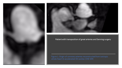
Pulmonary venous and coronary artery visualization from isotropic whole-heart kt-accelerated 3D cardiac CINE MRI
Did Not Present
Raluca Chelu, Hye-Jeong Lee, Tara Retson, Albert Hsiao
Isotropically-acquired 3D cine cardiac MRI has potential for quantification of cardiac size and function, which has been previously studied. We observed that pulmonary veins and coronary artery origins can be seen with this technique, and sought to further evaluate diagnostic visualization of these vessels. Two observers scored the coronary artery origins and pulmonary veins using a 5-point Likert score. Pulmonary veins were more readily visualized than coronary arteries. Isotropically-acquired 3D cine cardiac MRI enables depiction of vascular anatomy, and potentially may be used for pulmonary venous mapping and depicting coronary anomalies.
|
|
3355.
 |
41 |
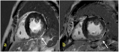
Comparison of SPGR Phase-Sensitive Inversion-Recovery and FIESTA Phase-Sensitive Inversion-Recovery MRI at 3.0T for the assessment of Late Gadolinium Enhancement in Patients with Hypertrophic Cardiomyopathy
Did Not Present
Huimin Yin, Ying Wang, Lizhi Xie
Cardiovascular magnetic resonance(CMR) imaging is now accepted as a valuable tool for the evaluation of many cardiac disease. It is particularly useful for the assessment of cardiomyopathies because it can depict different myocardial enhancement patterns on inversion-recovery(IR) late gadolinium-enhanced(LGE) images. This study would like to compare breath-holding SPGR PSIR with free-breathing FIESTA PSIR sequences and evaluate the feasibility of FIESTA for the assessment of LGE in patients with hypertrophic cardiomyopathy.
|
|
3356.
 |
42 |
Highly Accelerated 3D MR Angiography Using Multi-Channel Blind Deconvolution
Peizhou Huang, Jingyuan Lyu, Hongyu Li, Yongsheng Chen, Saifeng Liu, Chaoyi Zhang, Ukash Nakarmi, E. Mark Haacke, Leslie Ying
In many clinical applications, the three dimensional (3D) MRA plays an important role because that the 3D MRA can provide plenty of details for more compact anatomic regions with various flow directions. However, the speed limitation of the 3D MRA reconstruction is still an unignorable problem due to the size of the dataset, especially when the dataset has multi channels. With our proposed method, the Multi-Channel Blind Deconvolution (MalBEC), the experiment demonstrate that this method can provide high quality reconstruction image with high acceleration factors using much less time.
|
|
3357.
 |
43 |
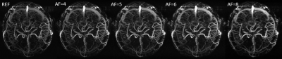
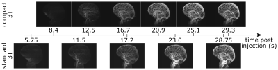
Comparison of Time-Resolved 3D Contrast-Enhanced MR Angiography on a Compact 3T Scanner with a Whole-Body 3T Scanner
Eric Stinson, Joshua Trzasko, Erin Gray, Eric Borisch, Jeffrey Gunter, Norbert Campeau, Matt Bernstein, John Huston III, Stephen Riederer
High spatiotemporal resolution contrast-enhanced MR angiography of the whole brain was performed on a compact 3T system with a 32 channel RF coil and compared to a spatial-resolution-matched study on a 60 cm bore whole-body 3T scanner. The quality of images from both scanners was excellent. Higher temporal resolution (4.18 s vs 5.75 s) on the compact 3T scanner was enabled by high performance gradients and increased PNS limits compared to the whole-body scanner.
|
|
3358.
 |
44 |
Efficacy of Gadoterate Meglumine enhanced MRA in evaluating thoracic aortic aneurysm and comparison with Gadobutrol enhanced MRA
Kaitlin Crawford, Ali Serhal, Olivia D. Reese, Pascale Aouad, Matthew Barrett, Monica Korell, Amir Rahsepar, Monda Shehata, Ahmadreza Ghasemiesfe, Jeremy Collins, James Carr
Contrast enhanced Magnetic resonance imaging plays an important role in the diagnosis and follow-up of patients with thoracic aortic aneurysm (TAA). Gadoterate Meglumine, which has recently become available in the US, is considered one of the safer gadolinium contrast agents with respect to tissue deposition and NSF, due its macrocyclic structure. In this study, we compare the qualitative image quality and quantitative aortic dimensions of Gadoterate Meglumine enhanced MRA and compare it to Gadobutrol enhanced MRA for evaluation of thoracic aortic disease. These preliminary results showed that Gadoterate Meglumine enhanced MRA has comparable image quality to Gadobutrol enhanced MRA and excellent correlation with respect to aortic diameter measurements.
|
|
3359.
 |
45 |
Truncation Artifact in Pulmonary Magnetic Resonance Angiography
Timothy Colgan, Scott Nagle, Scott Reeder
Pulmonary magnetic resonance angiography is a promising technique for the detection of pulmonary embolism but suffers from central vessel dropout (truncation artifact) that can mimics emboli in medium-sized vessels. Corner-cutting k-space acquisition strategies are suspected to exacerbate this artifact. Simulations and in vivo experiments were used to investigate the relationship between corner-cutting and truncation artifact. Our simulations suggest that eliminating corner-cutting reduces the symmetry and magnitude of the ringing with this artifact but we observed only minor differences in volunteers. We conclude that corner-cutting, which can be used to shorten scan times and/or improve spatial resolution, does not exacerbate the central vessel dropout artifact.
|
 |
3360.
 |
46 |
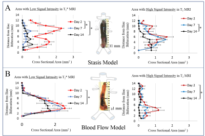
Multiparametric MRI Reveals Blood-Flow Dependent Spatial and Temporal Variations in Murine Venous Thrombosis
Olivia Palmer, Jie Ma, Jose Diaz, Joan Greve
There is a critical need for a noninvasive method to determine the optimal treatment for patients with deep vein thrombosis (DVT), the 3rd most common cardiovascular disease. We have implemented multiparametric MRI to evaluate spatial and temporal changes in thrombus composition using two murine models of DVT. We show that T2- and T2*-weighted MRI detects blood-flow dependent variations in thrombus composition. Classification in fully occlusive thrombi indicated an increased inflammatory response and more rapid thrombus organization when compared to thrombi developed in the presence of blood flow. This work provides foundational methodology that could eventually inform optimal DVT treatment planning.
|
|
3361.
 |
47 |
Quantification heterogeneous wall displacement and circumferential strain in the thoracic and abdominal aorta by spiral cine DENSE MRI
John Wilson, Xiaodong Zhong, John Oshinski
We used spiral cine DENSE (Displacement Encoding with Stimulated Echoes) MRI in the aortic wall to examine the heterogeneity of displacement and strain at three axial locations along the aorta. The major findings of this study were that spiral cine DENSE MRI is a viable technique for assessing patient-specific aortic wall kinematics in-vivo, that regional displacement and circumferential strain are heterogeneous and vary depending on aortic location, and neither mean nor maximum displacement co-localized with sections of peak circumferential strain.
|
|
Velocity & Flow
Electronic Poster
Cardiovascular
Monday, 18 June 2018
| Exhibition Hall |
14:45 - 15:45 |
| |
|
Computer # |
|
3433.
 |
1 |
Quantification of 3D aortic wall shear stress using k-t accelerated 4D flow MRI in under 2 minutes: a two center study
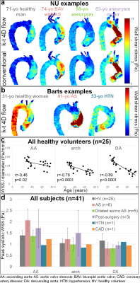
Emilie Bollache, Kristopher Knott, Redha Boubertakh, Ryan Dolan, Claudia Camaioni, Saadullah Ahmed-Villiers, Thomas Treibel, James Carr, Pim van Ooij, Jeremy Collins, Julia Geiger, James Moon, Alex Barker, Steffen Petersen, Michael Markl
Our aims were to study the feasibility at two centers of a newly developed k-t accelerated non-navigator gated 2-minute aortic 4D flow MRI sequence and to evaluate its wall shear stress (WSS) estimates. Eleven and 14 healthy volunteers, as well as 10 and 6 patients were scanned at Northwestern University and Barts Heart Centre, respectively. Despite an underestimation of distal aortic WSS in patients when compared to conventional 4D flow, our measurements were sensitive to expected aging and disease-related variations. We confirmed that aortic 4D flow MRI in 2 minutes is feasible and provides consistent WSS measurements.
|
|
3434.
 |
2 |
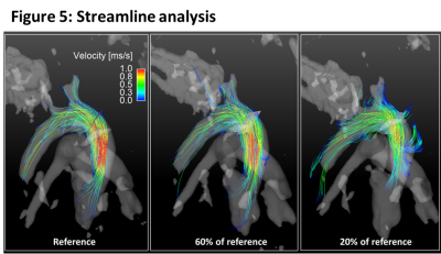
Quantitative evaluation of compressed sensing reconstruction in mouse phase contrast 4D-flow Magnetic Resonance Imaging
Moritz Braig, Marius Menza, Jochen Leupold, Li Feng, Pierre LeVan, Juergen Hennig, Axel Krafft, Dominik von Elverfeldt
Preclinical 4D-flow measurements remain challenging due to long acquisition times. This study presents a retrospective analysis of 4D-flow measurements with a radial 3D phase contrast sequence in combination with an advanced compressed sensing reconstruction. We evaluate the impact of different undersampling factors regarding peak velocities, flow, wall shear stress values and streamline analysis. We could show that high acceleration factors for preclinical phase contrast imaging can be used without substantially degrading the quantitative results. Our findings might enable high resolution 4D-flow acquisitions in less than one hour.
|
|
3435.
 |
3 |
Exploring vessel inward normal computation for 4D flow based wall shear stress estimation in complex vessel geometries.
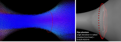
Judith Zimmermann, Daniel Demedts, Michael Markl, Christian Meierhofer, Heiko Stern, Anja Hennemuth
Wall shear stress (WSS) is a hemodynamic parameter which can be estimated from 4D flow MRI. The aim of this work was to advance the surface inward normal computation for complex (i.e. cone-shaped) vessel geometries and thus to improve the accuracy of wall shear stress estimates. We propose a Gauss gradient field approach to adapt to complex vessel courses and evaluate our method using synthetic flow data and selected patient data. Results show that correct inward normal definition is crucial for reliable WSS estimates, in particular in cases where complex vessel geometries are present.
|
|
3436.
 |
4 |
On the Influence of Intravoxel Velocity Distributions on the Noise of Phase Contrast Velocimetry
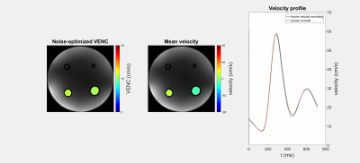
Simon Schmidt, Sebastian Flassbeck, Mark Ladd, Sebastian Schmitter
In this work we investigate the influence of intravoxel velocity distributions on the velocity noise in phase contrast velocimetry. Intravoxel velocity distributions are directly measured via Fourier velocity encoding and subsequently fitted by Gaussian distributions. Taking intravoxel dephasing due to a finite distribution width into account, a noise-optimized VENC is calculated for phantom flow measurements and in-vivo data at 7 Tesla.
|
|
3437.
 |
5 |
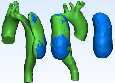
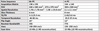
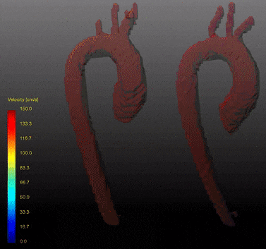
Whole-heart 4D phase-contrast MRI for clinical cardiovascular flow analysis: a comparison and validation on an imaging pulse sequence aspect
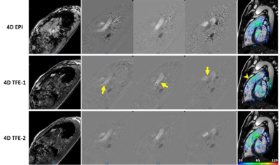
Shuo Zhang, Jun-Mei Zhang, Jennifer Ann Bryant, Bao Ru Leong, Pankaj Garg, Rob van der Geest, Ru San Tan, Liang Zhong
Whole-heart 4D phase-contrast magnetic resonance imaging (4D PC-MRI) provides qualitative and quantitative cardiovascular flow information. Recent technological advances in acquisition have rendered its clinical adoption without breath hold or respiratory gating. However, with different acquisition methods available their accuracy and influence in flow measurement are not well studied. We report our result in comparison of different commercially available imaging pulse sequences with validation to conventional 2D PC-MRI in healthy volunteers and patients with congenital heart disease.
|
|
3438.
 |
6 |
Feasibility of 4D phase-contrast MRI for the assessment of blood flow in the fetal aorta using Doppler ultrasound gating: preliminary results
Fabian Kording, Bjoern Schoennagel, Christian Ruprecht, Daniel Giese, Einar Heiberg , Manuela Tavares, Jin Yamamura
Fetal magnetic resonance imaging (MRI) is increasingly used as a second-line imaging tool for prenatal evaluation. The recet Doppler ultrasound trigger method enables the use of cardiovascular imaging techniques which may be usefull to improve prenatal cardiovascular imaging. This work investigates the use of 4D phase contrast measurements for the visualisation and quantification of fetal blood flow in the great vessels. In a small study group (n=2) it was shown for the first time that 4D flow measurements are feasible in the fetal vessels which may be beneficial for visualization and quantification of complex congenital cardiovascular malformations.
|
|
3439.
 |
7 |
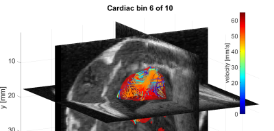
Magnetic Particle Imaging based 4D flow analysis technique using regional MRI data evaluation – initial in vivo results of a beating rodent heart
Jochen Franke, Heinrich Lehr, Volkmar Schulz
A dual-modal cardiovascular in vivo assessment in rodents was performed using a highly integrated Magnetic Particle Imaging – Magnetic Resonance Imaging hybrid system. 4D velocity flow field estimation of a beating rodent heart was extracted from the pulsed tracer information within the MPI dataset of a non-toxic tracer bolus. By means of co-registered morphological MRI data acquired at 0.5 T, an anatomical regional velocity flow field evaluation was performed for the four heart chambers individually.
|
|
3440.
 |
8 |
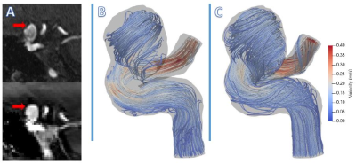
Dual-venc and single-venc 4D Flow MRI in cerebral aneurysms in comparison to image-based CFD modeling
Joseph Muskat, Sean Rothenberger, Ahmadreza Baghaie, Sameer Ansari, Craig Goergen, Susanne Schnell, Michael Markl, Vitaliy Rayz
Blood flow in two cerebral aneurysms was measured with 4D Flow MRI and simulated with image-based Computational Fluid Dynamics (CFD). A dual-venc 4D Flow MRI sequence with a shared reference scan was used in addition to a standard, single-venc 4D Flow acquisition in order to improve the dynamic range of measured velocities. Comparison of the MRI-measured and CFD-simulated flow fields showed that the 4D Flow and CFD methods can complement each other by eliminating modeling errors and augmenting imaging resolution. The dual-venc 4D Flow MRI provided valuable information on recirculating flow patterns that was not available from the single-venc data.
|
|
3441.
 |
9 |
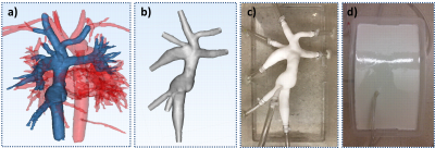
Modeling Physiological Flow Variation in Total Cavopulmonary Connection with Physical Model Experiments and 4D Flow MRI
David Rutkowski, Ryan Valk, Christopher François, Alejandro Roldán-Alzate
The total cavopulmonary connection (TCPC) is a successful treatment for single ventricle defect, however, long term complications, such as exercise intolerance still occur. To examine the effects of exercise conditions on TCPC fluid dynamics, in vitro experiments using 4D Flow MRI were conducted at high and low flow conditions. Significant difference in pulmonary flow distribution between conditions was found, and flow patterns and structures were characterized. After further development these models may provide a useful tool for analyzing and predicting changes in a variety of patient specific TCPC anatomy.
|
|
3442.
 |
10 |
Blood Velocity Measurement by RF Phase Gradient Differences between Receiver Coils: Initial Work towards Stenotic Jet Velocity
Jonathan Wagner, Peter Gatehouse, David Firmin
Fluid velocity was measured in vitro using the difference between spatial phase responses of array coils in 1993 by Famili, Wright and Porter. Aiming at application to cardiovascular stenotic jets, this abstract re-investigates their method with newer coil arrays and adds a multi-echo approach enabling aortic velocity measurement in normal subjects, with moderate results.
|
|
3443.
 |
11 |
Self-Gated Golden-Angle Spiral 4D Flow MRI
Rene Bastkowski, Kilian Weiss, David Maintz, Daniel Giese
A time efficient fully self-gated 4D flow sequence is presented that operates at predictable scan times and allows for a retrospective binning into an arbitrary number of cardiac and/or respiratory states. The acquisition time is fixed independently of the subjects’ physiology. Data is reconstructed using conjugate-gradient-SENSE. Feasibility is shown in 10 healthy volunteers and results are compared to a standard Cartesian 4D flow sequence.
|
|
3444.
 |
12 |
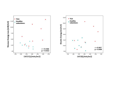
Evaluation of aortic viscous energy loss, kinetic energy and association with pumping function in congenital patients with transposition of great arteries using time-varying aortic geometry: Volumetric 4D Flow MRI Analysis
Covadonga Terol Espinosa de los Monteros, Roel Van der Palen, Arno Roest, Pieter Van den Boogaard, Lucia Kroft, Westenberg Jos, Mohammed Elbaz
Aortic hemodynamic energetics including kinetic energy (KE) and non-turbulent viscous energy loss (EL) and the association with cardiac function were evaluated in 8 TGA patients after arterial switch operation (ASO) and in 8 healthy individuals by 4D flow MRI. EL was significantly increased in TGA compared to healthy volunteers and aortic regions of highest levels indicate influence of complex ASO-related aortic geometry on blood flow efficiency. Significant positive correlation between aortic EL and cardiac index was found. Understanding the impact of ASO on aortic blood flow efficiency might enable insights on ways to improve operative procedure for TGA in future.
|
|
3445.
 |
13 |
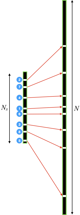
CArtesian sampling with Variable density and Adjustable temporal resolution (CAVA)
Adam Rich, Ning Jin, Yingmin Liu, Lee Potter, Orlando Simonetti, Rizwan Ahmad
We present a variable density Cartesian sampling method that allows retrospective adjustment of temporal resolution, providing added flexibility for real-time applications where optimal temporal resolution may not be known in advance. This method, called CArtesian sampling with Variable density and Adjustable temporal resolution (CAVA), is validated using real-time, free-breathing phase-contrast MRI data from four volunteers. Diagnostic quality images were successfully recovered at different temporal resolutions. Also, flow quantification based on CAVA was in good agreement with the breath-held segmented acquisition. In summary, CAVA provides a Cartesian alternative to Golden Angle-based radial sampling and can benefit a wide range of 2D real-time applications.
|
|
3446.
 |
14 |
4D Flow MRI velocity encoding effects on in vitro aortic dissection false lumen velocity distribution
Sylvana García-Rodríguez, Philip Corrado, Jon Wrobel, Timothy Ruesink, Alejandro Roldán-Alzate, Christopher François
Selecting the appropriate velocity sensitivity is critical in assessing flow within the false lumen of aortic dissections, where low velocities are dominant. This study compares effects of two VENC settings on velocity distribution within the false lumen of 3D printed aortic dissection models. We observed significant changes in velocity distributions depending on the VENC selected for the 4D Flow MRI acquisition. This difference tends to be accentuated at lower flow rates and would have important implications on calculations of other hemodynamic parameters, including wall shear stress, kinetic energy and vorticity, which may be predictors of outcomes in aortic dissection patients.
|
|
3447.
 |
15 |
Rapid, real-time phase-contrast MRI using a combination of radial k-space sampling and compressed sensing with spatially varying regularization weights
Hassan Haji-valizadeh, Bradley Allen, Roberto Sarnari, Matthew Barrett, Daniel Kim
We sought to develop a compressed sensing reconstruction method for radial k-space derived real-time phase contrast that uses spatially varying regularization to reduce flickering artifacts without significant loss in quantified flow accuracy, and evaluate its performance with respect to clinical breath-hold phase contrast in patients undergoing aortic valve evaluation with cardiovascular MRI.
|
|
3448.
 |
16 |
Pseudo Spiral Compressed Sensing for Aortic 4D flow MRI: a Comparison with k-t Principal Component Analysis
Lukas Gottwald, Eva Peper, Qinwei Zhang, Bram Coolen, Gustav Strijkers, R. Planken, Aart Nederveen, Pim van Ooij
In this study, 8-fold pseudo spiral compressed sensing (CS) accelerated aortic 4D flow MRI was compared with 8-fold k-t principal component analysis (k-t PCA) acceleration. Scan times were approximately 7 minutes at 50% respiratory navigator efficiency. Image quality of the peak systolic phase contrast magnitude images was scored slightly higher for CS than for k-t PCA and time-resolved velocity pathline trajectories were similar. Quantitative hemodynamic differences in velocity and wall shear stress were found but these were small and can be attributed to a combination of acquisition strategy and physiological variation. CS can be used to accelerate 4D flow MRI.
|
|
3449.
 |
17 |
Vortex Flow in Left Ventricle Interrupts Efficient Ejection: Demonstration by Vortex Flow Map of Cardiac Cine Magnetic Resonance Imaging
Video Permission Withheld
Masateru Kawakubo, Kenji Fukushima, Risako Nakao, Eri Watanabe, Yamato Shimomiya, Yasuhiro Goto, Hitoshi Tadenuma, Masami Yoneyama, Michinobu Nagao
Quantitative characterization of vortex flow might be a novel objective tool for evaluating left ventricular (LV) function. We developed the novel technique of vortex flow map (VFM). The VFM based on MR feature tracking can calculate the temporal displacement of the pixels on standard cine MRI. In this study, we analyzed the association with the VFM in LV and ejection fraction. As a result, it is indicated that the strong 3-dimensional vortex flow appears and impairs efficient LV ejection in severe heart failure. And the VFM is a useful tool for the evaluation of the efficiency of LV ejection.
|
|
3450.
 |
18 |
A dedicated MRI flow laboratory for quantitative flow measurements and method development
Did Not Present
Martin Bruschewski, Sven Grundmann
A unique MRI flow laboratory is currently being commissioned at the University of Rostock. The laboratory is specifically designed for quantitative measurements in technical flows. The MRI system, a whole-body Magnetom Trio (Siemens, Erlangen, Germany), will be integrated into various flow circuits with water and other fluids. This MRI system is not intended for clinical trials. Instead, the research focuses on the development and validation of flow quantification methods for medical and technical applications. The long-term aim is to make MRI more available to the field of fluid mechanics research and flow engineering.
|
|
3451.
 |
19 |
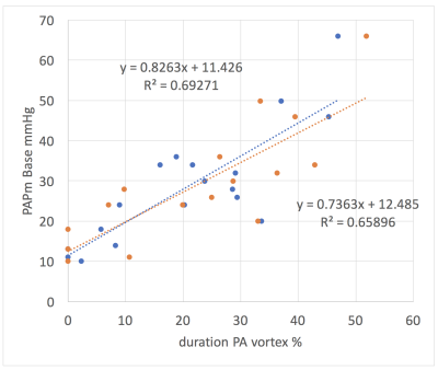
Assessment of Pulmonary Artery Mean Pressure with MRI and 4D Flow Vortex Assessment.
Lindsey Crowe, Anne-Lise Hachulla, Gabriel Guglielmi, Maurice Beghetti, Frederic Lador, Jean-Paul Vallée
Flow vortices have been observed in patients with elevated pulmonary artery pressure. We investigated 4D flow MRI and software packages to quantify vortex duration. This parameter was assessed compared to other MRI derived measurements to provide a potential non-invasive alternative to right heart catheterization.
|
|
3452.
 |
20 |
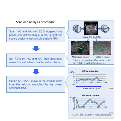
Effect of gravity on inferior vena cava and abdominal aortic flow: evaluation using multi-posture MRI
Yoshisuke Kadoya, Tosiaki Miyati, Naoki Ohno, Satoshi Kobayashi, Toshifumi Gabata
Inferior vena cava flow (IVCF) and abdominal aortic flow (AAF) are seems to be affected by gravity, ie., it depends on the body posture. We validated the effect of gravity on IVCF and AAF in supine and upright positions using an original multi-posture MRI. IVCF/AAF mean velocity, IVCF/AAF maximum velocity, mean IVCF/AAF, maximum IVCF/AAF in the upright position were significantly lower than those in the supine position. The cross-sectional area of IVC was significantly lower than those in the supine position, but that of AA was not significantly changed. Both IVCF and AAF decrease in the upright position.
|
|
3453.
 |
21 |
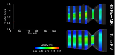
Experimental Validation of 4D Flow MRI for the Assessment of Recirculation and Acceleration using Tomographic Particle Image Velocimetry
Rafael Medero, Alejandro Roldán-Alzate
4D flow MRI has shown to be a feasible tool for the assessment of hemodynamics in different vascular territories, however reliable validation using gold standard fluid dynamics experiments is needed for improvement of its accuracy and precision. Particle image velocimetry (PIV) is an experimental technique widely used in engineering analysis of fluids. PIV measures flow velocity by optically tracking the movement of laser-illuminated particles. The purpose of this study was to validate 4D Flow MRI for the assessment of flow recirculation and acceleration using tomographic PIV.
|
|
3454.
 |
22 |
4D flow MRI: Preliminary in vivo results comparing two commercially available analysis software platforms
Christopher François, Lindsay Griffin, Niti Aggarwal, Mark Schiebler
Main pulmonary artery (MPA) and ascending aorta flow was quantified from 4D flow MRI using two commercially available software programs. Flow measurements made in three locations in each of these vessels were internally consistent with negligible bias for both programs. Furthermore, differences in mean MPA and aorta flow between programs were also negligible. These preliminary results are encouraging in affirming the reproducibility and reliability of flow measurements from clinical 4D flow MRI acquisitions.
|
|
3455.
 |
23 |
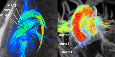
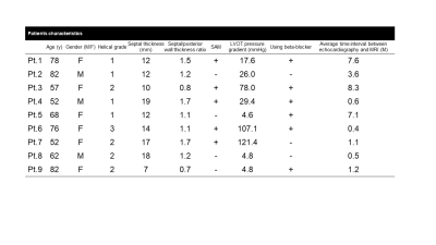
4D Flow MRI assessment of hypertrophic cardiomyopathy –the comparison with pressure gradient measured by Doppler ultrasound-
Kotomi Iwata, Tetsuro Sekine, Masaki Tachi, Yoichi Imori, Minako Takeda, Yasuo Amano, Makoto Obara, Yoshio Matsumura, Shin-ichiro Kumita
The purpose of this study was to compare the flow abnormality based on 4D Flow MRI data with the PG measured by Doppler ultrasound. we enrolled 9 patients who underwent echocardiography followed by 4D Flow MRI. Helical grade of HOCM group was higher than that of HNCM group (2.33±0.47 vs. 1.33±0.47, p=0.032). There was no significant difference between HG and each characteristic of HCM (PG, septal thickness, septum/free wall ratio and the presence of SAM). 4D Flow MRI can visualize abnormal helical flow of ascending aorta in patients with hypertrophic obstructive cardiomyopathy.
|
|
3456.
 |
24 |
Regional distribution of Local 3D Pulse Wave Velocity with and without Aortic Arch Replacement in Pediatric Patients with Single Ventricle
Amol Pednekar, Matthew Goette, Prakash Masand, Cory Noel
PC-CMR permits precise discrete 3D aortic pulse wave velocity (PWV) measurements as a surrogate for changes in vascular stiffness and ventricular-vascular interaction. In this preliminary study, global PWVs are 50% higher in 3 single ventricle (SV) patients with reconstructed arch (RA) than 3 patients with native arch (NA). Furthermore, the regional distribution of PWVs in SV-RA indicates rapid deceleration (50-70%) from proximal to distal aorta that is absent in SV-NA. The abnormal shape, large caliber change, and prosthetic material of reconstructed aortas may be additional explanatory variables. Regional distribution of local PWVs could potentially predict development of abnormal ventricular-vascular interaction.
|
|
Atherosclerosis & Vessel-Wall Imaging
Electronic Poster
Cardiovascular
Monday, 18 June 2018
| Exhibition Hall |
14:45 - 15:45 |
| |
|
Computer # |
|
3457.
 |
25 |
High Spatiotemporal Resolution Dynamic Contrast Enhanced (DCE) For Assessing Vascular Inflammation: Initial Clinical Experience
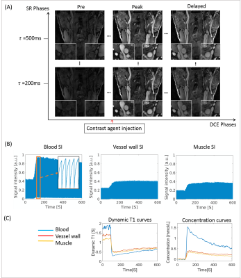
Nan Wang, Anthony Christodoulou, Yibin Xie, Zixin Deng, Bill Zhou, Zhaoyang Fan, Wei Yu, Debiao Li
Dynamic contrast enhanced (DCE) MRI is a promising technique for quantitatively assessing the inflammation of atherosclerosis. However, current applications are facing demanding sampling challenges, and compromises have to be made among spatial resolution, coverage and temporal resolution. We recently proposed a 3D DCE protocol based on Low Rank Tensor (LRT) framework to achieve high spatiotemporal resolution, adequate anatomical coverage and dynamic T1 mapping. In this work, we demonstrated the in vivo feasibility on both healthy subjects and patients with known atherosclerosis.
|
|
3458.
 |
26 |
An inverse association between microvasculature and intraplaque hemorrhage in atherosclerotic carotid lesions: an MR imaging study
Geneviève Crombag, Raf van Hoof, Floris Schreuder, Martine Truijman, Sylvia Heeneman, Paul Nederkoorn, Werner Mess, Robert van Oostenbrugge, Jan-Willem Daemen, Mat Daemen, Joachim Wildberger, Eline Kooi
The presence of intraplaque haemorrhage (IPH) has been related to plaque rupture, plaque progression, and predicts cerebrovascular events. However, the mechanisms leading to IPH are not fully understood. The dominant view is that IPH is caused by leakage of erythrocytes from immature microvessels. 101 patients underwent MRI of the symptomatic carotid plaque for detection of IPH and dynamic contrast-enhanced MRI for assessment of plaque microvasculature. A decreased vessel wall Ktrans was found for IPH positive patients. No difference in adventitial Ktrans was found in patients with and without IPH. Not only leaky plaque microvessels, but additional factors may contribute to IPH development.
|
|
3459.
 |
27 |
Decreased plaque microvasculature in symptomatic carotid plaques: a DCE-MRI study
Geneviève Crombag, Raf van Hoof, Floris Schreuder, Martine Truijman, Tobien Schreuder, Narender van Orshoven, Werner Mess, Paul Hofman, Robert van Oostenbrugge, Joachim Wildberger, Eline Kooi
Rupture of a vulnerable atherosclerotic plaque can lead to thrombus formation and, subsequently, to ischemic events. Intraplaque microvessels are thought to play an important role in atherogenesis, since they may facilitate entrance of red blood cells and inflammatory cells into the plaque tissue due to increased endothelial permeability. Symptomatic patients underwent DCE-MRI to assess plaque microvasculature. A significantly lower vessel wall Ktrans was found in the symptomatic carotid plaque compared to the contralateral asymptomatic side. The decrease in vasa vasorum in the symptomatic plaques might be due to a higher amount of necrotic tissue on this side.
|
|
3460.
 |
28 |
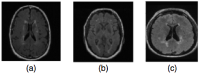
Feature Extraction using Convolutional Networks for Identifying Carotid Artery Atherosclerosis Patients in a Heterogeneous Brain MR Dataset
Mariana Bento, Luis Souto Maior Neto, Marina Salluzzi, Yunyan Zhang, Richard Frayne
Analysis of pathology in patients from heterogeneous datasets using machine learning techniques provide valuable information for identifying patients with carotid artery atherosclerosis disease. We propose and evaluate a method to automatically identify these patients based only on MR brain imaging findings in a dataset also containing multiple sclerosis patients and healthy control subjects. The features extracted using convolutional networks were discriminative, showing high accuracy rates (>96%) to distinguish between the three classes: atherosclerosis patients, multiple sclerosis patients or healthy controls. The method may help specialists in the diagnosis (specially in critical cases), and evaluation of disease activity.
|
|
3461.
 |
29 |
Investigating the estimated intracranial wall thickness on MRI vessel wall images: what voxel size do we need?
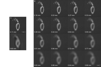
Kees van Hespen, Jaco Zwanenburg, Anita Harteveld, Peter Luijten, Jeroen Hendrikse, Hugo Kuijf
We investigated the influence of voxel size on the accuracy and precision of intracranial vessel wall thickness measurements on MR images. Circle of Willis specimens were scanned at ultra-high resolution (0.11mm). Downsampling these images showed that distinguishing thin (0.35-0.45mm), medium (0.65-0.75mm) and thick (0.95-1.05mm) vessel walls requires voxel sizes below 0.55-0.66mm, although thickness measurements showed considerable bias at those resolutions. Unbiased measurements required a voxel size of 0.2mm or less. A clinically used MRI protocol (0.8mm), could only correctly measure vessel walls thicker than 0.9mm. In summary, current intracranial vessel wall MRI protocols provide limited quantification of vessel wall thickness.
|
|
3462.
 |
30 |
Precision and Accuracy of Coronary Cross-Sectional Area MRI Measurements Used to Measure Coronary Endothelial Function
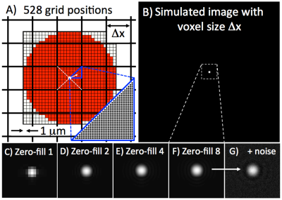
Michael Schär, Sahar Soleimanifard, Gabriele Bonanno, Jérôme Yerly, Allison Hays, Robert Weiss
Coronary endothelial function (CEF) can be measured noninvasively with MRI by quantifying changes in coronary artery cross-sectional area in response to isometric handgrip exercise. Those area changes are only a few imaging pixels because of MRI’s limited spatial resolution. Here we show with both numerical simulations and phantom measurements that 8-fold Fourier interpolation enables sub-pixel area measurement precision. Second, area measurement precision and accuracy can be further improved with smaller acquisition voxels as long as the signal-to-noise ratio remains above 30. Third, the currently used CEF-MRI protocol distinguishes area-changes of less than 5% at SNR measured in vivo.
|
|
3463.
 |
31 |
3D dual-contrast vessel wall imaging of the aortic arch
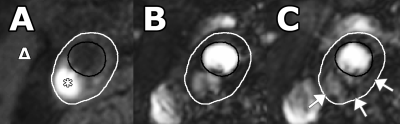
Zhaoyang Fan, Qi Yang, Guoxi Xie, Xiaoming Bi, Shlee Song, Marcel Maya, Debiao Li
Atherosclerotic disease of the aortic arch has been considered a potential cause of cryptogenic stroke. Vessel wall MR imaging can directly probe atherosclerotic lesions to provide assessment of plaque burden and instability. However, its clinical adoption for the aortic arch is hindered primarily by long imaging time that is associated with the needs for large spatial coverage, high spatial resolution, compensation of motion, and multiple image contrasts. In this work, we proposed a fast 3D dual-contrast vessel wall MR technique that is potentially useful for detecting intraplaque hemorrhage and calcification as well as measuring plaque burden at the aortic arch.
|
|
3464.
 |
32 |
Quantitatively monitoring therapeutic response in patients with symptomatic intracranial atherosclerotic disease using 3D MR plaque imaging
Zhaoyang Fan, Qi Yang, Feng Shi, Shlee Song, Konrad Schlick, Mercel Maya, Nestor Gonzalez, Debiao Li
Intracranial atherosclerotic disease (ICAD) is one of the most common causes of ischemic stroke worldwide. Despite intensive medical management, which is the current standard of care, the rate of recurrent stroke is 13% in the first year and as high as 35% in certain populations by 2 years. Initial and follow-up evaluations of these patients rely exclusively on assessments of clinical risk factors and, in some circumstances, the degree of luminal stenosis on imaging, which may overlook subtle non-luminal changes within ICAD lesions. In the present work, we sought to assess the feasibility of quantitatively monitoring regression or progression of intracranial atherosclerotic plaques using 3D VWI.
|
|
3465.
 |
33 |
Quantitative and Noninvasive MRI of the Endothelial Permeability and Function in Carotid Atherosclerosis
Alkystis Phinikaridou, Justinas Silickas, Begoña Lavin, Marcelo Andia, Alberto Smith, Prakash Saha, René Botnar
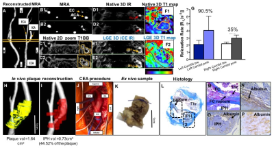
Over the past two decades, the central role of the endothelium in the initiation, progression, and clinical sequelae of atherosclerosis has been recognized. Increased endothelial permeability and impaired function precedes and portends the development of atherosclerotic lesions and their clinical manifestations. We have previously shown that quantitative assessment of albumin leakage into the vessel wall, using a clinically approved albumin-binding contrast agent, and endothelial-dependent dysfunction associated with lesion progression and instability in animal models. Here, we translated this technique in man to test whether endothelial permeability and dysfunction associate with carotid atherosclerosis risk in patients undergoing endarterectomy.
|
|
3466.
 |
34 |
Automatic segmentation of the aortic arterial wall in a pre-clinical rabbit model of atherosclerosis: preliminary experience with a convolutional neural netwoek
Daniel Samber, Claudia Calcagno, Edmund Wong, Venkatesh Mani, Cheuk Tang, Zahi Fayad
The task of manually evaluating medical images can be onerous, plagued by subjective bias, and subject to human error. In this study we apply a convolutional neural network (CNN) for automated image segmentation of the atherosclerotic vessel wall, a notoriously challenging and time consuming segmentation task. Our CNN shows a classification accuracy of 90% on testing data, and a intersection over union (IoU) weighted by the number of pixels in each class of 86%, indicating excellent segmentation. Our results suggest that, if appropriately optimized this method has the potential deliver faithful and automatic segmentation of the arterial vessel wall.
|
|
3467.
 |
35 |
Head and Neck Vascular Calcification Imaging using Zero Echo Time
Jianmin Yuan, Florian Wiesinger, Pascal Ruetten, Ilse Patterson, Martin Janich, Ana Beatriz Solana, Gaspar Delso, Scott Reid, Jonathan Gillard, Martin Graves
Calcification is an important factor in carotid plaque development and rupture. Current standard method for detecting plaque calcification is CT angiography (CTA). The purpose of this study is to introduce a new MRI method using zero echo time sequence for detection of carotid vessel wall calcification with high sensitivity. The protocol was optimized in volunteers and then applied to patient scan. The results demonstrate that carotid plaque calcification can be detected with high accuracy using proposed protocol compared with contrast-weighed images.
|
|
3468.
 |
36 |
Quantitative Assessment of Carotid Artery Atherosclerosis by Three-Dimensional Magnetic Resonance Imaging and 2D Ultrasound: A Comparison Study
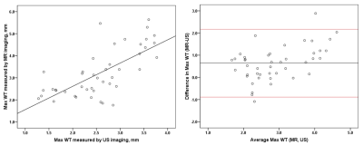
Huiyu Qiao, Ying Cai, Qiang Zhang, Lingyun Huang, Manwei Huang, Chun Yuan, Xihai Zhao
The size of carotid atherosclerotic plaques is associated with ischemic cerebrovascular events. Both 3D MR vessel wall imaging and 2D ultrasound can measure carotid plaques. To improve the work flow of screening for subclinical carotid atherosclerosis, this study sought to compare the quantitative measurements of carotid plaque between 3D MR and 2D ultrasound imaging. Excellent agreement was found between MR and ultrasound imaging in measuring carotid artery maximum wall thickness. Although there was moderate to strong correlation between MR and ultrasound imaging, the plaque area measured by MR imaging was more than two folds than that measured by ultrasound imaging.
|
|
3469.
 |
37 |
PD-T2-Shuffled Volumetric ISotropic Turbo spin echo Acquisition (VISTA) for 3D Simultaneous Multi-contrast Intracranial Vessel Wall Imaging
Shuo Chen, Zechen Zhou, Haikun Qi, Chun Yuan, Rui Li
The aim of this study was to develop a PD-T2-shuffled VISTA technique, which can provide co-registered 3D high multi-contrast intracranial vessel wall images with high scan efficiency. Phantom study and healthy volunteers study were performed to validate the feasibility of the proposed method. The results showed that middle cerebral artery vessel wall was clearly depicted on different weighed images of PD-T2-shuffled VISTA.
|
|
3470.
 |
38 |
Imaging uptake of plasma macromolecules in the arterial wall
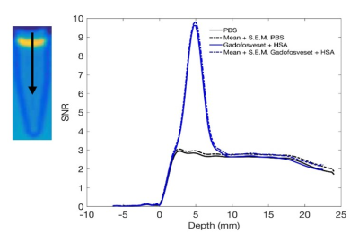
Marta Dazzi, René Botnar, Peter Weinberg
Elevated endothelial permeability is a precursor to atherosclerosis. Imaging macromolecule uptake in the artery wall can be used to detect and investigate early structural and functional endothelial dysfunction. Current techniques make use of destructive post-mortem analysis of tissue, limiting studies to animal models only. MR imaging of the transport of an albumin-binding contrast agent (Gadofosveset) could be used instead. We employed a mathematical model to differentiate between the bound and unbound fraction of the contrast agent thus making this method a promising non-invasive technique to measure permeability in humans for the first time.
|
|
3471.
 |
39 |
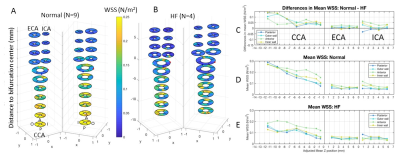
Quantification of Wall Shear Stress in the Carotid Arteries of Naïve and Dyslipidemic Non-Human Primates
Smita Sampath, Weiwei Luo, Ying-Hua Chu, Fa-Hsuan Lin, Michael Klimas, Elaine Manigbas, Willy Gsell, Kirsten Jacobsen, Eric Gifford, Asad Abu Bakar Ali, Jeffrey Evelhoch, Chih-Liang Chin
Herein, we quantified regional carotid wall shear stress (WSS) in 9 naïve NHPs on a normal diet and 4 dyslipidemic NHPs on a high fat diet. A custom-built carotid coil was used to achieve high-resolution imaging. Image analysis was performed including 3D structural and velocity interpolation, contour extraction and through-plane velocity projection. Velocity gradients at the contours were computed to quantify WSS. Animals on high fat diet showed enlarged common carotid arteries (diameter: 2.717 ± 0.195 mm) and lower WSS (0.1478 ± 0.0522 N/m2) compared to animals on a normal diet (diameter: 2.132 ± 0.203 mm, 0.2209 ± 0.0817 N/m2).
|
|
3472.
 |
40 |
A Phantom Study to Compare the Theoretical Accuracy and Precision of CT Angiography versus Radial MRI for the Assessment of Coronary Endothelial Function
Video Permission Withheld
Jerome Yerly, Fabio Becce, Danilo Gubian, Ruud van Heeswijk, Francis Verdun, Reto Meuli, Matthias Stuber
CT angiography (CTA) and MRI are non-invasive imaging modalities to assess coronary endothelial function (CEF). However, the performance of these techniques has yet to be quantitatively compared. Images of an in vitro phantom that simulates a physiological range of coronary artery cross-sectional areas (CSAs) were acquired using both CTA and MRI. CSAs were automatically measured and compared to the known nominal values. Statistical analysis suggests that MRI is capable of detecting significantly smaller CSA differences than CTA (2.25±0.80% vs. 7.42±0.63% for a 3-mm baseline diameter; p<0.0001), which is well within the range of physiological vasomotor responses of proximal coronary arteries.
|
|
3473.
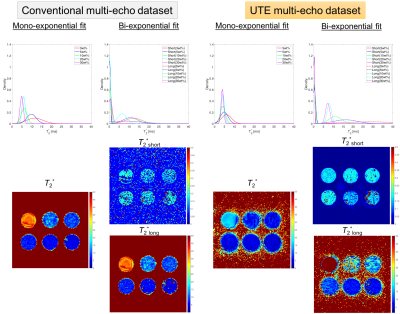 |
41 |
Does the amount of signal change depend on calcium concentration in lipid-rich core plaque?
Yuki Kanazawa, Kotaro Baba, Tosiaki Miyati, Masafumi Harada, Hiroaki Hayashi, Ikuho Kosaka, Mitsuharu Miyoshi, Michael Carl, Yuki Matsumoto
The purpose of this study was to quantitatively detect calcification in LRNC plaques using multi-component analysis with UTE imaging. A phantom experiment was performed using a four echo-UTE sequence. The phantom consists of six different concentrations of hydroxyapatite (as calcification) and mayonnaise (as a lipid). The bi-exponential analysis with UTE enabled a split into two components of short- and long T2*successfully. R2* derived from each dataset increased with increasing concentrations of hydroxyapatite. In conclusion, multi-component T2* analysis with UTE makes it possible to evaluate calcification in atherosclerotic plaques.
|
|
3474.
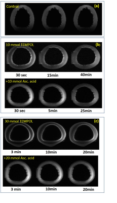 |
42 |
Dynamic MRI of Nitroxide Radical for TEMPOL kinetics and Redox State Assessment in Porcine Aortic Wall
Maxim Terekhov, Mihaela Pali, Christian Wittke, David Lohr, Anja Schroeder, Sueleyman Erguen, Alma Zernecke-Madsen, Heike Walles, Laura Schreiber
Reactive oxygen species (ROS) plays a key role in vascular disease. The physiological mechanisms regulating vascular local oxidative stress (LOS) are not completely understood. Nitroxide radicals like TEMPOL have been used in basic science NMR studies of ROS and LOS. Therefore, we developed a technique for dynamic imaging of nitroxide radicals to assess their kinetics and redox state of tissue.
|
|
3475.
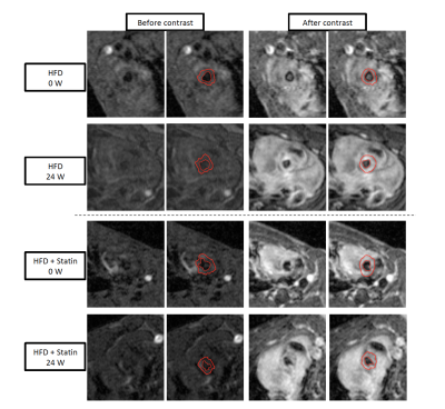 |
43 |
Quantification of endothelial permeability in the mouse aortic root with 3D self-gated T1 mapping: disease progression and treatment with statins
Raphael Soler, Zahi Fayad, Christopher Faries, Gustav Strijkers, Claudia Calcagno
Quantification of endothelial permeability in the mouse aortic root with 3D self-gated T1 mapping: disease progression and treatment with statins
|
|
3476.
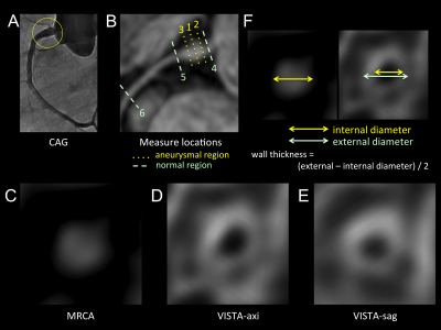 |
44 |
Feasibility of coronary artery wall imaging using 3D turbo spin-echo (black blood -T1-VISTA) in children with Kawasaki disease
Koji Matsumoto, Hajime Yokota, Hiroki Mukai, Naoki Saito, Yoshitada Masuda, Takashi Uno, Tosiaki Miyati
We evaluate feasibility of three-dimensional turbo spin echo imaging (BB-T1-VISTA) as coronary artery wall imaging for Kawasaki disease. BB-T1-VISTA could visualize the proximal regions of the coronary artery appropriately. BB-T1-VISTA with axial orientation produced the highest reproducibility to magnetic resonance coronary angiography (MRCA). Although BB-T1-VISTA showed significantly smaller diameter than MRCA, the limits of agreement were small in the proximal regions with normal diameter. By contrast, the larger aneurysms were, the larger bias became between MRCA and BB-T1-VISTA. BB-T1-VISTA has a potential to evaluate wide range of the coronary artery at once and was feasible in regions with small diameter.
|
|
3477.
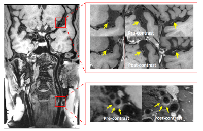 |
45 |
Infarct Pattern and Large Artery Atherosclerosis Features Revealed by Combined Intra- and Extra-cranial Vessel Wall Imaging in Patients with Non-cardiac Acute and Sub-acute Ischemic Stroke
Na Zhang, Jinhao Lyu, Lei Zhang, Lin Jia, Wenxiao Jia, Hairong Zheng, Xin Liu
Infarct pattern referring single or multiple infarction lesions of patients with ischemic stroke is a feasible imaging marker to predict future stroke recurrence. The aim of this study was to detect large artery atherosclerosis features resulting in single or multiple infarction lesions in patients with non-cardiac acute and sub-acute ischemic stroke using the combined intra- and extra-cranial vessel wall MR imaging method. The results demonstrated that in large artery atherosclerosis, infarct pattern may be associated with the vulnerability of atherosclerotic plaques, and this combined imaging method can be used to explore the vascular pathology and predict recurrence of ischemic stroke.
|
|
3478.
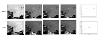 |
46 |
DCE-MRI of Intracranial Artery Athoersclerosis: Quantitative Change After 6-month Statin Therapy.
Huijun Chen, Jiaqi Dou, Le He, Chunyao Wang, Haikun Qi, Qiang Zhang, Xihai Zhao
Dynamic contrast-enhanced MRI (DCE-MRI) has been proven to be able to quantify the inflammation and neovasculature in the atherosclerotic plaque of carotid artery. However, few studies has focus on the DCE-MRI of intracranial artery atherosclerosis, which is another major cause of cerebral ischemic events. This study found that the Ktrans of intracranial artery calculated from DCE-MRI was significantly lower at follow-up comparing with baseline after 6-month statin therapy, suggesting DCE-MRI a good tool for therapeutic response evaluation for intracranial artery atherosclerosis
|
|
3479.
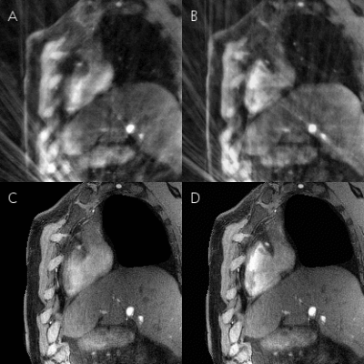 |
47 |
“3D-Stars” Cine MRI for the Coronary Arteries: Initial Steps towards Volumetric Endothelial Function Assessment
Video Permission Withheld
Gabriele Bonanno, Davide Piccini, Jérôme Yerly, Sahar Soleimanifard, Li Pan, Xiaoming Bi, Allison Hays, Matthias Stuber, Robert Weiss, Michael Schär
Recently, 2D coronary cine MRI has been shown to noninvasively assess coronary endothelial dysfunction, which is an early manifestation of atherosclerosis and a predictor of future acute events. However, atherosclerosis is a diffuse process, whereas this 2D approach provides local functional measures. Here, we present a free-breathing golden-angle 3D stack-of-stars cine sequence with isotropic spatial resolution combined with respiratory self-gating and 5D-GRASP reconstruction to image the proximal and mid segments of the right coronary artery. We call this new method “3D-Stars” and show feasibility to obtain volumetric cross-sectional area measures that can be used in future endothelial function studies.
|
|
3480.
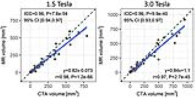 |
48 |
Influence of Field Strength on the Appearance of Peripheral Vascular Calcifications using Magnetic Resonance Imaging
Ali Serhal, Ioannis Koktzoglou, Pascale Aouad, James Carr, Shivraman Giri, Omar Morcos, Robert Edelman
A major deficiency of MR angiography compared with CT angiography (CTA) has been its inability to detect and characterize peripheral vascular calcifications. Recently, MRI using a proton density-weighted, in-phase stack of stars technique proved capable of identifying these calcifications. However, the diamagnetic susceptibility and short T2* of calcifications have the potential to cause clinically relevant, field strength-dependent changes in lesion appearance with MRI. Since the impact of field strength on the appearance of vascular calcifications is unknown, we performed a two-center clinical study which demonstrated that MRI accurately depicts peripheral vascular calcifications at both 1.5 Tesla and 3.0 Tesla.
|
|
| Back |
| The International Society for Magnetic Resonance in Medicine is accredited by the Accreditation Council for Continuing Medical Education to provide continuing medical education for physicians. |
