|
Electronic Poster Session
Neuro |
Monday, 18 June 2018
Electronic PosterNeuro
3601 -3624 Neurodegeneration
3625 -3648 Parkinson's Disease
3649 -3672 Genes, Their Correlates & Other Biomarkers
3721 -3744 Neurodegeneration
3745 -3767 Alzheimer's Disease & Other Dementias
3768 -3791 Neuroimaging: Clinical Studies |
| |
Neurodegeneration
Electronic Poster
Neuro
Monday, 18 June 2018
| Exhibition Hall |
16:15 - 17:15 |
| |
|
Computer # |
|
3601.
 |
49 |
Disrupted topological organization of brain structural network associated with prior overt hepatic encephalopathy in cirrhotic patients
Did Not Present
Hua-Jun Chen
Disrupted topological organization of brain structural network associated with prior overt hepatic encephalopathy
|
 |
3602.
 |
50 |
 Resting-State fMRI Low-Frequency Fluctuations in Temporal-Lobe Epilepsy Patients and The Relationship with Hemodynamic Correlates Resting-State fMRI Low-Frequency Fluctuations in Temporal-Lobe Epilepsy Patients and The Relationship with Hemodynamic Correlates
Chantelle Lim, Baxter Rogers, Victoria Morgan
Amplitude of low-frequency fluctuations (ALFF) of blood oxygenation is a marker of resting-state functional magnetic resonance imaging (fMRI) used to measure local spontaneous activity of the brain. However, interpretation of ALFF results is still unclear. Comparing ALFF values in left and right temporal-lobe epilepsy (TLE) patients with controls showed an increase in seizure-related regions. Increases in cerebral blood flow (CBF) were also found to be partially responsible for the increase in ALFF in another cohort of controls. Therefore, the development of ALFF measures may potentially provide a non- invasive perfusion measure in the presurgical evaluation of TLE.
|
|
3603.
 |
51 |
Effect of repetitive head trauma on diffusion MRI derived measures of diffusivity and free water
Virendra Mishra, Zhengshi Yang, Karthik Sreenivasan, Xiaowei Zhuang, Sarah Banks, Dietmar Cordes, Charles Bernick
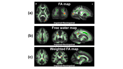
In this study, we utilized the diffusion MRI (dMRI) data of cognitively impaired and nonimpaired active professional fighters from the Professional Fighters Brain Health Study and studied the effect of repeated head trauma on conventional and advanced dMRI derived measures of diffusivity and free-water fraction (fiso). Our study revealed increased fiso and weighted FA in cognitively impaired active fighters, in addition to increased conventional radial and mean diffusivity, which was further associated with years of fighting in impaired fighters. Our study opens new avenues to explore advanced dMRI measures to understand the effect of repetitive head trauma on cognition.
|
|
3604.
 |
52 |
Exploratory Group Independent Components Analysis of resting state fMRI data reveals widespread brain function impairments in Gulf War Illness
Kaundinya Gopinath, Unal Sakoglu, Bruce Crosson, Robert Haley
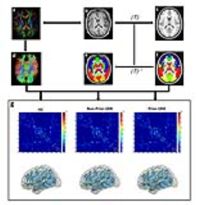
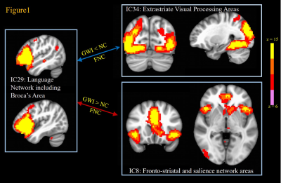
Around 200,000 veterans (up to 32% of those deployed) of the 1991 Gulf War (GW) suffer from GW illness (GWI), which is characterized by multiple deficits in cognitive, emotion, sensory and interoception domains. In this study we examined 23 GWI patients and 30 age-matched controls with resting state fMRI (rsFMRI) in order to map impairments in brain function networks in GWI with group independent components analysis. The results show that GWI veterans exhibit impaired, or abnormally increased functional connectivity in a lot of brain function networks consistent with their self-reported symptoms.
|
|
3605.
 |
53 |
Measuring the effect of soman, a seizure-inducing chemical warfare nerve agent, with simultaneous perfusion and brain oxygenation measurements
Kevin Lee, Sara Bohnert, Matthew Bouchard, Ying Wu, Cory Vair, John Mikler, Jeff Dunn
Chemical warfare nerve agents induce neurological damage through seizures. It is widely accepted in the field of nerve agent research that excitotoxicity is the main contributor to the neuropathology. Growing evidence suggests the involvement of hypoxia in seizure-related neuropathology. However, it is difficult to detect hypoxia without directly measuring oxygenation. We applied a method we developed to simultaneously quantify tissue oxygenation and cerebral blood flow to detect hypoxia following soman-induced seizures.
|
|
3606.
 |
54 |
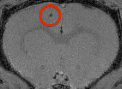
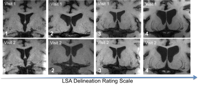
High Resolution 3D T1-weighted Black Blood MRI of Human Lenticulostriate Arteries as Biomarker for Small Vessel Diseases
Samantha Ma, Lirong Yan, Lei Cao, Giuseppe Barisano, Marlene Casey, John Ringman, Meng Law, Arthur Toga, Danny Wang
The early microvascular changes related to cerebral small vessel disease are still unclear. In this study, we applied a T1-weighted turbo spin echo sequence with variable flip angles for high resolution black-blood MRI to delineate the lenticulostriate arteries (LSA) at 3T. The vessel delineation was rated using a 4-point scale, and then correlated with clinical vascular risk factors as well as measures of executive function, cognitive flexibility and attention. LSA ratings were found to be reliable. Subjects with >4 visible LSA that were minimally tortuous exhibited positive trends with executive function, and male subjects tended to have poorer LSA delineation.
|
|
3607.
 |
55 |
Investigation of differences in the connectivity between the insular cortex and brain activity and metabolites between male smokers and non-smokers in their 20s
Seung-Man Yu
The purpose of the experiment was to investigate differences in the connectivity between the insular cortex out of the brain areas and other brain areas and the concentration of neuro-metabolites between male adult smokers and non-smokers in their 20s so that the results can be utilized as basic data for smoking cessation programs. The Cr concentration in the right insular cortex area of non-smokers is higher than that of smokers. In addition, it could be seen that smokers had stronger connectivity between their right insular cortex area and Gyrus Right, Occipital Fusiform, and Gurus Right areas than non-smokers and that non-smokers had stronger connectivity between their left insular cortex area and the frontal role right than smokers.
|
|
3608.
 |
56 |
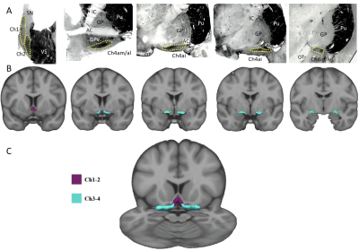
Iron accumulation in the striatonigral pathway of patients with Fabry Disease
Camilla Russo, Giuseppe Pontillo, Antonio Pisani, Francesco Saccà, Eleonora Riccio, Antonio Macera, Giovanni Rusconi, Arnaldo Stanzione, Pasquale Borrelli, Vincenzo Brescia Morra, Enrico Tedeschi, Arturo Brunetti, Sirio Cocozza, Giuseppe Palma
In Fabry Disease (FD) patients, compared to healthy controls, a significant increase in magnetic susceptibility has been observed in the substantia nigra and in the striatum, associated to a significant volume loss limited to the single substantia nigra. These findings probably reflect neurodegenerative phenomena due to pathological iron deposition in these particular extrapyramidal relay stations. This evidence supports the current hypothesis of a permeative cerebral involvement in FD that goes further the pure cerebrovascular association, thus shedding new light on this condition.
|
|
3609.
 |
57 |
Pre-operative DTI of white matter tracts in patients with idiopathic normal pressure hydrocephalus (iNPH) correlates with changes in clinical findings after shunt surgery
Johanna Mårtensson, Katarina Laurell, Johan Virhammar, Elna-Marie Larsson
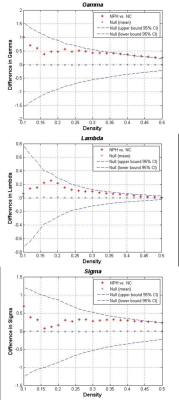
Preoperative prediction of shunt surgery outcome is difficult. A test to predict outcome is missing. The use of diffusion tensor imaging (DTI) as a bioimaging marker was investigated. DTI parameters in white matter tracts were compared between patients and healthy controls, and also correlated with changes in clinical findings after shunt surgery. Significantly differences between healthy controls and patients were found. Correlations were found between preoperative DTI results and changes in clinical findings after shunt surgery, suggesting DTI to be a supportive tool for prediction of clinical outcomes from shunt surgery in patients with idiopathic normal pressure hydrocephalus (iNPH).
|
|
3610.
 |
58 |
Abnormal Gray Matter Structural networks in Idiopathic Normal Pressure Hydrocephalus
Did Not Present
Lekang Yin, Yanmei Yang, Jianding Ye, Hong Yu
Idiopathic normal pressure hydrocephalus (iNPH) is a neurological disorder, the structural networks changes were never studied. We examined changes in gray matter structural network of patients with iNPH comparing with normal elderly people. Global network modularity was significantly larger in the iNPH network compared with the NC network (P<0.05). Eight nodes with significantly decreased betweenness were found in right frontal, temporal, insula lobe and right posterior cingulate region of iNPH network, while only one node was detected with significantly larger betweenness. Hubs of the iNPH network were mostly located in temporal areas and limbic lobe, while hubs of NC network were mainly located in frontal areas. We found some abnormalities in gray matter structural network that may relate to the occurrence of iNPH.
|
|
3611.
 |
59 |
Structural and functional assessments of eye, brain and visual field in glaucoma using optical coherence tomography, magnetic resonance imaging and perimetry
Vivek Trivedi, Yue Chen, Carlos Parra, Ahmel Arshad, Ji Won Bang, Mengfei Wu, Ian Conner, Kevin Chan
We used 3-Tesla anatomical MRI, DTI, optical coherence tomography, and perimetry to assess structural and functional changes in the eye and brain in glaucoma patients across disease stages. Both optic nerve and optic chiasm volumes were found to decrease from early to advanced staged glaucoma, and were associated with inner retinal thinning and visual field functional loss. Fractional anisotropy, mean diffusivity, and radial diffusivity in optic radiation were significantly different from early to advanced staged patients, but not when comparing control to early disease states. These results suggest anatomical MRI and DTI may be useful in monitoring glaucomatous brain damages non-invasively across stages.
|
|
3612.
 |
60 |
Dose-dependent effects of citicoline on the visuomotor response and white matter integrity in the visual pathway after chronic intraocular pressure elevation
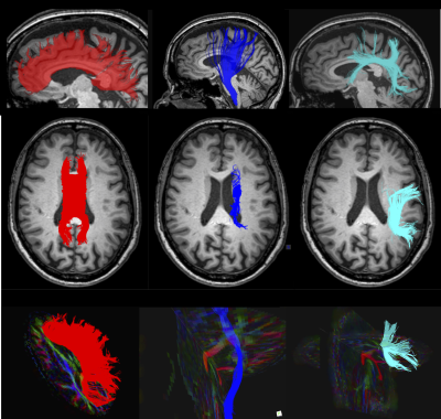
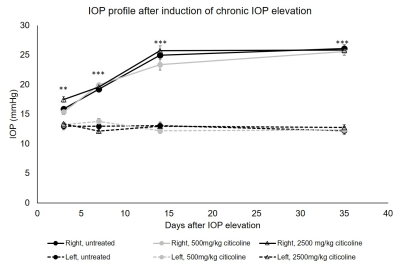
Yolandi van der Merwe, William Kohler, Michael Krawchuk, Xiaoling Yang, Leon Ho, Yu Yu, Ying Chau, Christopher Leung, Kevin Chan
Glaucoma is a neurodegenerative disease that can cause irreversible vision loss. Elevated intraocular pressure is a major risk factor for the glaucoma; however, the disease may still progress in some patients after lowering IOP. Citicoline has been suggested as a potential therapeutic to ameliorate damage caused by neurodegenerative diseases, including glaucoma, but its neuroprotective effects remain incompletely studied. In this study, we analyzed the dose-dependent effect of oral citicoline on visual behavior response and white matter integrity in a rodent model of glaucoma. The results show citicoline preserves visual behavior response and visual system integrity in a dose dependent manner.
|
|
3613.
 |
61 |
Microstructural and metabolism changes in the longitudinal progression of white matter hyperintensity
Did Not Present
Yeerfan Jiaerken, Xiao Luo, Minming Zhang
To evaluate the microstructural and metabolism property in the white matter that later become white matter hyperintensity (WMH), and the property of WMH that later disappeared. And we discovered that there is a dynamic change in microstructural and metabolism in WMH. Metabolism in NAWM will start to decline rapidly to a point where microstructure will then start to deteriorate.
|
|
3614 .
 |
62 |
Neurological symptoms and pathology in patients with newly-diagnosed, “classical” coeliac disease
Iain Croall, Marios Hadjivassiliou, Panagiotis Zis, David Sanders, Pascale Aeschlimann, Richard Grünewald, Paul Armitage, Dan Connolly, Daniel Aeschlimann, Nigel Hoggard
Coeliac disease is known to cause neurological problems, although these are not well recognised by healthcare professionals. In this prospective study of 100 newly-diagnosed patients with classical coeliac disease, we show 67% to have neurological symptoms, and 46% to have abnormal NAA/Cr values by MR Spectroscopy investigation. Further, we demonstrate how participants with positivity to transglutaminase 6, an auto-antibody suggested to be involved in neuro-pathology of gluten-related disorders, are more likely to suffer these. Finally, we present a volumetric analysis showing subcortical atrophy in this sub-group. This study highlights the prevalence, and potential mechanisms, of neurological involvement in coeliac disease.
|
|
3615.
 |
63 |
Paravermal sign: new MR imaging features of cerebellum in neuronal intranuclear inclusion disease
Video Permission Withheld
Noriko Sato, Atsuhiko Sugiyama, Yukio Kimura, Yuko Saito, Hiroshi Matsuda
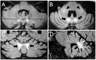
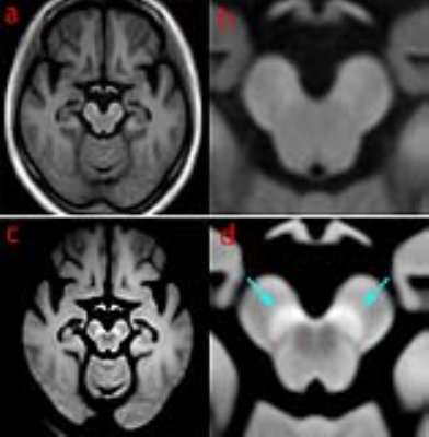
Neuronal intranuclear inclusion disease (NIID) is a neurodegenerative disorder with characteristic high signals along the corticomedullary junction on MR DWI. However, cerebellar findings have not been fully evaluated on MRI and we reviewed them in a series of ten NIID patients. MRI results showed cerebellar atrophy (10/10), high intensity signal on FLAIR images in medial part of the cerebellar hemisphere immediately beside the vermis (paravermal area) (7/10), and in the middle cerebellar peduncle (6/10). The paravermal abnormal signals could be a clue for diagnosis of NIID even in the past MR studies in which DWI was not examined.
|
|
3616.
 |
64 |
Regional cortical folding morphometry in Friedreich ataxia using Laplace Beltrami based gyrification index
Rosita Shishegar, Imis Dogan, Martin B. Delatycki, Gary F. Egan, Elsdon Storey, Louise A. Corben, Nellie Georgiou-Karistianis
Friedreich ataxia (FRDA) is an inherited neurodegenerative disorder mainly affecting the spinal cord and dentate nuclei of the cerebellum. Although there is growing evidence of cerebral atrophy and cortical thinning in FRDA, no research has investigated the pattern of cortical folding (gyrification) in the disorder. We have proposed a new MRI analysis technique, Laplace Beltrami based gyrification index (LB-GI), and validated its use in individuals with FRDA. Preliminary results reveal significantly increased regional gyrification in the motor cortex in individuals with FRDA, compared to healthy controls. Overall, our results demonstrate that LB-GI is a sensitive technique which requires further investigation as a potential neuroimaging marker of disease progression in FRDA.
|
|
3617.
 |
65 |
Multi-delay Simultaneous-Multi-Slice pCASL Imaging of Acute Treatment Effects of Ketamine in Major Depression
Kai Wang, Xingfeng Shao, Katherine Narr, Amber Leaver, Randall Espinoza, Danny Wang
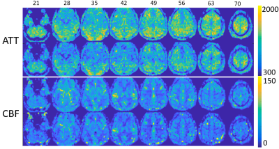
Sub-anesthetic dose of ketamine elicit acute and robust antidepressant effects, however the mechanism is still less understood. With a state-of-the-art Simultaneous Multislice EPI pCASL sequence, whole-brain rCBF and Arterial Transit Time (ATT) maps were acquired pre and post intravenous infusion of ketamine. Eight clusters were detected with significant CBF change, two of which (one in left inferior frontal gyrus, the other in right parietal lobe) were significantly correlated ( r=-0.7, p=0.04; r=-0.74, p=0.03 respectively) with Hamilton Depression rating score. This study suggests that possible mechanism of the antidepressant effects of ketamine is to increase CBF of certain mood- and attention-related brain regions.
|
|
3618.
 |
66 |
Impaired microstructural integrity of the callosum forceps minor in type 2 diabetes mellitus affect bilateral frontal functional connectivity
Did Not Present
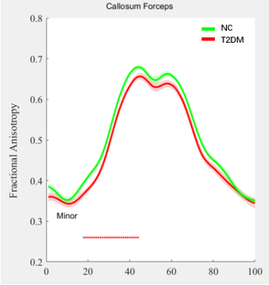
Xin Wang, Zhou Zhang, Jiaming Lu, Xin Zhang, Zhao Qin, Yan Bi, Bing Zhang
Type 2 diabetes mellitus (T2DM) is a risk factor for cognitive impairment. While its mechanism remains to be explored. In this study, using Automating Fiber-Tract Quantification (AFQ) analysis, we found that the fractional anisotropy (FA) in callosum forceps minor decreased in patients with T2DM, which indicated transverse white matter tracts connecting bilateral frontal cortex, was damaged. Meanwhile, functional connectivity between multiple brain regions within bilateral frontal cortex was decreased. The changes in the tract of callosum forceps minor might be the microstructural basis for functional changes of frontal cortex and help us understand the mechanism of T2DM related cognition decline.
|
|
3619.
 |
67 |
Quantitative Magnetic Resonance Imaging in diabetes: inflammation, oedema and neurodegeneration
Ana-Maria Oros-Peusquens, Ricardo Loucao, Elene Iordanishvili, Melissa Schall, Markus Zimmermann, Svenja Caspers, N. Jon Shah
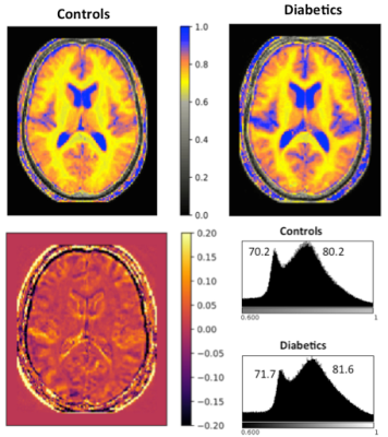
Type II diabetes is one of the most important metabolic disorders for public health with around 8% prevalence in European population (11% in the US). We report here for the first time a generalized increase in brain water content of ~ 2% in type II diabetics compared to age- and gender-matched controls, supporting the presence of neuroinflammation in diabetes. Several other quantitative measures are investigated (T1, T2*, MT parameters, magnetic susceptibility and diffusion kurtosis) as well as region-based volume, area and cortical thickness. Regions with significant changes in a large number of quantitative parameters are identified.
|
|
3620.
 |
68 |
Perfusion Pattern Scores Associates with Disease Severity in Type 2 Diabetes
Yuheng Chen, Wenna Duan, Parshant Sehrawat, Vaibhav Chauhan, Freddy Alfaro, Anna Gavrieli , Vera Novak, Weiying Dai
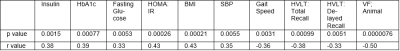
Type 2 diabetes mellitus (T2DM) is associated with alterations in the blood brain barrier, neuronal damage, and arterial stiffness, thus affecting cerebral metabolism and brain perfusion. We develop a machine learning method to investigate T2DM-related covariance pattern and its association with cognitive performance/disease severity. Our pipeline is superior to the traditional method and the pattern-related individual scores are associated to diabetes severity variables, mobility and cognitive performance at baseline. Besides, the longitudinal score change is associated with change of HbA1c, and baseline cholesterol, indicating that this score is a promising biomarker for tracing the disease progression of individual T2DM patients.
|
|
3621.
 |
69 |
Distinct mechanism underlying patients who might benefit from edaravone or those may not - a combined quantitative susceptibility mapping and DTI study
Qiuli Zhang, Haining Li, Ming Zhang, Dandan Zheng, Lijun Bai
The coexistence of multiple complicated pathological mechanisms, and lack of valid biomarker for treatment effects, remains to be main challenges that retarded ALS clinical trials. The approve of edaravone in treating ALS confirmed the importance of oxidative stress in ALS pathology. However, only a subgroup of patients would benefit with edaravone. We used QSM and DTI to explore the different mechanism that underlying those two cohorts. Our study revealed that neuroinflammation-prominent pathology and age-dependent oxidative stress might a feature for patients benefit with eradavone. While for patients not fulfill edaravone therapy, iron-mediated injury, like ferroptosis might accelerate disease progression.
|
|
3622.
 |
70 |
Structural analysis of m.3243A>G patients using 3D anatomical MRI brain scans
Catherine Hossain, Julie Hall, Yi Ng, Alexandra Bright, Andrew Blamire, Douglass Turnbull, Robert McFarland, Grainne Gorman
Symptoms associated with mitochondrial disease are heterogeneous and unpredictable. 17 m.3243A>G patients and 24 controls had 3D anatomical MRI brain scans, which were analysed to test the hypothesis that there are structural differences in patients that may potentially be used as biomarkers to predict symptoms. Results indicate head size is a possible surrogate biomarker for susceptibility to seizure-mediated strokes, but more detailed analysis did not provide any specific markers. Thinning of the cortex in the temporal poles appears to be directly related to the presence of the m.3243A>G point mutation and is an area to investigate further.
|
|
3623.
 |
71 |
CORRELATION OF IMMUNE ACTIVATION BIOMARKERS AND MRS MEASURES IN ACUTE HIV INFECTION
Napapon Sailasuta, Michelle D’Antoni, Philip Chan, Thep Chalermchai, Pasiri Sithinamsuwan, Somporn Tipsuk, Suteeraporn Pinyakorn, Bonnie Slike, Shelly Krebs, Vedbar Khadka, Eugene Kroon, Robert Paul, Serena Spudich, Victor Valcour, Jintanat Ananworanich, Lishomwa Ndhlovu
Proton MRS was performed in 51 acute HIV participants. We observed Inverse correlations between sCD163 and markers of neuronal integrity (BG and PCG NAA) in select brain regions suggests regional variability to neuronal injury in AHI. Further, correlations between CSF sCD163 and BG Cho and mI reveal that membrane turnover activity and glia activation that occur early in infection remain susceptible to CNS neuroinflammation. Our observation suggests select brain injury linked to myeloid activation persists even early ART in AHI arguing for additional interventions to halt detrimental neuroinflammation.
|
|
3624.
 |
72 |
Whole-brain MRSI for Quantification of Metabolite Markers of Inflammation and Neuronal Integrity in HIV-1 Clade C Infection
Varan Govind, Sameer Vyas, Vivek Gupta, Aman Sharma, Sulaiman Sheriff, Mahendra Kumar, Niranjan Khandelwal
HIV-1 enters the brain early in the course of infection and its replication continues despite use of combination anti-retrovirals (cART), causing chronic neuroinflammation, resulting in mild-to-moderate HIV-associated neurocognitive disorders in up to 50% of infected individuals. HIV-1 virus can be found throughout the brain of infected individuals, however, its maximum viral loads were found in the basal ganglia, frontal and medial temporal lobes, and hippocampus.1 We evaluated the use of a whole-brain proton MR spectroscopic imaging (MRSI) method at 3Tesla to better characterize the metabolite changes within the whole brain as a result of HIV infection.
|
|
Parkinson's Disease
Electronic Poster
Neuro
Monday, 18 June 2018
| Exhibition Hall |
16:15 - 17:15 |
| |
|
Computer # |
|
3625.
 |
73 |
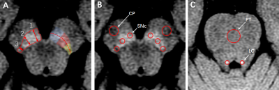
Overlap of R2* map based SN and SNpc defined by neuromelanin-sensitive MRI in Parkinson’s Disease: A promising diagnostic biomarker
Naying He, Jason Langley, Shengdi Chen, Chunlei Liu, Yong Zhang, Fuhua Yan, Xiaoping Hu
There is an urgent need for developing diagnostic imaging biomarkers for Parkinson’s Disease (PD). In this work, we applied a standardized substantia nigra pars compacta (SNpc) mask based on neuromelanin-sensitive MR images from healthy subjects to investigate the diagnostic performance of the SNpc overlap percentage and R2* in the SNpc overlap in PD. R2* in the SNpc overlap volume was increased in PD patients as compared to controls. Furthermore, it was significantly positively correlated with the disease duration in PD. We found an excellent diagnostic accuracy for the SNpc overlap percentage (AUC, 0.927) in PD.
|
|
3626.
 |
74 |
Visualization of Substantia Nigra Pars Compacta: MPRAGE vs. DANTE T1-SPACE
Sonoko Oshima, Yasutaka Fushimi, Tomohisa Okada, Takuya Hinoda, Takayuki Yamamoto, Hikaru Fukutomi, Yusuke Yokota, Akira Yamamoto, Tsutomu Okada, John Grinstead, Sinyeob Ahn, Kaori Togashi
Neuromelanin-sensitive magnetic resonance techniques have been developed for depicting neuromelanin-rich structures such as substantia nigra pars compacta (SNpc). We compared visualization of SNpc between magnetization-prepared rapid gradient-echo imaging (MPRAGE) and delay alternating with nutation for tailored excitation-prepared T1-weighted variable flip angle turbo spin echo (DANTE T1-SPACE) in 21 healthy volunteers. DANTE T1-SPACE provided much better delineation of SNpc and showed higher signal intensity than MPRAGE. DANTE T1-SPACE can be used for evaluating SNpc.
|
|
3627.
 |
75 |
Automated White Matter Lesion Quantification Correlates With Gait and Cognitive Dysfunction In Parkinson’s Disease
Eric Fang*, Mário Fartaria*, Chu-Ning Ann, Bénédicte Maréchal, Tobias Kober, Jie-Xin Lim, Celeste Chen, Soo-Lee Lim, Julian Gan, Eng-King Tan, Ling-Ling Chan
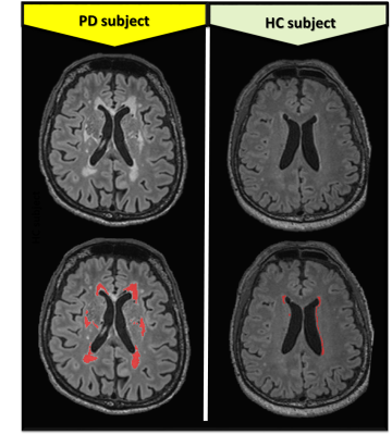
White matter lesions (WMLs) have an impact on neuronal connectivity; and consequently affect balance, mobility and cognition in both normal aging and disease states. Using a fully automated segmentation algorithm and multi-modal images, we estimated WMLs volumes to predict the clinical severity in a cohort of Parkinson’s disease (PD) patients and healthy controls (HC). Increased WMLs volume is strongly associated with both motor/gait and cognitive dysfunctions in PD. Lobar WMLs are found to have differential impact on distinctive cognitive domains. Automated volumetric quantification of WMLs load, particularly within the frontal and prefrontal regions can predict severity of symptoms in PD.
|
|
3628.
 |
76 |
White Matter Alterations in Parkinson’s Disease Analyzed with Combined Diffusion Magnetic Resonance Imaging and Magnetization Transfer Saturation Imaging: A Tract-Based Spatial Statistics Analysis
Christina Andica, Koji Kamagata, Taku Hatano, Asami Saito, Yuki Takenaka, Akifumi Hagiwara, Masaaki Hori, Ryusuke Irie, Akihiko Wada, Kanako Kumamaru, Nobutaka Hattori, Shigeki Aoki
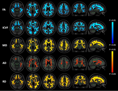
Our study combined three in-vivo imaging modalities (diffusion tensor imaging, neurite orientation dispersion and density imaging, and magnetization transfer saturation imaging) to provide a deeper understanding of white matter (WM) pathologies in Parkinson`s disease (PD). The tract-based spatial statistics analysis showed significantly decreased fractional anisotropy and intracellular volume fraction, and increased mean diffusivity, axial diffusivity, and radial diffusivity in an extensive area of WM in PD patients. Meanwhile, non-significantly increased myelin volume fraction was observed in limited areas of WM. The findings of this study might indicate that PD predominantly affects axons rather than myelin in WM.
|
|
3629.
 |
77 |
Brain Structural Differences in Healthy LRRK2 G2019S Mutation Carriers: An MR Radiomics Study
Moran Artzi, Avner Thaler, Avi Orr Urterger, Talma Hendler, Nir Giladi, Anat Mirelman, Dafna Ben Bashat
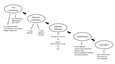
Structural brain differences in healthy LRRK2-G2019s mutation carriers who are at risk of developing Parkinson's disease (PD) were studied using radiomics analysis of DTI and T1-weighted images. 83 subjects were included: 43 healthy-carriers (HC) and 40 healthy-non-carriers (HNC). 18 statistical parameters were extracted for each modality in 14 subcortical brain regions. Various machine-learning classifiers were tested. The best classification results were obtained using RUSBoosted classifier, with average accuracy 73%, sensitivity 68% and specificity 79%. Radiomics analysis revealed brain differences in HC in comparison to HNC. These results together with the preliminary results among converters support the hypothesis of utilization of structural compensatory mechanisms in this "at risk" cohort.
|
|
3630.
 |
78 |
Contribution of basal forebrain damage to cognitive deficits in Parkinson’s disease
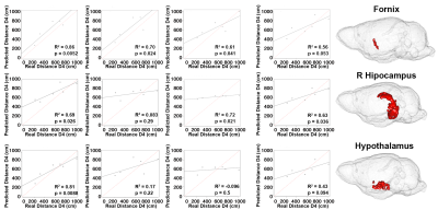
Fatma Gargouri, Cécile Gallea, Marie Mongin, Nadya Pyatigorskaya, Romain Valabregue, Marie Sarazin, Lydia Yahia-Cherif, Marie Vidailhet, Stéphane Lehéricy
We investigated the contribution of basal forebrain damage in the cognitive dysfunction of 52 non-demented patients with Parkinson’s disease (PD) and 25 age-matched healthy controls using diffusion and resting state functional MRI. Patients showed diffusion changes in the basal forebrain and the fornix. They also showed reduced functional connectivity between the septal area and the temporal lobe including the hippocampi and parahippocampal gyri, and between the basal nucleus of Meynert and frontal areas and bilateral thalami. Structural and functional changes correlated with memory and executive functions.
|
|
3631.
 |
79 |
Automatic analysis of multi-echo GRE and DTI data of Parkinson’s disease patients and control subjects using atlas-based subcortical regions
Gerd Melkus, Santanu Chakraborty, Goncalo Santos, Donghoon Kim, David Grimes, Tiago Mestre
Multi-parametric brain MRI longitudinal studies, which result in multiple quantitative parameter maps, can require intense post-processing work and time if several sub-cortical structures need to be analyzed individually on a region of interest (ROI) basis. In this study, we used a brain co-registration process together with a structural subcortical probability atlas for an automated analysis of quantitative MRI data. Patients with a recent diagnosis of Parkinson’s disease (<1 year) and healthy controls underwent brain MRI in a longitudinal study design. Quantitative maps derived from multi-echo GRE and DTI acquisitions were analyzed using this automated post-processing procedure at 3 different time points.
|
|
3632.
 |
80 |
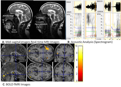
Clinical diagnosis based on speech mechanics with dynamic (real time) MRI, functional MRI and acoustics of speech (spectrogram)
Sunita Gudwani, S. Senthil Kumaran, Madhuri Behari
In evidence based clinical practice, mapping speech production problems is a challenge, further with cognitive impairment in degenerative disease makes the examination and assessments difficult. Visualization of the flexible vocal tract (dynamic MRI), cognitive planning (BOLD activation) and automated acoustic analysis (spectrogram) of speech production in motor degenerative diseases (like Parkinson’s Disease) might reframe the diagnostic evaluations. Empirically dynamic MRI is important technique for articulatory movements when adjunct with cognitive planning (fMRI) and spectrogram analysis may team as sensitive measures in clinical diagnosis. Thus this pilot study was planned to observe the significance.
|
|
3633.
 |
81 |
White Matter Differences in Parkinson’s Disease: a 7Tesla Tract-Based Spatial Statistics Study
Remi Patriat, Jacob Jacob Niederer, Sommer Huffmaster, Matthew Petrucci, Noam Harel, Colum MacKinnon, Christophe Lenglet
We used tract-based spatial statistics (TBBS) of 7Tesla fractional anisotropy (FA) measures to find differences between a cohort of early PD patients (N=30) and healthy controls (N=28). We examined the evolution of these differences by adding a PD cohort with longer disease duration (N=21). TBSS analysis revealed increased FA in the early PD group in the portions of the superior longitudinal fasciculus and the corticospinal tracts near M1, pre-motor and SMA. Subsequent analyses revealed increased FA in both PD groups compared to the control group. Radial diffusivity was significantly lower in the early and late PD groups compared to controls.
|
|
3634.
 |
82 |
Investigating differences in laterality and novel diffusion-derived metrics between freezing of gait (FOG) and non-FOG early Parkinson’s disease patients
Virendra Mishra, Zhengshi Yang, Karthik Sreenivasan, Xiaowei Zhuang, Dietmar Cordes, Brent Bluett
In this study, we utilized the diffusion MRI (dMRI) data of self-reported freezing of gait early Parkinson’s disease (PD-FOG) patients and PD-nonFOG patients from the Parkinson’s Progressive Markers Initiative (PPMI) database and investigated laterality along with the sensitivity of the conventional and advanced dMRI derived measures to classify the two groups. Our study revealed an asymmetric left laterality in early PD-FOG and a significant association of weighted fractional anisotropy with UPDRS scores in PD-FOG patients. Our study opens a window to understand disease severity using a potential new imaging biomarker for PD-FOG.
|
|
3635.
 |
83 |
Concurrent assessment of perfusion and functional connectivity in Parkinson’s disease
Maria Marcella Lagana, Laura Pelizzari, Niels Bergsland, Alice Pirastru, Giuseppe Baselli, Mario Clerici, Pietro Cecconi, Raffaello Nemni, Francesca Baglio
We aimed to assess if resting state functional connectivity (FC) changes were related to hypoperfusion in a group of Parkinson’s disease (PD) patients. Independent component analysis was performed to identify common spatial patterns of FC and of arterial spin labeling perfusion separately, in the whole group of PD and healthy controls. Concurrent FC and perfusion group differences were assessed. The observed FC alteration in the visual network may be influenced by the significantly reduced cerebral blood flow in the lateral occipital cortex, and vice-versa. The cross-talk between functional and perfusion findings should be considered when interpreting the results.
|
|
3636.
 |
84 |
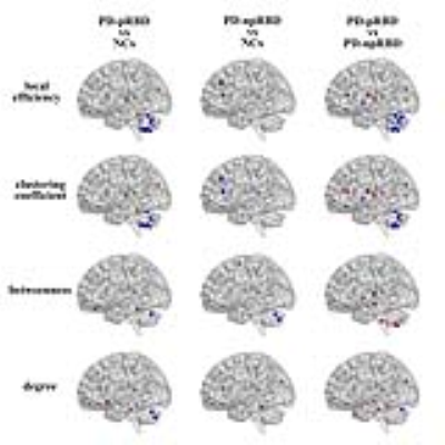
Alteration of brain structure network in Parkinson’s disease with and without rapid eye movement sleep behavior disorder
Did Not Present
Tao Guo, Xiaojun Guan, Qiaoling Zeng, Min Xuan, Quanquan Gu, Xiaojun Xu, Minming Zhang
We detected the alteration of structure correlation network in Parkinson’s disease (PD) patients with and without Rapid eye movement sleep behavior disorder (RBD). 191 PD patients including 51 possible RBD (pRBD) and 140 non-possible RBD (npRBD) and 76 normal controls were included. Structure brain networks were constructed by thresholding gray matter volume correlation matrices and analyzed using graph theoretical approaches. Significant enhanced nodal properties and hub recruitment were found mainly in limbic system while decreased nodal parameters were observed in cerebellum in PD-pRBD. This study may contribute to understand the pathophysiology of PD-RBD.
|
|
3637.
 |
85 |
White Matter Property of the Reward Circuits in Impulse Control Disorders in Parkinson’s Disease
Ru-Jen Lin, Weng-Ming Liu , Yung-Chin Hsu, Yu-Chen Wei, Pin-Yu Chen , Joshua Goh, Wen-Yih Tseng, Ruey-Meei Wu
Approximately 10-20% of Parkinson’s disease (PD) patients treated with dopamine agonist may develop impulse control disorder (ICD). We aimed to investigate microstructural characteristics of the white matter (WM) tracts in the reward circuits in PD-ICD by DSI images. ReMAP (Mean Apparent Propagator) was used for DSI reconstruction and TBAA (tract-based automatic analysis) for further tract analysis. Among the frontostriatal tracts (FS) and amygdala-related tracts, we found larger AD and/or RD in FS_motor cortex and amygdala-related tracts in ICD subjects, which may indicate a less healthy WM microstructure, and may further underlie the vulnerability to dopamine stimulation and result in hyper-gambling behavior.
|
|
3638.
 |
86 |
Differential diagnosis in Parkinsonism using diffusion tensor as measured from multiple cortical regions
Fan Huang, Sung-han Lin, Chin-Song Lu, Yi-Hsin Weng, Yao-Liang Chen, Shu-Hang Ng, Yi-Ming Wu, Chih-Chien Tsai, Yi-peng Liu, Jiun-Jie Wang
Parkinsonism is a long-term degenerative disorder of the central nervous system that mainly affects the motor system. The cortical parcellation algorithm was applied to evaluate the cortical involvement in the patients with PD by using diffusion tensor image. The fractional anisotropy is feasible in the brain of patients with Parkinsonism. Therefore fractional anisotropy could be a potential image based biomarker for monitoring Parkinson progression.
|
|
3639.
 |
87 |
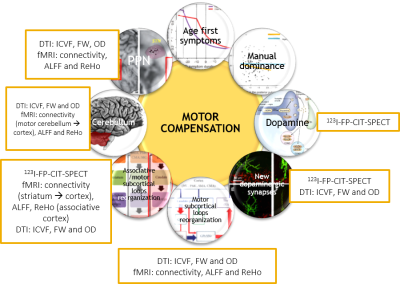
Motor compensation in Parkinson's disease (PD): a multimodal neuroimaging study
Did Not Present
Nicolas Villain, Stéphane Lehéricy, Nadya Pyatigorskaya, Romain Valabrègue, Sara Fernandez-Vidal, Rahul Gaurav, Olivier Jaubert, Marie-Odile Habert, Graziella Mangone, Jean-Christophe Corvol, Marie Vidailhet, David Grabli
Motor compensation mechanisms in PD rely on very distinct levels of evidence and have never been assessed altogether. They were tested among 68 early PD patients who underwent dopaminergic imaging and MRI (T1 neuromelanin, fMRI, DTI). We created an adjusted motor severity index (ratio between akinetic motor severity and neuromelanin substantia nigra alteration) and performed correlations between this index and the hypothesized compensation mechanisms. As a result, new dopaminergic synapses or more active dopaminergic synapses, reorganization of motor and cognitive subcortical loops and of the associative areas of the cerebellum are the main motor compensation mechanisms in early PD.
|
|
3640.
 |
88 |
The roles of sensorimotor and default mode-memory retrieval networks in staging PD cognitive decline
Meng-Hsiang Chen, Weiyan Yin, Wei-Che Lin, Weili Lin
In addition to motor disability, the development of cognitive decline is one of the most notorious symptoms in Parkinson’s disease (PD). Yet, a biomarker capable of accurately diagnosing cognitive impairments in PD patients remains elusive. Our results, based on resting-state functional MRI, suggested that functional connectivity in sensorimotor together with default mode-memory retrieval networks may serve a promising biomarker either for staging cognitive decline or neuropsychiatric scores.
|
|
3641.
 |
89 |
Cerebellum and Sensory Motor Resting State Network Behaviors in tremor- and akinetic rigid -predominant patients with Parkinson’s disease
Jiaming Lu, Prasanna Karunanayaka, Eunyoung Lee, Qing Yang, Mechelle Lewis, Paul Eslinger, Xuemei Huang
The striatal dopamine depletion in Parkinson's disease (PD) explains clinical symptoms such as bradykinesia and rigidity, but not resting tremor thought to be associated with cerebellothalamic (CTC) circuit dysfunction. In this study we used resting state fMRI to investigate differences in the CTC network in akinetic-rigid (PDAR) and tremor predominant (PDT) PD patients with closely matched demographic and cognitive variables. The results support a type dependent functional disruption in the cerebellum and SMA rs-networks in PD patients with otherwise comparable disease severity, neurocognitive performance, and overall brain morphology.
|
|
3642.
 |
90 |
Neuromelanin sensitive MRI features of substantia nigra and locus coeruleus in de novo Parkinson’s disease and its phenotypes
Lirong Jin, Jian Wang, Yuanfang Li, Zhen Huang, Yong Zhang, Kai Liu, Mengsu Zeng
This current study provides in vivo evidence that width and high signal were significantly decreased in SNc and LC of de novo PD patients using NM-MRI, compared with controls. We also observed that the neuromelanin changes in substantia nigra were across both motor phenotypic expressions and non-motor (with vs. without depressive symptoms) subtypes, while LC neuron reduction is more notable in PD with depressive symptoms. Our finding implies that the NM-MR imaging have potential applications to improve the clinical diagnosis and detect the heterogeneityof PD.
|
|
3643.
 |
91 |
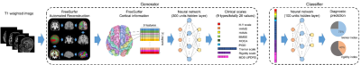
Deep Learning on Anatomical Brain MRI to Classify Motor Dysfunction in Parkinson's Disease
Yu-Hsueh Wu, Yasi Jiang, Yu-Chun Lo, Yumei Yue, Ting Shen, Fu-Shan Jaw, You-Yin Chen*, Baorong Zhang*, Hsin-Yi Lai*
We introduced an innovative two-staged deep artificial neural network (DNN) model focusing on diagnostic prediction of Parkinson’s disease (PD) using T1-weighted images, given a training set consisting of cortical thickness, surface area, grey matter volume and corresponding clinical scales, our proposed model was trained to classify the PD with different motor symptoms and performed the diagnostic prediction on basis of generated clinical scales. Results showed our DNN classifier and generator reached the averaged accuracy of 100% and 97.9%, respectively. To our knowledge, our technique was the first to tackle the classification of motor dysfunction in PD from anatomical brain MRI.
|
|
3644.
 |
92 |
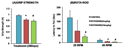
1H-[13C]-NMR Evaluation of Neurometabolism in Rotenone Mouse Model of Parkinson’s disease
Anant Patel, Shoumik Roy, Varadarajan Komanduri
Parkinson’s disease (PD) is a common progressive neurodegenerative disorder, which affects motor coordination and movement. Currently there is no biomarker for the early diagnosis of disease. In the current study we have evaluated the neurometabolism in rotenone mouse model of PD using 1H-[13C]-NMR NMR spectroscopy in conjunction with an infusion of [1,6-13C2]glucose. Our results show that the neurometabolism is compromised in rotenone treated mice suggesting that both the excitatory and inhibitory neurotransmission are impaired in PD.
|
|
3645.
 |
93 |
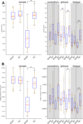
Non-invasive Characterization of the Cerebellar Nuclei in Degenerative Cerebellar Ataxias (SCA3, SCA6) with Quantitative Susceptibility Mapping (QSM) at 3 Tesla
Andreas Deistung, Dominik Jäschke, Sophia Göricke, Dae-In Chang, Katharina Steiner, Andreas Thieme, Jens Claaßen, Ellen Uslar, Markus Gerwig, Mark Ladd, Jürgen Reichenbach, Dagmar Timmann
We investigated non-invasively the cerebellar nuclei of spinocerebellar ataxia type 3 (SCA3) and SCA6 patients by using quantitative susceptibility mapping (QSM) at 3 T. Absolute volumes, relative volumes (with respect to the total intracranial volume), and magnetic susceptibilities of the cerebellar nuclei were found to be significantly lower for the dentate between SCA6 patients and healthy controls but did not significantly differ between SCA3 and healthy controls. Univariate linear correlations of relative nuclei volumes with ataxia rating scales revealed statistically significant correlations for the dentate and globose nuclei in SCA6 but not in SCA3.
|
|
3646.
 |
94 |
Multi-modal multi-centre MRI study of rare spinocerebellar ataxia, type 14
Farida Grinberg, Ezequiel Farrher, Peter Pieperhoff, Elena Schlapakow, Vincent Gras, Tanja Schmitz-Hübsch, Silke Lux, Ute Kopp, Hanna Gärtner, Eylem Kirlangic, Dagmar Timmann, Matthis Synofzik, Peter Bauer, Friedemann Paul, Matthias Endres, Thomas Klockgether, N. Jon Shah, Katrin Amunts, Sarah Doss, Martina Minnerop
Diffusion MRI studies reveal significant white matter degeneration in cerebellar and cerebral regions of spinocerebellar ataxia patients, however, only common pathology types have been included. Here, we present the first combined deformation-based morphometry (to depict regions of volume loss), and diffusion tensor/kurtosis imaging study to analyse microstructural changes in the largest reported sample of rare spinocerebellar ataxia, type 14, enrolled in a coordinated multi-centre study. Beyond expected strong changes observed for all metrics in cerebellar regions, axial diffusion kurtosis exhibited additional extracerebellar alterations in compliance with observed extracerebellar symptoms. Our results also suggest that patients might develop compensatory mechanisms.
|
|
3647.
 |
95 |
Phase Sensitive Inversion Recovery (PSIR) spinal cord imaging as a potential biomarker for Motor Neuron Disease
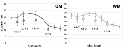
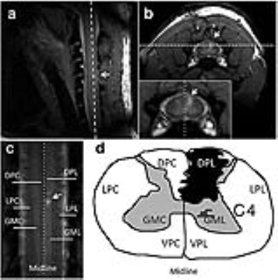
Antje Bischof, Nicholas Olney, Howard Rosen, Eduardo Caverzasi, William Stern, Catherine Lomen-Hoerth, Bruce Miller, Roland Henry, Nico Papinutto
With this study we investigated whether spinal cord gray and white matter atrophy could be detected in vivo in patients with a diagnosis within the motor neuron disease spectrum using PSIR imaging. Gray and white matter were successfully delineated in ten patients. Gray and white matter atrophy was detected in the majority of patients even if the lower or upper motor neuron was clinically unaffected. Patients with predominantly bulbar symptoms did not show relevant spinal cord abnormalities. This suggests a possible role of PSIR imaging as a biomarker for the spatial and temporal distribution of pathological changes in motor neuron disease.
|
|
3648.
 |
96 |
Intracortical T1-weighted/T2-weighted ratio signal changes in Huntington’s Disease
Christopher Rowley, Sarah Tabrizi, Blair Leavitt, Raymund Roos, Alexandra Durr, Nicholas Bock
Huntington’s disease (HD) is a genetic neurodegenerative disorder that is characterized by motor and cognitive dysfunction. Previous imaging studies have shown cortical thickness is reduced in HD, and here we investigated potential changes in cortical tissue composition in Huntington’s based on previously acquired T1-weighted (T1W) and T2-weighted (T2W) images. We analyzed T1W/T2W ratios formed using images collected in 321 subjects from the TRACK-HD dataset representing various stages of HD and healthy controls. Intracortical T1W/T2W signal analysis revealed significant changes in the most advanced HD group. This may reflect HD related increases in myelin and/or iron in the cortex or a change in cytoarchitecture.
|
|
Genes, Their Correlates & Other Biomarkers
Electronic Poster
Neuro
Monday, 18 June 2018
| Exhibition Hall |
16:15 - 17:15 |
| |
|
Computer # |
|
3649.
 |
97 |
Genome-wide association studies of brain structure and function from UK Biobank data
Did Not Present
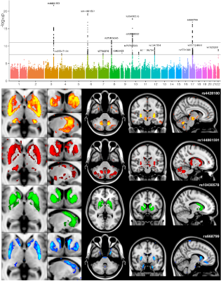
Gwenaelle Douaud, Lloyd Elliott, Kevin Sharp, Fidel Alfaro-Almagro, Sinan Shi, Karla Miller, Jonathan Marchini*, Steve Smith*
The genetic basis of brain structure and function is largely unknown. We carried out genome-wide association studies of 3,144 distinct brain imaging derived phenotypes in UK Biobank. Notable significant associations include: iron-related genes linked to T2* in subcortical regions; extracellular matrix associated with white matter microstructure and lesion volume; genes regulating midline axon guidance related to pontine crossing tract organisation. More broadly, effects were mainly seen in imaging measures associated with genes involved in brain development and transport of nutrients. Genes implicated in neurodegenerative disorders were largely related to iron and cardiovascular traits, and to brain development for psychiatric disorders.
|
|
3650.
 |
98 |
The Protective Effects of Ultramicronized Palmitoylethanolamide on the glutamatergic system in a triple transgene mouse model of Alzheimer disease
Rossella Canese, Giulia Carpinelli, Gianmauro Palombelli, Maria Brunzuoli, Silvio Calcagnini, Luca Steardo, Tommaso Cassano, Caterina Scuderi
We investigate the effects of a chronic treatment with an endogenous lipid mediator palmitoylethanolamide on the onset and progression of AD in 3xTg-AD mice. Behavioural tests showed improvements in learning and memory, and in both the depressive and anhedonia-like phenotype in PEA-treated 3×Tg-AD mice. MRI/MRS in vivo analysis, microdialysis and western blot, RT-PCR, and immunofluorescence show that PEA normalizes astrocytic function, rebalances glutamatergic transmission, and restrains neuroinflammation. The efficacy of PEA is particularly potent in younger mice, suggesting its potential as an early treatment.
|
|
3651.
 |
99 |
Relationship between brain and gut in autism spectrum disorder using diffusion MRI and intestinal bacteria gene analysis
Ting-Chun Lin, Ssu-Ju Li, Ching-Wen Chang, Hui-Ching Lin, Yin-Chieh Liu, Han-Fang Wu, Ming-Chia Chu, You-Yin Chen, Yu-Chun Lo
Brain-behavior-gut-microbiome interaction, a bidirectional communication, was proposed as an important role in autism spectrum disorders (ASD). However, the correlation among gut microbiota, behavioral performance, and brain microstructure in ASD are remained unclear. We chose a VPA-exposed rat model which performed autistic behaviors to investigate their brain-behavior-gut interaction. Diffusion MRI, behavioral tests, and intestinal bacteria gene analysis were applied in this study. The findings implied that the altered brain microstructure and atypical distribution of the gut microbiota associate with the severity of the autistic behavior in ASD compared to the control group.
|
|
3652.
 |
100 |
Rapamycin treatment increases cerebral blood flow and attenuates anxiety in pre-symptomatic APOE4 mice: effects of sex and APP transgene
Did Not Present
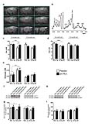
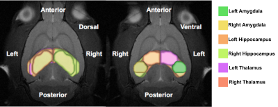
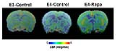
Ishita Parikh, David Ma, Jared Hoffman, Amy Wang, Ai-Ling Lin
APOE4 is the strongest genetic risk factor for Alzheimer’s disease (AD). Pre-symptomatic APOE4 carriers have developed neurovascular deficits decades before amyloid beta (Aβ) aggregation. Here we show that with Rapamycin treatment for 16 weeks, pre-symptomatic APOE4 mice had restored cerebral blood flow (CBF) and attenuated anxiety, compared to those of APOE3 mice. The CBF restorations were particularly significant in female mice or those with APP transgene. As Rapamycin and MRI and are readily to be used in humans, the findings may provide valuable information for future clinical trials to prevent AD for APOE4 carriers.
|
|
3653.
 |
101 |
Genetic influence on Brain Microstructure and Cognitive Function and Change in an Ageing Cohort
Video Permission Withheld
Kiyana Zarnani, Jayachandra Raghava, Naja Hansen, Erik Mortensen, Merete Osler, Martin Lauritzen, Egill Rostrup
The notion that the brain’s early-life retains hints to its end is an evolving avenue explored by basic and clinical researchers alike. Implementing neuroimaging, genetics and neuropsychological tests, our focus is to unravel the complex process of brain ageing through characterisation of normal brain development in a longitudinal ageing cohort. This approach has the power to unveil robust candidate determinants responsible for the vulnerability of the ageing brain to late-life pathology. We demonstrate statistically significant correlations between common genetic variants, white-matter integrity and cognitive ability and change, highlighting biological risk-factors as mediators of differential trajectories of ageing.
|
|
3654.
 |
102 |
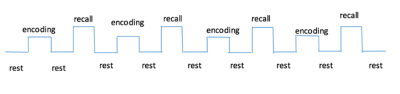
Differences in Metabolite Concentration and fMRI Activation in Subjects with Low and High Genetic Risk During Face-Name Paired-Associates Encoding and Retrieval Task in Healthy Adults
H Zhang, PW Chiu, SWH Wong, T Liu , GHY Wong, Q Chan, HKF Mak
APOE-ε4 is an important genetic risk factor of early onset of AD. To investigate the possible differences in metabolite concentration and fMRI activation in subjects with high risk of developing AD (APOE-?4 positive) compared to those with low risk, we employed Face-Name Paired-Associates (FN-PA) Encoding and Retrieval Task and evaluated the absolute concentrations of Glx in bilateral hippocampi and whole brain BOLD signal changes. Significant metabolic and activation differences were observed in the left hippocampus of pre-symptomatic subjects with high genetic risk of developing AD compared to subjects with low risk.
|
|
3655.
 |
103 |
Deep learning convolutional neural networks accurately classify genetic mutations and survival in gliomas
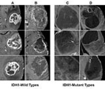
Peter Chang, Melissa Khy, Yang Zhang, Min-Ying Su, Daniel Chow
Gliomas represents a heterogeneous group of tumors with variable response to therapy despite sharing overlapping morphologic features. These differing outcomes partly relates to the multiple genetic mutations. For example, mutations in isocitrate dehydrogenase (IDH1) demonstrate significantly better survival compared to their wild counterparts1,2. Therefore, an obstacle in glioma imaging analysis is that radiographic interpretation fails to account for the tumoral genetic variance, making it difficult to integrate clinically relevant biological activities. The primary objective of this abstract is to use a convolutional neural network (CNN) approach to discover specific imaging patterns predictive of the underlying genetic alterations of gliomas.
|
|
3656.
 |
104 |
Mapping the functional and anatomical signatures of chemogenetically modulated neurons in the salience network.
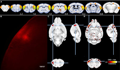
Joanes Grandjean, Francesca Mandino, Ling Yun Yeow, Teoh Chai Lean, Chris Jun Hui Ho, Amalina Attia, Lai Guan Ng, Malini Olivo, Fu Yu, Akhila Balachander
BOLD functional magnetic resonance imaging (fMRI) provides crucial information about the large-scale organisation and function of the healthy and diseased brain. Despite its widespread use, identifying the neuronal-basis for specific functional imaging-based signatures, identified in human disorders and animal models, remains a mostly unmet challenge. Presently, we combine chemogenetic neuromodulation with whole-brain resting-state mouse fMRI and tissue clearing, to reveal both the functional contribution and spatial localization of a targeted neuronal population on resting-state functional connectivity. This approach enables researchers to examine the functional role played by selected neuronal populations on distributed neuronal networks.
|
|
3657.
 |
105 |
Clinico-genetic-anatomical comparisons of paroxysmal kinesigenic dyskinesia with and without PRRT2 mutations
Lei Li, Xueling Suo, Qiyong Gong
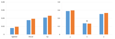
Paroxysmal kinesigenic dyskinesia (PKD) is a rare movement disorder characterized by sudden, brief attacks of involuntary movements. This study aims to detect the topological organization of white matter structural connectivity in PKD with and without PRRT2 mutations using graph theoretical approaches. Compared with non-PRRT2 mutation carriers, PRRT2 mutations carriers are significantly associated with a younger age of onset, a complicated form of PKD, combined phenotypes of dystonia and chorea, and a tendency for a family history of PKD in our population, as well as showed topological trends for randomization.
|
|
3658.
 |
106 |
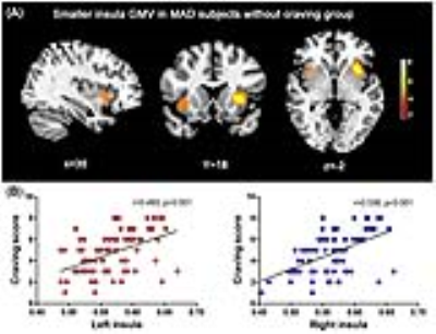
Neuroimaging based biomarkers for detecting craving and predicting relapse in methamphetamine dependence
Chang Qi, Xiaobing Fan, Sean Foxley, Yanhui Liao, Qiuxia Wu, Jingsong Tang, Wei Hao, Tieqiao Liu
This study investigates whether insula cortex abnormalities of methamphetamine-dependent subjects (MADs) can detect craving state and predict relapse susceptibility. Voxel based morphometry and statistical parametric mapping were used on structural MRI of MADs. Total 142 MADs were divided into two groups: model-group (n=112) and validation-group (n=30) from follow-up MADs. The results showed that MADs without craving had significantly smaller insula volume. Optimal insula volume determined from Youden index cut-off point on ROC analysis could be used as MRI bio-markers with acceptable accuracy for detecting craving state. Our results could help guide optimally timed intervention, prevention, and treatment strategies for MADs.
|
|
3659.
 |
107 |
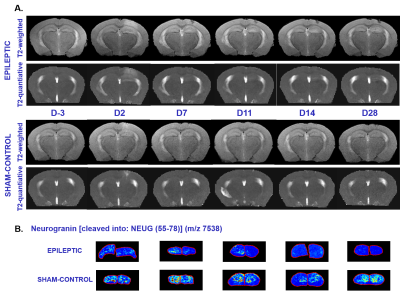
Epileptogenesis Imaging biomarker identification in the Mesial Temporal lobe epilepsy (MTLE) mouse model
Yvan Dietrich, Gabriel Dieuset, Mélanie Lagarrigue, Hervé Saint-Jalmes, Benoit Martin, Charles Pineau, Fabrice Wendling, Pierre-Antoine Eliat
This work presents preliminary promising results on Mesial temporal lobe epilepsy (MTLE) mouse model suggesting that MRI/MRS-based biomarkers could be of great interest to detect early changes in the lesioned brain and to characterize epileptogenesis after traumatic brain injury.
|
|
3660.
 |
108 |
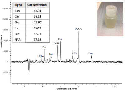
Quantifying acrolein with MRS: a viable biomarker to assess location and severity of TBI
Nicole Vike, Jonathan Tang, Riyi Shi, Joseph Rispoli
Currently, only invasive methods exist to study the molecular effects of traumatic brain injury (TBI). By instead using magnetic resonance spectroscopy (MRS), a non-invasive, quantitative method can be used to safely assess the severity and location of injury. Acrolein, a biomarker of TBI, increases in rat brain tissue following TBI. Acrolein shows signature peaks downfield of water at 6.5 and 9.4 ppm. We have successfully measured whole-brain phantom-injected acrolein using Bruker 7T MRS sequences and determined T1 for acrolein, using NMR, so MRS parameters can be adjusted to maximize acrolein signal. Quantifying acrolein with MRS could provide a viable method to assess injury location and severity.
|
|
3661.
 |
109 |
BIOmarkers of DEPression (BIODEP) study: Raised peripheral inflammation associated with changes in striatal microstructure
Did Not Present
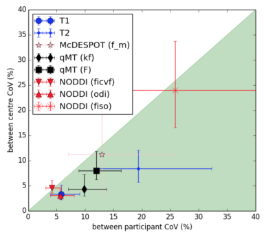
Charlotte Clarke, Manfred Kitzbichler, Gareth Barker, Marta Correia, Nick Dowell, Samuel Hurley, John Mclean, Tobias Wood, NIMA Consortium, Mara Cercignani, Ed Bullmore, Neil Harrison
Inflammation is implicated in the aetiology of major depressive disorder (MDD) and is known to affect brain microstructure: e.g. the striatum as found with neurite orientation dispersion and density imaging (NODDI) and quantitative magnetization transfer (qMT) imaging. Further, individuals with high inflammation (C-reactive protein (CRP)>3mg) respond less well to antidepressants. The BIOmarkers of DEPression (BIODEP) multi-centre study is aiming to characterise the inflamed MDD phenotype. We report across-site data harmonization and interim results demonstrating a negative correlation between the water fraction content (NODDI) and CRP within the ventral striatum. Multi-modal imaging results (qMT, multi-component relaxometry) across the full cohort will be reported.
|
|
3662.
 |
110 |
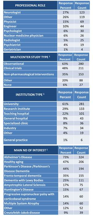
A survey of best practice guidelines to harmonize neuroimaging biomarkers for neurodegenerative diseases
Jorge Jovicich, Frederik Barkhof, Claudio Babiloni, Karl Herholz, Christoph Mullert, Bart van Berckel, Giovanni Frisoni, SRA-NED JPND Working Group
Current challenges in neurodegenerative diseases (ND): need for early and accurate markers for differential diagnosis, prognosis, progression tracking and intervention assessments. Big-data neuroimaging studies might help addressing these challenges. However, large-sample neuroimaging data still lacks standardization (multi-vendor acquisition, analysis) and cross –validation (across markers, across large populations), in particular with state-of-the-art MRI hardware (≥32 head RF channel coils and powerful gradients).
Here we: i) evaluate current barriers perceived by the broad neuroimaging research/clinical/industry communities for the large-scale harmonization of MRI/PET-SPECT/EEG neuroimaging methods in the context of ND studies, and ii) propose actions that may help addressing these barriers.
|
|
3663.
 |
111 |
Development of an MR-based Biomarker of Arteriolar Sclerosis
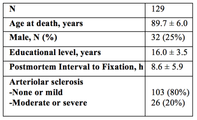
Nazanin Makkinejad, Arnold Evia, Ashish Tamhane, Chantal Sopacua, Sue Leurgans, Julie Schneider, David Bennett, Konstantinos Arfanakis
Arteriolar sclerosis is often present in the brain of older adults and has been associated with lower cognitive performance and higher risk of dementia. There is currently no standard biomarker for arteriolar sclerosis. Future clinical trials of therapeutic interventions targeting arteriolar sclerosis will require an approach for participant selection with high positive predictive value (PPV). The purpose of this work was to develop a classifier with high PPV for arteriolar sclerosis based on information about regional white matter hyperintensities, magnetic susceptibility, cerebral microbleeds, and demographics.
|
|
3664.
 |
112 |
Parameters From Dynamic Contrast-Enhanced Magnetic Resonance Imaging Are Biomarkers Predicting Response after Radiotherapy to Brain Metastases
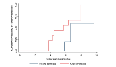
Zhuo Shi, Lizhi Xie, Peng Wang, XinMing Zhao, Han Ou-Yang
Dynamic contrast-enhanced (DCE) MRI provides additional information regarding blood-brain barrier integrity, and Ktrans is directly proportional to the level of permeability of the blood-brain barrier. In our study, we found demonstrates that SRS of cerebral metastasis is associated with a reduction of Ktrans values in the early post-treatment period. DCE-MRI derived parameters of may be a promising imaging biomarker of tumor aggressiveness.
|
|
3665.
 |
113 |
23Na-MRI demonstrates a sodium gradient within gliomas as a biomarker of tumor heterogeneity
Video Permission Withheld
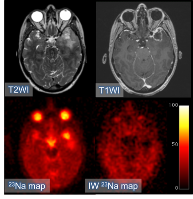
Fulvio Zaccagna, Frank Riemer, Mary McLean, James Grist, Joshua Kaggie, Rolf Schulte, Sarah Hilborne, Tomasz Matys, Jonathan Gillard, Colin Watts, Stephen Price, Martin Graves, Ferdia Gallagher
Glioma grade and the extent of local infiltration are important for guiding management. Imaging tumor heterogeneity may improve diagnosis and therapy planning. 23Na-MRI has been used here to demonstrate a gradient in sodium concentration across gliomas: necrosis > viable tissue > edema. This gradient was evident in all the tumors analyzed and is consistent with the expected underlying cellular microstructure where the sodium concentration is dominated by the extracellular fluid in edema and by an absence of cells in the necrotic core. The study provides evidence that 23Na-MRI represents an imaging biomarker of tumor heterogeneity and tissue microstructure in glioma.
|
|
3666.
 |
114 |
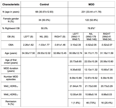
Is occipital bending a structural biomarker of risk for depression and sensitivity to treatment?
Karen Fullard, Jerome Maller, Thomas Welton, Matthew Lyon, Fraser Callaghan, Leanne Williams, Mayuresh Korgaonkar, A. Rush, Evian Gordon, Stephen Koslow, Stuart Grieve
Occipital Bending (OB) was investigated as an MRI imaging biomarker for major depressive disorder (MDD) using data from large, well characterized, international, randomised study recruiting non-geriatric adult participants (iSPOT-D, n=68 control, 231 MDD). The presence of OB and the angle of occipital bending (OBA) was correlated with a repeated battery of neuropsychiatric assessments, and response to 6 weeks of antidepressant treatment. A greater proportion of rightward bending was present in MDD in comparison to control individuals. Underlying association between OB and MDD is likely.
|
|
3667.
 |
115 |
Multivariate MR Biomarkers Predict Cognitive Decline in Mouse Models of Alzheimer’s Disease
Alexandra Badea, Robert Anderson, Russell Dibb, Yi Qi, Natalie Delpratt, Hongjiang Wei, Chunlei Liu, William Wetsel, Brian Avants, Carol Colton
We propose a multivariate approach for characterizing mouse models of Alzheimer’s disease (AD), which integrates imaging and behavior in a joint analysis. We used manganese enhanced MRI (MEMRI) to identify brain areas associated with reduced performance in a spatial memory task. We quantified genotype differences based on morphometry, T1 weighted (T1W) signal and quantitative susceptibility maps (QSM). We find that the integration of multiple imaging biomarkers is a better predictor of cognitive decline, relative to using single biomarkers in isolation.
|
|
3668.
 |
116 |
Is MRI all we need? Prediction of conversion between normal cognitive function, mild cognitive impairment and Alzheimer’s disease
Shiyang Chen, Ke Qi, Deqiang Qiu
. In this study, we aimed to use machine learning methods to establish the quantitative value of MRI alone in the prediction of changes between disease states such as from normal cognitive function (NC) to mild cognitive impairment (MCI), and MCI to AD, and compare with the combined predictive power of MRI, PET, neuropsychological evaluations and CSF analysis. Very high overall accuracy can be achieved using both RF and DNN methods. Interestingly, predictive power of MRI features is very close to all features combined, suggesting MRI might contain much of the information provided by neuropsychological evaluations, PET scans among others combined. The methodology adopted in this study also provides a framework for evaluating the value of different imaging techniques in a quantitative manner.
|
|
3669.
 |
117 |
Rapid and robust high-resolution mapping of proton pool size ratio in spinal cord after injury in squirrel monkeys
Feng Wang, Tung-Lin Wu, Ke Li, Li Min Chen, John Gore
High-resolution quantitative magnetization transfer (qMT) MRI provides a noninvasive means to detect and characterize myelination before and after neural injury and during repair. This study aims to systematically evaluate the accuracy and precision of pool size ratio (PSR) measurements using either 5-, 2- or 1-parameter modeling for assessing injury-associated changes in spinal cords in squirrel monkeys in order to optimize a rapid, sensitive, and high-resolution PSR mapping protocol for applications in primates at high field. In addition, the sensitivity of PSR to demyelination in the dorsal pathway rostral and caudal to an injury site has been evaluated.
|
|
3670.
 |
118 |
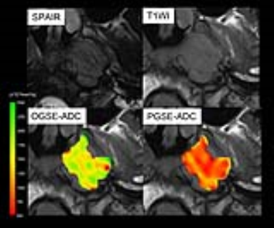
Time makes the difference: Comparison of ADC values obtained with OGSE and PGSE sequences for differentiation of human head and neck tumors
Mami Iima, Akira Yamamoto, Ichiro Tateya, Morimasa Kitamura, Thorsten Feiweier, Koichi Omori, Kaori Togashi
The correlation of ADC values with different diffusion times, as obtained from OGSE and PGSE, was investigated in patients with head and neck tumors. A significant decrease of ADC values in head and neck cancers was noted with the increase of diffusion time (p < .0.05). Care needs to be taken when interpreting OGSE-ADC and PGSE-ADC values, as flow effects will contribute to these values, which was suspected in light of the (artefactual) increase of ADC values using PGSE compared to OGSE.
|
|
3671.
 |
119 |
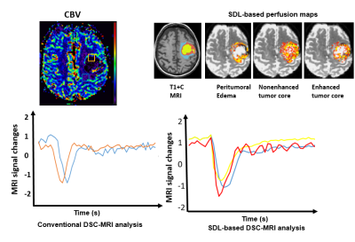
Improving classifications of brain tumor tissue with Sparse Dictionary Learning based analysis of dynamic susceptibility contrast enhanced MRI data
Silun Wang, Shu Zhang, Liya Wang, Bing Ji, Tianming Liu, Hui Mao
We analyzed the DSC MRI signals based on patterns of descriptive DSE-MR parameters by using Sparse Dictionary Learning (SDL) coding method. We successfully decomposed DSC MRI signals into linear combinations of multiple components based on sparse representation of DSC MRI signals in the tumor region of tumor core and peritumoral edema which might be represent multiple heterogeneity component in brain tumors. Assessment of diagnostic performance of SVM classification after cross validation revealed that the combination of conventional DSC temporal characteristics and dictionary learning based DSC temporal features would result in the best classification accuracy between tumor core and peritumoral edema (with total diagnostic accuracy of 77%, AUC 0.78).
|
|
3672.
 |
120 |
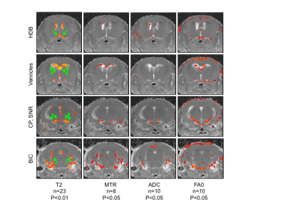
The effects of LIS1 deletion in the adult mouse brain and partial rescue by LiCl: A T2 mapping, magnetization transfer and diffusion MRI study
Hagit Dafni, Liraz Kaidar, Inbal Biton, Aditya Kshirsagar, Orly Reiner
LIS1, a gene mutated in lissencephaly (“smooth brain”) have been investigated mainly in the developing brain. Initial studies demonstrated distinct ataxia and rapid lethality following Lis1 conditional deletion in adult mice. Therefore, our aim was to investigate the postnatal roles of LIS1 and the underlying mechanism. Conditional Lis1knockout mice studied by MRI pre, and 5 days post, tamoxifen-induced Lis1 deletion, showed increase in T2 and ADC and decrease MTR and FA0, in the lateral ventricles and in brain regions related to motion and auditory functions. These alterations and changes in the Wnt pathway were partially rescued by LiCl treatment.
|
|
Neurodegeneration
Electronic Poster
Neuro
Monday, 18 June 2018
| Exhibition Hall |
17:15 - 18:15 |
| |
|
Computer # |
|
3721.
 |
49 |
Brain microstructural abnormalities in cirrhotic patients without overt hepatic encephalopathy: A voxel-based diffusional kurtosis imaging study
Did Not Present
Hua-Jun Chen
Brain microstructural change in cirrhotic patients without overt hepatic encephalopathy: A DKI study
|
|
3722.
 |
50 |
Altered Dynamic Functional Connectivity in the Default Mode Network in Patients with Cirrhosis and Minimal Hepatic Encephalopathy
Did Not Present
Hua-Jun Chen
Altered Dynamic Functional Connectivity in Default Mode Network in Minimal Hepatic Encephalopathy
|
|
3723.
 |
51 |
Quantitative MRI comparison of neurological and hepatic forms of Wilson disease
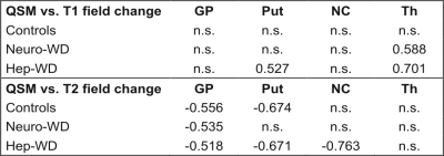
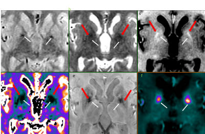
Monika Dezortova, Vit Herynek, Petr Dusek, Julio Acosta-Cabronero, Radan Bruha, Filip Jiru, Milan Hajek
We examined 40 patients with neurological and hepatic form of Wilson disease by susceptibility mapping at 3T and T1 and T2 relaxometry at 1.5T and 3T. We were able to distinguish patients with neurological form from hepatic form and healthy controls based on quantitative relaxometry and susceptibility mapping. Higher susceptibility observed in the globus pallidus, putamen, and caudate nucleus in patients with neurological form corresponds to higher iron content while higher susceptibility in the thalamus corresponds rather to demyelination.
|
|
3724.
 |
52 |
Global Cerebral Metabolic Rate of Oxygen in Older People: Gender Differences
Ana Rodríguez-Soto, John Detre, David Wolk, Joseph Harrison, Felix Wehrli
Cerebral metabolic rate of oxygen (CMRO2) has been reported to vary as a function of age and gender. Here, we re-examined possible gender differences as part of an ongoing study in people over the age of 60 years by means whole-brain MR oximetry. Data corroborate earlier findings that cognitively normal females appear to have higher CMRO2 than their male peers. This difference may be largely due to greater oxygen extraction rather than changes in cerebral blood flow. Possible biological causes of this gender bias will need detailed scrutiny, as do the underlying physiologic assumptions that may impact the results.
|
|
3725.
 |
53 |
Tract based spatial statistics analysis of DKI metrics in type 2 diabetes patients
Zaheer Abbas, Ezequiel Farrher, Elene Iordanishvili, Nino Kobalia, Krzysztof Dzieciol, Farida Grinberg, Nadim Jon Shah
In addition to cardiovascular risk factors, Type 2 diabetes mellitus (T2DM) is associated with microstructural, structural, functional, and metabolic changes in the brain. Patients with T2DM tend to develop cognitive impairment and dementia. In this study, we report differences between groups of T2DM patients and age-matched healthy volunteers by comparing diffusion tensor and diffusion kurtosis metrics using the tract-based spatial statistics (TBSS) method. We found that all diffusivities are larger and the axonal water fraction is reduced in the T2DM group compared to the controls. This suggests presence of more water in the extra-axonal space.
|
|
3726.
 |
54 |
Gray matter cortical thickness changes in Hypothyroid patients: A study using high-resolution structural imaging
Did Not Present
Mukesh Kumar, Poonam Rana, Pooja Rathore, Deepak Sharma, Prabhjot Kaur, Tarun Sekhri, Subash Khushu
The aim of our study was to assess changes in cortical thickness, cortical area and cortical volume of gray matter (GM) for hypothyroid patients. We had acquired high-resolution 3D T1 weighted structural data for both control (24) and hypothyroid subjects (22). Reduced gray matter cortical thickness was observed in lateraloccipital cortex, postcentral gyrus, medialorbitofrontal cortex, lingual gyrus, superior and inferior parietal cortex in hypothyroid patients as compare to controls. These findings of reduced gray matter (CTh) suggest abated activities of motor, attention, working memory and executive cognitive function in hypothyroid patients.
|
|
3727.
 |
55 |
On the Importance of Using High-Resolution Atlases for Voxel-Based Morphometry of High-Resolution MRI Data: A Case-Control Study on Essential Tremor
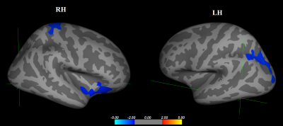
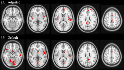
Eric Cameron, Jonathan Dyke, Elan Louis, Ulrike Dydak
The use of a high resolution atlas for segmentation and normalization greatly improves the accuracy of voxel-based morphometry analysis of magnetic resonance images. An adjusted method including the high resolution atlas was compared to the default method with the standard resolution atlas in a case-control study on essential tremor to demonstrate the impact of higher resolution segmentation. After multiple comparison correction using extent cluster thresholding, the adjusted method showed bilaterally consistent results, while the default method showed some false positive results in peripheral regions of the brain. A high resolution atlas should be used to segment equally high resolution images.
|
|
3728.
 |
56 |
Developmental alteration of mean diffusivity in striatum of transgenic Huntington’s disease monkey
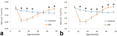
Yuguang Meng, Jocelyne Bachevalier, Anthony Chan, Xiaodong Zhang
Huntington Disease (HD) is a neurodegenerative disorder with a primary etiology of striatal pathology. Abnormal mean diffusivity changes have been seen previously in HD patients. However, it remains not fully understood how the diffusivity property of striatum evolves during the development of the disease. This study examined the progressive changes of striatum of rhesus monkey brains with HD gene mutation using diffusion tensor imaging (DTI), and it was found that there was significant MD difference from the control animals in striatum at very early age.
|
|
3729.
 |
57 |
Radiological Imaging of Brain Iron Deposits and T1 quantification feasibility using 3D UTE Cones.
Piotr Wielopolski, Elene vroegindeweij, Agnita Boon, Janneke Langendonk, Juan Hernandez-Tamames
In this work, we demonstrate that 3D UTE cones enables the possibility of T1 contrast and T1 relaxometry of iron deposits in the brain. We present radiological images of brain iron and T1 quantification of iron deposits in a pathology with an exacerbated amount of iron in the brain. We demonstrate that even for conventional T2 and T2* mapping is challenging to obtain relaxometry. However, it is feasible for a 3D UTE cones sequence.
|
|
3730.
 |
58 |
Longitudinal Diffusion Tensor Imaging in the brain in Friedreich’s Ataxia: follow-up at 12 and 24 months
Pierre-Gilles Henry, James Joers, Dinesh Deelchand, Diane Hutter, Christophe Lenglet
We report 12-month and 24-month longitudinal diffusion tensor imaging (DTI) data in the brain of subjects with Friedreich’s ataxia (FRDA). Significant longitudinal changes were observed in several brain areas (including the corpus callosum, internal capsule and superior corona radiata) in a group of 13 patients over 24 months. Our data suggest that diffusion MRI of the brain could be useful to better understand the impact of FRDA on brain microstructure and connectivity, and to assess the effect of potential treatments on neurodegeneration in upcoming clinical trials in FRDA.
|
|
3731.
 |
59 |
Magnetic susceptibility of the dentate in a longitudinal study of Friedreich ataxia
Phillip Ward, Ian Harding, Parnesh Raniga, Tom Close, Louise Corben, Martin Delatycki, Monique Stagnitti, Elsdon Storey, Nellie Georgiou-Karistianis, Gary Egan
We performed in-vivo measurements of the magnetic susceptibility in the dentate nucleus in individuals with Friedreich ataxia and healthy controls over a two-year longitudinal study using quantitative susceptibility mapping. The results show a significant susceptibility difference between individuals with Friedreich ataxia and control subjects, and a strong correlation with disease severity in the Friedreich ataxia cohort. These findings may lead to the development of a sensitive biomarker of disease severity and progression in Friedreich ataxia.
|
|
3732.
 |
60 |
Delivery of mHTT to the rhesus macaque brain leads to a reduction in caudate volume in a new monkey model of Huntington’s disease
Zheng Liu, Alison Weiss, Christopher Kroenke, Jodi McBride
Huntington’s disease (HD) is a genetic, neurodegenerative disorder caused by CAG repeat expansion in mutant huntingtin gene (mHTT) on Chromosome 4 and is characterized by degeneration of several brain regions, with the caudate nucleus and the putamen being the most heavily affected. Structural MRI was used to investigate the longitudinal striatal atrophy in a new rhesus macaque HD model, especially the reduction of rostral caudate volume. According to the longitudinal volumetric analysis, the delivery of 82Q mHTT to rhesus macaque striatum leads to a significant reduction in caudate volume. However, no such significant differences following delivery of 16Q mHTT model was observed. These data indicate that viral delivery of 82Q mHTT serves as a nonhuman primate model of Huntinton’s disease that reproduces neuropathological characteristics of the disease.
|
|
3733.
 |
61 |
MRI Connectivity Impairment Associated with Timing of Seizure Recurrence After Surgery in Temporal Lobe Epilepsy
Victoria Morgan, Dario Englot, Baxter Rogers, Adam Anderson, Bennett Landman, Bassel Abou-Khalil
Surgical resection of the seizure focus in the mesial temporal lobe is a common treatment of drug-resistant temporal lobe epilepsy (TLE) with an approximately 80% success rate. Our previous work showed that presurgical MRI-based functional and structural network connectivity can identify those TLE patients with the most unfavorable seizure outcomes. The goal of this work was to increase specificity of our prediction by characterizing those with seizure free and favorable outcomes. The results suggest that when impairment in functional connectivity of the seizure propagation network extends to the contralateral hemisphere, patients will experience rare post-surgical seizures sooner.
|
|
3734.
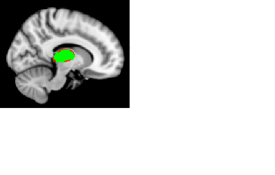 |
62 |
Vertex-based thalamus morphometry and tract-based spatial statistics of the white matter show differences between patients with chronic low back pain and fibromyalgia
Huiling Peng, Alyssa Smith, Kelly Boland, Jason Craggs
Chronic low back pain (CLBP) is now considered a disease of the central nervous system. Two-thirds of individuals with CLBP also have fibromyalgia (FM) which is a chronic pain syndrome characterized by widespread deep musculoskeletal pain and cognitive deficits. The aim of this study was to investigate gray and white matter changes between FM and CLBP groups using vertex analysis of thalamus and tract-based spatial statistics (TBSS). Significant surface depression was detected in right thalamus of patients with FM compare to CLBP. TBSS analysis showed significantly reduced FA in several white matter tracts of patients with FM compared to CLBP.
|
|
3735.
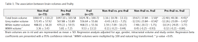 |
63 |
The association between brain volumes and physical frailty in older individuals
Video Permission Withheld
Ilse Kant, Jeroen de Bresser, Simone van Montfort, Ellen Aarts, Ilona Bader, Yarit Wiggerts, Georg Winterer, Claudia Spies, Arjen Slooter, Jeroen Hendrikse
Physical frailty develops with increasing age and is a chronic state of vulnerability that is associated with disability and cognitive decline. Few studies have assessed the underlying structural brain abnormalities of physical frailty. We therefore examined the association between brain volumes on MRI and physical frailty in a group of 214 non-demented elderly participants. Frail participants showed a lower total brain volume and a lower grey matter volume compared to pre-frail and non-frail participants. Furthermore, pre-frail participants showed more cortical infarcts compared to non-frail participants. These brain abnormalities could be the underlying substrate of the physical frailty phenotype.
|
|
3736.
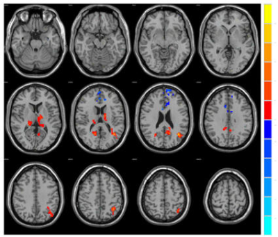 |
64 |
Changes in brain connectivity of the first-episode idiopathic epilepsy patients before and after treatment: A comparative study of resting-state fMRI
Did Not Present
Pengfei Qiao, Guang-ming Niu, Yang Gao, Lizhi Xie
This study investigates the fractional amplitude of low frequency fluctuation (fALFF) of the first-episode complex partial seizures epilepsy patients before and after treatment by using resting state fMRI (rfMRI). It is found that fALFF can be used to detect interictal epileptiform change before and after treatment.
|
|
3737.
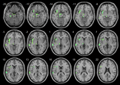 |
65 |
Investigation of cognition associated cerebral perfusion changes in Alzheimer's disease and mild cognitive impairment using a novel 3D arterial spin labeling technique
Yong Zhang, Hua-Wei Lin, Bei Ding, Fu-Hua Yan, Bing Wu
This preliminary study investigated cognition associated cerebral perfusion changes in Alzheimer’s disease (AD) and mild cognitive impairment (MCI) measured with a novel 3D arterial spin labeling technique. The voxel-wise regression analysis against MMSE scores was performed using whole brain CBF maps. Cognition associated CBF deficits in the hippocampus and insular cortex might cause the typical AD symptoms including memory loss and emotion issues. CBF decrease in MCI patients in the lingual gyrus could affect vision and wording functions. CBF increase in the frontal lobe might serve as an early indicator for high-level cognitive function disturbance.
|
|
3738.
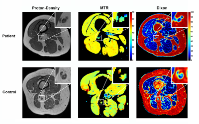 |
66 |
Multi-parametric MRI of the Proximal and Distal Nerves of the Leg: Longitudinal Findings in Patients with Inherited Neuropathies
Michael Pridmore, Richard Dortch, Jun Li
This project proposes a multi-parametric set of MRI tools for assaying human inherited neuropathies in vivo, with the longer-term goal of establishing biomarkers of disease progression for future clinical trials. Previous research shows magnetization transfer ratio (MTR) values, which assay myelin content changes from demyelination and axonal loss, relate to disability in neuropathy patients. Here, we proposed additional fat-water (Dixon) imaging to assay fat replacement following deinnervation. MTR/Dixon data were collected in the sciatic/tibial nerves in patients with primary dysmyelinating inherited neuropathies. Longitudinal results showed lower that MTR/Dixon were responsive to disease progression.
|
|
3739.
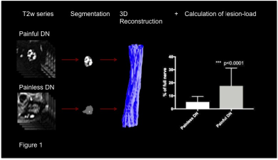 |
67 |
Structural remodeling of the sciatic nerve differs between painful and painless diabetic polyneuropathy: an in vivo study using magnetic resonance neurography.
Johann Jende, Jan Groener, Stefan Kopf, Tim Hilgenfeld, Sabine Heiland, Mirko Pham, Peter Nawroth, Martin Bendszus, Felix Kurz
Diabetic polyneuropathy (DN) is one of the most severe complications of diabetes. It is yet uncertain why patients either suffer from painful (PDN) or painless (NPDN) diabetic polyneuropathy. We prospectively performed magnetic resonance neurography of the sciatic nerve in 120 patients suffering from DN of varying severity and correlated the results with clinical symptoms and electrophysiological data. We found a higher load of lesions to the sciatic nerve in PDN compared to PDN (p<0.0001), extending over longer distances (p<0.0001). This indicates that proximal nerve damage is one of the main contributors to the development of PDN.
|
|
3740.
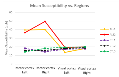 |
68 |
Quantitative Susceptibility Mapping of post-mortem ALS brains at 7T
Chaoyue Wang, Benjamin Tendler, Menuka Pallebage-Gamarallage, Olaf Ansorge, Ricarda Menke, Martin Turner, Sean Foxley, Karla Miller
Amyotrophic lateral sclerosis (ALS) is a fatal neurodegenerative disease of the motor system and its wider cortical connections. Progress in therapeutic development in ALS is compromised by a lack of specific biomarkers. In this work, we describe a platform for acquiring QSM data in post-mortem brains and propose a protocol for post-mortem QSM reconstruction. Preliminary results have shown that ALS brains had substantially greater mean susceptibility in motor cortex than control brains, which indicates that QSM has the potential to accurately quantify iron concentration and thus serve as an imaging biomarker for ALS.
|
|
3741.
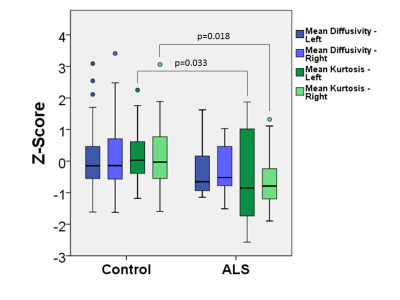 |
69 |
Diffusion Kurtosis Imaging of the Motor Cortex in Amyotrophic Lateral Sclerosis
Thomas Welton, Fraser Callaghan, Jerome Maller, Matthew Middione, R Marc Lebel, Ek Tan, Dominic Rowe, Ajit Shankar, Stuart Grieve
Amyotrophic lateral sclerosis (ALS) is characterised by degeneration of the motor neurons. Diffusion kurtosis imaging (DKI) may be a suitable biomarker for neuronal injury in ALS. We imaged 30 people with ALS and 63 controls with a 140-direction DKI protocol and explored group differences diffusion and kurtosis scalar values within the motor cortex. Our preliminary data show that significant microstructural changes are measurable by DKI in people with early ALS. DKI may represent a viable biomarker of early ALS progression.
|
|
3742.
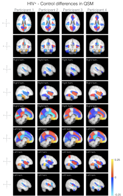 |
70 |
Initial Experience of Cerebral Quantitative Susceptibility Mapping in the HIV Neurocognitive Disorder
Sara Dupont, Yan Li, Ycheng Chen, Janine Lupo, Felicia Chow, Jared Narvid
This study investigates the patterns of tissue susceptibility within brains of older HIV+ participants using quantitative susceptibility mapping (QSM) as marker of iron deposition in the brain. Four HIV+ participants and one healthy control were recruited, and QSM and T1w (for cortical parcellation) data were acquired on a 3T MRI scanner. Comparison of QSM values by brain region for each HIV+ participant with the control shows a pattern of increased susceptibility in frontal regions, especially in the frontal medial and subcallosal cortices. Although these results warrant further investigation in a larger cohort, this is the first study investigating changes in QSM in the HIV+ population.
|
|
3743.
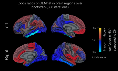 |
71 |
The Abnormal Cerebral Perfusion Profile in older adults with HIV-Associated Neurocognitive Disorder: Discriminative Power of Arterial Spin Labelling MRI
Sara Dupont, David McCoy, Andrew Callen, Duygu Tosun, Joanna Hellmuth, Victor Valcour, Jared Narvid
This study aimed at evaluating the pattern of cerebral blood flow (CBF) abnormalities in older adults with HIV-associated neurocognitive disorder (HAND) compared to cognitively healthy controls. Pulsed ASL (for CBF quantification) and T1-weighted images (for registration to cortical parcellation atlas) were collected in a retrospective cohort of 19 HIV+ participants and 15 age and education matched controls. To investigate differences in CBF patterns between HIV+ and controls, CBF values were used to train generalized linear models (GLMnet) to predict patient diagnosis. Older HIV+ exhibited lower CBF values in the temporal and occipital lobes compared to controls suggesting a specific pattern of CBF in individuals aging with HAND.
|
|
3744.
|
72 |
Structural connectivity alterations in amyotrophic lateral sclerosis are modulated by the topology of the anatomical brain connectome
Video Permission Withheld
Silvia Basaia, Federica Agosta, Nilo Riva, Edoardo Spinelli, Yuri Falzone, Adriano Chiò, Andrea Falini, Giancarlo Comi, Massimo Filippi
In this study, we used graph theory and connectomics to test whether the spatial patterning of structural brain alterations in amyotrophic lateral sclerosis (ALS) is modulated by the topology of the anatomical brain network. In the healthy subject connectome, brain regions of subsequent stages of ALS pathology are shown to be more closely interconnected (shorter topological distance) with the primary motor cortex (ALS epicenter) than regions of more distant stages. Altered structural connectivity was greater between closely connected regions. Axonal connections may influence the spatial spreading of pathology in ALS.
|
|
Alzheimer's Disease & Other Dementias
Electronic Poster
Neuro
Monday, 18 June 2018
| Exhibition Hall |
17:15 - 18:15 |
| |
|
Computer # |
|
3745.
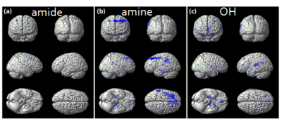 |
73 |
Evaluation of the Full Z Spectrum Obtained from Demented Patients
Video Permission Withheld
Geon-Ho Jahng, Bo Guem Choi, Hak Young Rhee, Chang-Woo Ryu, Dal Mo Yang, Wook Jin
To evaluate the characteristics of chemical exchange proton pools using a CEST MRI technique in dementia patients, 19 demented and 22 non-demented subjects were scanned a full Z spectrum data using a 3D GRASE sequence. The 6 pool maps with the Lorentzian fitting and MTR asymmetry map pool were calculated and compared between the two groups. The amide, amine, hydroxyl, DWS, NOE, MT, and MTR asymmetry values at 1 ppm, 3ppm, and 3.5 ppm were higher in the demented group than in the non-demented group.
|
|
3746.
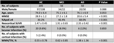 |
74 |
MRI based assessment of cerebrovascular pathology in Alzheimer’s disease
Ying Xia, Nawaf Yassi, Pierrick Bourgeat, Parnesh Raniga, Patricia Desmond, Jurgen Fripp, David Ames, Paul Maruff, Chris Fowler, Ralph Martins, Victor Villemagne, Collin Masters, Christopher Rowe, Olivier Salvado
Cerebrovascular disease often coexists with Alzheimer’s disease (AD) and its contribution to the development of AD remains unclear. This study assessed cerebrovascular pathology in terms of brain infarction and white matter hyperintensities (WMH) in the AIBL study, and investigated its impact on amyloid deposition. Our data revealed significantly greater WMH burden and higher prevalence of cerebrovascular pathology in AD patients. No correlation was found between amyloid load and WMH in either healthy controls, mild cognitive impairment or AD. Moreover, the presence of cerebrovascular pathology showed no association with amyloid deposition in subjects with or without AD.
|
|
3747.
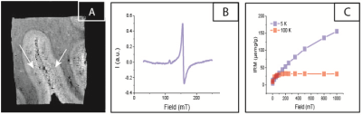 |
75 |
Assessment of different molecular iron forms in the brain tissue of Alzheimer patients
Lucia Bossoni, Marjolein Bulk, Wico Breimer, Andrew Webb, Jelle Goeman, Martina Huber, Tjerk Oosterkamp, Louise van der Weerd
Magnetic Resonance Imaging (MRI) has the potential to measure iron accumulation in the brain of Alzheimer patients. However, current MRI techniques cannot differentiate between different molecular iron forms. We have recently developed a combination of MRI, EPR and SQUID magnetometry, to quantify specific molecular iron forms in brain tissue. In this current study of AD patients and controls we found significant differences in R2*, ferritin concentration, and magnetic moment of magnetite nanoparticles between patients (N=22) and controls (N=14). Additionally, we identified correlations between some iron forms.
|
|
3748.
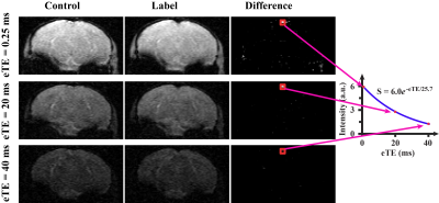 |
76 |
Oxygen extraction fraction is differentially affected in aging and Alzheimer’s disease: cross-sectional and longitudinal investigations in mice at 11.7T
Zhiliang Wei, Lin Chen, Zixuan Lin, Jiadi Xu, Siyuan Cheng, Zheyu Wang, Peter van Zijl, Hanzhang Lu
Quantification of oxygen extraction fraction (OEF) is traditionally challenging and often requires the use of radiotracers or other invasive procedures, but has become feasible with recent advances in MR oximetry techniques. Here, we investigated the OEF in aging and Alzheimer’s disease (AD) with the T2-Relaxation-Under-Spin-Tagging (TRUST) technique. Based on the cross-sectional and longitudinal investigations, we demonstrated that normal aging and AD have different effects on brain OEF, which may reflect the dynamic nature of changes in brain energy homeostasis during these two processes. TRUST MRI may be a useful tool in elucidating physiological mechanisms of brain diseases in mouse models.
|
|
3749.
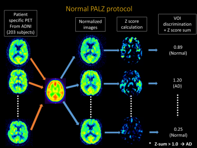 |
77 |
The estimation of diagnostic accuracy of FDG-PET/MR imaging for Alzheimer's disease— simulation study using ADNI-data
Tetsuro Sekine, Alfred Buck, Gaspar Delso, Edwin ter Voert, Martin Huellner, Patrick Veit-Haibach, Geoffrey Warnock
The purpose of this study was to estimate the impact of commercial MRAC on the evaluation of dementia, especially in Alzheimer’s disease (AD). We combined ADNI-data and patients data. We multiplied 14 error map derived from commercial Atlas-based MRAC method and each 203 ADNI data after the same nomalization and smoothing. To clarify the statement of prediction of AD, PET score was calculated by using PALZ. The accuracy, sensitivity and specificity for the discrimination of AD-patients from the normal control was not so impaired by MR-AC (Original vs. Error; 83.2%, 83.3% and 83.1% vs. 81.3% [range 77.6-83.2], 82.6% [range 79.2-85.4%,] and 80.3% [range71.2-86.4%]).
|
|
3750.
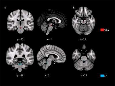 |
78 |
In-vivo mapping of monoaminergic network disruption in Alzheimer’s disease: implications for neuropsychiatric symptoms
Laura Serra, Marcello D'Amelio, Carlotta Di Domenico, Camillo Marra, Nicola Mercuri, Carlo Caltagirone, Mara Cercignani, Marco Bozzali
Some recent animal models and neuropathological evidence highlighted the loss of noradrenergic neurons in the locus coeruleus and dopaminergic neurons in the ventral tegmental area (VTA) as important pathophysiological events in Alzheimer's disease. Here we use resting-state functional MRI to show reductions in VTA connectivity in patients with Alzheimer's disease and mild cognitive impairment, which are more prominent in patients with neuropsychiatric symptoms.
|
|
3751.
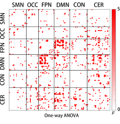 |
79 |
Loss of Intra- and Inter-Network Resting State Functional Connectivity in Patients with Mild and Moderate Alzheimer’s Disease
Video Permission Withheld
Yiqiu Liu, Man Sun, Yuanyuan Chen, Xiuwei Fu, Tianyi Qian, Hongyan Ni
This study aimed to investigate the differences in the alteration of the entire brain network functional connectivity of mild and moderate AD patients based on fMRI, taking the cerebellum network into consideration. There have seldom been reports regarding the cerebellum and cerebral connectivity alteration during the progression of clinical AD. Moreover, the cerebellum network functional connectivity is significantly changed in moderate AD, and we further suspected that the cerebellum network is a pivotal one in the progression to the moderate AD stage.
|
|
3752.
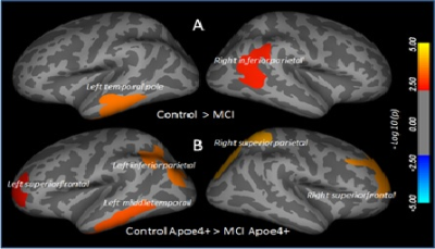 |
80 |
Apolipoprotein E e4 allele is associated with higher structural brain changes in patients with mild cognitive impairment
Sheema Hashem, Santosh Yadav, Asma Al-Sulaiti, Ajaz Bhat, Tooba Ahmed, Deepak Kaura, Ari Borthakur, Mohammad Haris
The aim of this study was to measure the difference in cortical thickness, subcortical volume and CSF biomarkers between MCI patients who carry or lack ApoE ε4 allele. High-resolution T1-weighted images were used for the measurement of cortical thickness and subcortical volume using FreeSurfer. MCI patients who carry ApoE ε4 showed significant reduction in cortical thickness and subcortical volume in multiple brain regions than non-carriers. This suggests that having an ApoE ε4 allele could be a risk factor for the larger tissue damage in the brain of MCI, and these patients may have higher chance of developing Alzheimer’s or other dementia.
|
|
3753.
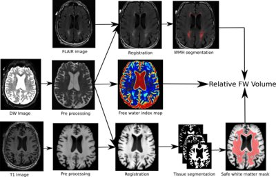 |
81 |
White matter free water content at different stages of Alzheimer’s disease
Matthieu Dumont, Koene Van Dijk, Felix Morency, Jean-Christophe Houde, Pierre-Marc Jodoin, Zhiyong Xie, Cici Bauer, Tarek Samad, Maxime Descoteaux, James Goodman
Recent evidence shows that neuroinflammation plays a role in many neurological diseases including Alzheimer’s disease (AD), and that free water (FW) diffusion imaging can be sensitive to this phenomenon. We processed MRI data from the ADNIGO and ADNI2 databases with a state-of-the-art processing pipeline that uses T1 weighted (T1w), diffusion weighted (DW), and fluid-attenuated inversion recovery (FLAIR) scans to extract FW fractional volume in normal appearing white matter. We call this metric relative FW volume (rFW volume). We demonstrate for the first time that rFW volume in white matter is higher for subjects with greater cognitive impairments.
|
|
3754.
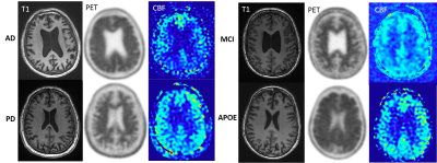 |
82 |
Semiautomatic Quantification of Cortical Amyloid with 18F-florbetaben PET/MRI in Alzheimer's disease and Other Neurodegenerative Disorders
JeeYoung Kim, Audrey P. Fan, Fabiola Bezerra de Carvlho Macruz, Jia Guo, Athanasia Boumis, Michael Greicius, Sharon Sha, Minal Vasanawala, Michael Moseley, Michael Zeineh, Greg Zaharchuk
Amyloid PET is a useful quantitative biomaker in patients with dementia. We compared cortical amyloid deposition in Alzheimer’s disease (AD), Parkinson’s disease (PD), mild cognitive impairment (MCI) and cognitively normal elderly subjects and correlated it with cortical CBF and volume using simultaneous 18F-florbetaben PET/MRI. Total cortical SUV is significantly higher in AD than PD and MCI. Precuneus, superior temporal, and calcarine cortices were identified as significant regions on amyloid PET to discriminate between AD and other neurodegenerative diseases.
|
|
3755.
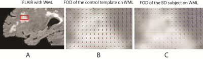 |
83 |
Fixel-based fiber-specific analysis of white matter lesions in Binswanger disease
Rajikha Raja, Josef Ling, Gary Rosenberg, Arvind Caprihan
Binswanger's disease (BD) is a form of vascular dementia which is prevalent in older population. BD is characterized by the presence of white matter lesions due to injured small vessels in the brain. Magnetic resonance imaging findings related to BD reported large white matter lesions in FLAIR and high blood-brain barrier permeability in dynamic contrast enhanced MRI. Changes in water diffusion in white matter lesions and axonal damage were reported using diffusion tensor imaging metrics. While all the studies focused on exploring the voxel level measures as BD markers, we aimed to quantify the WM integrity through measures estimated for fibers within a voxel. In this study, we apply fixel-based analysis (FBA) to multi-shell diffusion data to evaluate the fiber specific measures such as fiber density (FD) and fiber cross-section (FC) in regions of white matter lesions in BD subjects and compared the measures with healthy controls. Reduced FD and FDC are revealed in areas of white matter lesions in BD subjects as compared to those in control group.
|
|
3756.
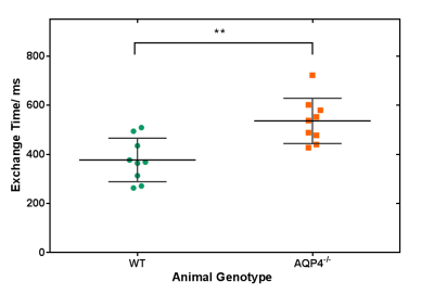 |
84 |
Non-Invasive Imaging of Brain Clearance Pathways using Multiple Echo Time ASL: An Aquaporin-4 Study
Yolanda Ohene, Ian Harrison , Payam Nahavandi , Ozama Ismail , Ole Ottersen , Erlend Nagelhus, David Thomas , Mark Lythgoe , Jack Wells
We have developed the first non-invasive technique that is able to detect changes in brain AQP4-mediated clearance pathways. Our multi-TE ASL technique measures the exchange of vascular water into cortical brain tissue of mouse brain. We report a significant increase in the cortical exchange time between WT (377 ± 89ms) and AQP4-deficient (536 ± 92ms) mice. While measured CBF, ADC and δa did not detect differences, suggesting preserved haemodynamic and energetics between groups. This highlights the novelty of the technique being targeted method to assess water transport in brain clearance pathways, to help better understand neurodegenerative diseases.
|
|
3757.
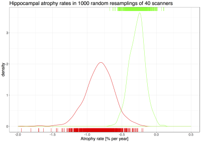 |
85 |
What reference for reference ranges? How scanner and subject data heterogeneity impact MR hippocampal volumetry statistics in Alzheimer’s Disease
Jonas Richiardi, Bénédicte Maréchal, Alexis Roche, Reto Meuli, Tobias Kober
The volume of specific brain structures is of clinical interest in many brain diseases. By using a volumetric reference range for healthy subjects, radiologists can contribute to refining diagnosis. However, both scanner and subject characteristics impact the construction and use of these reference ranges. Using a diverse dataset with 80 MRI scanners and 302 subjects, we show Alzheimer’s disease detection from hippocampal volume is robust to mismatch between training (development) and testing (deployment) environments despite showing some influence, but that estimates of atrophy rates can vary considerably depending on the training set used. Radiologists should interpret volumetry statistical results accordingly.
|
|
3758.
|
86 |
Automatic classification of patients with Alzheimer’s disease (AD) and mild cognitive impairment (MCI) who will convert to AD using deep neural networks
Video Permission Withheld
Federica Agosta, Silvia Basaia, Luca Wagner, Giuseppe Magnani, Massimo Filippi
We built and validated a deep learning algorithm that predicts the individual diagnosis of Alzheimer’s disease (AD) and the development of AD in mild cognitive impairment (MCI) patients based on a single cross-sectional brain structural MRI scan. The deep neural network (DNN) procedure discriminated AD and heathy controls with an accuracy up to 98%, and MCI converters and MCI stable with an accuracy up to 75%. DNNs provide a powerful tool for the automatic classification of AD and MCI prognosis.
|
|
3759.
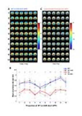 |
87 |
Quasi-Periodic patterns contribute to rsfMRI functional connectivity in a mouse model of Alzheimer’s disease
Michaël Belloy, Disha Shah, Behnaz Yousefi, Anzar Abbas, Amrit Kayshap, Annemie van Der Linden, Georgios A. Keliris, Shella Keilholz, Marleen Verhoye
We show the detection of Quasi-Periodic patterns (QPPs) in a mouse model of Alzheimer’s disease and illustrate that QPP detection was altered between wild-type and transgenic animals. We show that QPPs contributed to BOLD functional connectivity (FC) within groups and to FC differences between groups. Regression of QPPs diminished FC in co-active regions within the QPP, while anti-correlated regions became correlated. Regression of similar QPPs in wild-type and transgenic animals altered between-group FC differences by 30-50%. These findings shed light on how QPPs contribute to FC and are promising for the application of QPPs as a new pre-clinical tool.
|
|
3760.
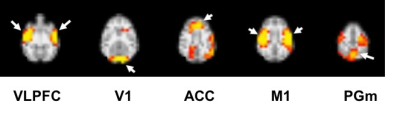 |
88 |
Evaluate the Methanol-induced Alzheimer’s Disease Monkey Model by Resting-state Functional MRI
Yin-Chieh Liu, Chaoyang Tian, Ssu-Ju Li, Xudong Zhao, Ting-Chun Lin, Jianjun Li, Ching-Wen Chang, Kaimin Wang, Yingshan Yang, Yu-Chun Lo, YuanYe Ma, You-Yin Chen*, Feng Chen*, Hsin-Yi Lai*
Alzheimer’s disease (AD) is the most comment neurodegenerative disorder that results in the irreversible loss of neurons, especially in the cortex and hippocampus. The aim of this study is to evaluate the changes of brain functional connectivity in methanol-induced AD monkey. To find the corresponding response areas of AD, five brain regions defined by independent component analysis (ICA) were analyzed using seed-based correlation analysis (SCA). Results of resting-sate fMRI showed that the AD Monkey presented abnormal functional connectivity in anterior cingulate cortex (ACC), primary visual cortex (V1) and ventrolateral prefrontal cortex (VLPFC), those were also shown in AD patient.
|
|
3761.
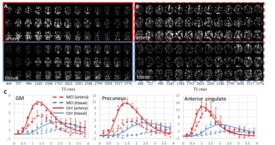 |
89 |
Assessment of cerebrovascular alterations in Alzheimer’s disease with simultaneous multislice Look-Locker arterial spin labeling
Tae Kim, Oscar Lopez, James Becker
The ASL dynamics, the transit time to arterial and capillary, CBVa and CBF, were measured in control and mild cognitive impairment subjects to access cerebrovascular alteration in AD progression. The MCI subjects had prolonged transit time, lower CBVa and CBF in comparison to control subjects. Detailed aseesements of cerebrovascular alterations can provide better characterization of AD pathophysiology.
|
|
3762.
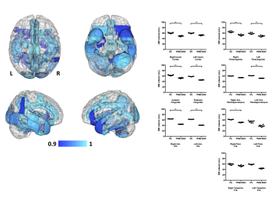 |
90 |
Bilateral salpingo-oophorectomy prior to natural menopause is associated with Alzheimer’s disease-like reductions in gray and white matter
Video Permission Withheld
Suzanne Witt, Åsa Rydmark Kersley, Elisabet Classon, Nina Lykke, Magrit Shildrick, Cecilia Åsberg, Cheryl Grady, Elvar Theordorsson, Jan Ernerudh, Elisabeth Åvall Lundqvist, Preben Kjølhede, Gillian Einstein
Women with Breast Cancer Gene mutations (BRCAm) are recommended to undergo prophylactic bilateral salpingo-oophorectomy prior to natural menopause (PNM-BSO) in order to reduce the risk of ovarian cancer. This surgery is associated with an increased risk of developing Alzheimer's disease (AD). In comparison with healthy women, BRCAm women who had undergone PNM-BSO exhibited decreased gray matter volume across frontal and medial-temporal regions, as measured using the quantitative QRAPMASTER sequence. These regions included many previously linked with AD including, parahippocampal gyrus, cingulate cortex, and inferior temporal gyrus. White matter reductions were only observed in a limited number of superior longitudinal tracts.
|
|
3763.
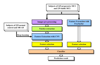 |
91 |
Deep Learning-based MRI Image Analysis for the Prediction of Conversion from Mild Cognitive Impairment to Alzheimer's Disease
Weiming Lin, Min Du, Di Guo, Xiaofeng Du, Yonggui Yang, Gang Guo, Xiaobo Qu
Accurate prediction of the conversion from mild cognitive impairment (MCI) to Alzheimer’s disease (AD) is critically important to slow down the progression to AD with early clinical trials. In this work, this prediction for 3 years is conducted on MRI images shared in Alzheimer’s Disease Neuroimaging Initiative (ADNI) dataset. Two powerful image analysis tools, including convolutional neural networks in deep learning and FreeSurfer in brain MRI analysis, are introduced to learn image features which are used for further classification. Cross validation results demonstrate that the proposed approach achieves more accurate and robust prediction comparing with the state-of-the-art grading biomarker method.
|
|
3764.
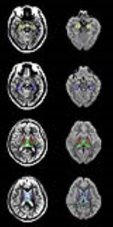 |
92 |
Correlation between Apparent Diffusion Coefficient at Ultrahigh b-values and cognitive impairment: a pilot study in early stage Alzheimer’s disease
Qiong Ye, Zhenhua Zhang, Caiyun Wen, Zhao Zhang, Jiance Li
Non-Gaussian diffusion showed the potential to advance the understanding of microstructure alternation in Alzheimer’s disease (AD). Apparent Diffusion Coefficient at Ultrahigh b-values (ADC_uh) demonstrated improved sensitivity to white matter degeneration in AD. In this study we compared the quantitative change of diffusion characteristics including conventional ADC, Diffusion Kurtosis Imaging (DKI), and ADC_uh. Moreover the correlation between diffusion characteristic and cognitive impairment was investigated. The results of this pilot study showed a close link between ADC_uh in left thalamus and cognitive impairment in AD patients. But further study with larger sample size is required to draw a clear conclusion.
|
|
3765.
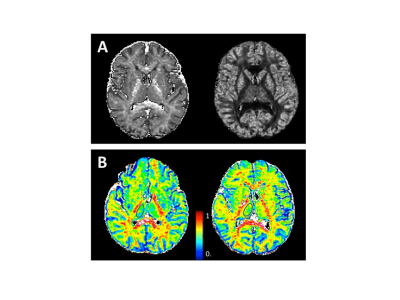 |
93 |
The Application of NODDI in Characterizing Brain Microstructural Changes in Type 2 Diabetes with Cognitive Impairment
Ying Xiong, Qiang Zhang, Yang Fan, Wenzhen Zhu
This study aims to investigate brain microstructural changes in white-matter and gray-matter of type 2 diabetes mellitus(T2DM) patients using the NODDI model. Thirty-three T2DM patients were divided into two sub-groups (impaired and normal cognition), together with ten healthy controls, were imaged at a 3T scanner. It was found that the T2DM patients with cognitive impairment had a lower ICVF value compared to healthy controls in WM regions and the thalamus. Decreased ICVF values in the genu of corpus callosum were correlated with HbA1c level. The NODDI model shows potential feasibility in characterizing brain microstructural alterations for patients with T2DM.
|
|
|
94 |
|
|
3766.
 |
95 |
Subregional structural alterations in deep gray matter structures in alcohol-dependent patients
Hyeon-Man Baek, Young-Tae Kim, Jae-Hyuk Shim
The aim of this study was to investigate whether the volumes of subcortical gray matter structures in alcohol-dependent patients (ADP) are different, and to localize the surface morphometric changes of these subcortical structures. Compared with healthy controls (HC), we found that the bilateral Accu, Hipp, Puta, and Thal showed significant atrophy in ADP. We also revealed that vertex analysis directly can measure localized changes in brain structures that showed significant volumetric change. It might have potential to precisely detect regional alterations of the subcortical gray matter structures.
|
|
3767.
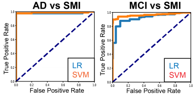 |
96 |
Brain Structural Connectome Accurately Classifies Alzheimer’s Disease Related Dementia
Jiook Cha, Yun Wang, Allen Liu, Jonathan Posner, Jong-Hun Kim, Shinjae Yoo, Hyoung-Seop Kim
There is an urgent, unmet need for biomarkers of risk for Alzheimer’s disease related dementia (ADRD) suitable for routine examination in naïve patients. Studies suggest MRI-derived brain connectome may contain salient information about brain health status. However, it has yet to be tested in sufficiently large samples whether connectome can be used to predict reliably diagnosis of ADRD. Here we performed high-throughput computational analysis using structure and diffusion MRI in a clinical cohort (N=211) to estimate morphometry and connectome. Our results show potential utility of data-driven machine learning models using large-scale MRI-derived brain phenotypes in classifying ADRD, particularly structural connectome.
|
|
Neuroimaging: Clinical Studies
Electronic Poster
Neuro
Monday, 18 June 2018
| Exhibition Hall |
17:15 - 18:15 |
| |
|
Computer # |
 |
3768.
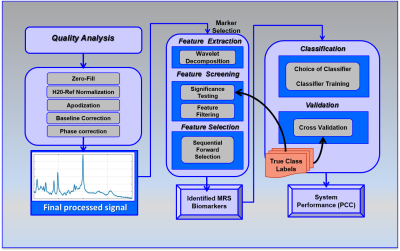 |
97 |
MR Spectroscopy Biomarker Discovery Algorithms (MBDA) to Discriminate PTSD from Blast Exposure and Healthy Controls
Video Permission Withheld
Nathan Tosh, Laura Mariano, Scott Quadrelli, Graham Galloway, Valerie Graves, Aaron Urquhart, Peter Malycha, Katie Trickey, Lisa Rich, Rosanna Tremewan, John Irvine, Carolyn Mountford
Single Voxel Point RESolved Spectroscopy (PRESS) was used to obtain neuro-metabolite and lipid levels from the Posterior Cingulate Gyrus (PCG) of 4 cohorts – Control, Civilian PTSD, Artillery and Close Combat soldiers. Using a wavelet based statistical analysis approach classifiers were developed to objectively diagnose and monitor neuro-deregulation associated with PTSD and blast injury to artillery and to close combat soldiers.
|
|
3769.
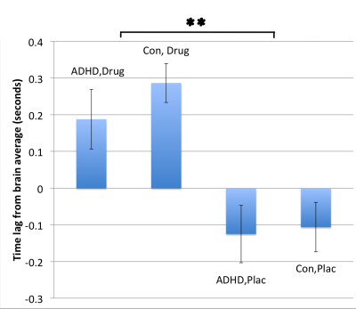 |
98 |
ADHD and stimulant medication disrupt the temporal lag structure within default mode and salience networks
Charlotte Clarke, Arjun Sethi, Mara Cercignani, Neil Harrison
Attention Deficit Hyperactivity Disorder (ADHD) is a neurodevelopmental disorder that has been associated with reduced Default Mode (DMN) (precuneus) and salience network (Anterior Cingulate) activity in conventional resting-state fMRI studies (rs-fMRI). However, these studies ignore the significant temporal structure in these data. Thirty ADHD patients and 30 matched controls underwent rs-fMRI on two occasions, 90-minutes after blindly administered stimulant medication and placebo. Using a novel ‘lag-thread’ analysis we demonstrate faster recruitment of the precuneus in ADHD which additionally correlated with severity of inattention. Stimulants slowed ACC recruitment, together suggesting potential importance of temporal activation of these regions in ADHD.
|
|
3770.
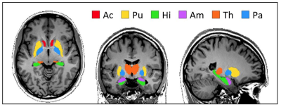 |
99 |
BMI correlates with tissue stiffness in deep gray matter regions controlling eating behavior
Stefan Hetzer, Martin Weygandt, Sebastian Hirsch, Jürgen Braun, Ingolf Sack
Cerebral MR elastography was applied to a group of healthy male subjects in the normal weight range up to overweight to explore a potential relationship between the in vivo mechanical properties of brain tissue and body-mass index (BMI). We observed a highly significant negative correlation between tissue stiffness and BMI in the globus pallidus and putamen – two regions identified in the literature as being linked to eating behaviour – while the stiffness of other brain regions did not correlate with BMI. This is the first report on in vivo mechanical properties of brain tissue related to BMI.
|
|
3771.
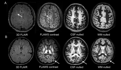 |
100 |
Fluid and White Matter Suppression sequence for detecting epileptogenic zones of focal cortical dysplasia
Did Not Present
Xin Chen, Tianyi Qian, Tobias Kober, Nan Chen, Kuncheng Li
The aim of this study was to evaluate a novel sequence, called the fluid and white matter suppression (FLAWS) sequence, for detecting focal cortical dysplasia (FCD) lesions. FLAWS provides two sets of 3D contrast images from one acquisition and subsequently calculates another set of images that can suppress the signal from both the cerebral spinal fluid and white matter. The detection rate of the FCD lesions on FLAWS was higher than on the conventional 2D MR scan and 3D fluid-attenuated inversion recovery scan. Additionally, the transmantle sign, which is widely believed to be speci?c for FCD type II, could also be observed in FCD type I on the FLAWS images.
|
|
3772.
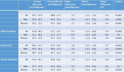 |
101 |
Cortical Cerebral Blood Flow in Aging: Effects of Haematocrit, Sex and Ethnicity.
Video Permission Withheld
Lorna Smith, Andrew Melbourne, David Owen, M. Cardoso, Carole Sudre, Therese Tillin, Magdalena Sokolska, David Atkinson, Nish Chaturvedi, Sebastien Ourselin, Alun Hughes, Frederik Barkhof, H. Jager
Cerebral blood flow (CBF) estimates using arterial spin labelling (ASL) show unexplained variability in older populations. We studied the impact of haematocrit (Hct) on CBF quantification in a tri-ethnic elderly population cohort. Hct was measured from blood samples and pseudo-continuous ASL performed on 3T MR. CBF was estimated using a fixed value of 43.5% (model 1) and individually measured Hct (model 2) to calculate the longitudinal relaxation time of blood in simplified Buxton equations. CBF estimates using individual Hct were lower than CBF estimates using a mean Hct in all ethnic and sex categories except white European men.
|
|
3773.
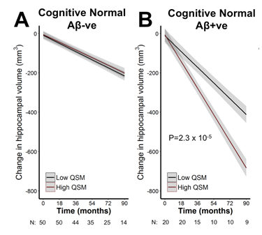 |
102 |
Predictive value of QSM for hippocampal atrophy in pre-clinical Alzheimer’s disease
Amir Fazlollahi, Scott Ayton, Ibrahima Diouf , Pierrick Bourgeat, Vincent Dore, Parnesh Raniga, Jurgen Fripp, Patricia Desmond, David Ames, Paul Maruff , Ralph Martins , Chris Fowler, Roger Ordidge, Colin Masters, Christopher Rowe, Victor Villemagne, Ashley Bush, Olivier Salvado, on behalf of the Australian Imaging Biomarkers and Lifestyle (AIBL) Research Group
One-third of cognitively normal people over the age of 65 exhibit β-amyloid plaques, a defining pathology of Alzheimer’s disease. The hippocampus also undergoes early and pronounced neurodegeneration in Alzheimer’s disease, which underlies the memory impairment. Cognitively normal people with high β-amyloid pathology are at risk of hippocampal neurodegeneration, but the rate of decline is variable between subjects. Here, we investigate whether the iron load of the hippocampus can be used to stratify risk for future hippocampal atrophy in cognitively normal people with and without β-amyloid. We applied Quantitative Susceptibility Mapping (QSM), a relatively new MRI modality that is sensitive to tissue iron levels, to 70 cognitively normal people who also had a PET scan for β-amyloid, and were monitored for brain volume changes in MRI scans performed every 1.5 years for up to 7.5 years. We found that QSM of the hippocampus was strongly predictive of future atrophy of this region in cognitively normal subjects who had high β-amyloid pathology (P=2.3x10⁻⁶), but not in cognitively normal subjects with low pathology. These data support a role for iron in contributing to neurodegeneration in Alzheimer’s disease, and QSM in combination of β-amyloid PET scans could be used to stratify patients at risk for cognitive decline in the pre-symptomatic phase.
|
|
3774.
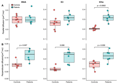 |
103 |
Biomarkers of dynamic energy metabolism in Huntington disease
Isaac Adanyeguh, Francesca Branzoli, Marie-Pierre Luton, Ben Cassidy, Emmanuel Brouillet , Caroline Rae, Pierre-Gilles Henry, Fanny Mochel
Huntington disease (HD) is a dominantly inherited neurodegenerative disease characterized by involuntary abnormal movements, cognitive and psychiatric symptoms. Evidence suggests that energy deficit plays a critical role in the disease pathophysiology. There is however a lack of robust biomarkers for testing therapeutic strategies targeting brain energy metabolism. This study aims to measure dynamic parameters of brain energy metabolism and identify novel functional biomarkers of for use in therapeutic trials in HD. This study showed altered creatine kinase rate in patients with HD as well as altered diffusion rates of several metabolites in the corpus callosum of patients with HD.
|
|
3775.
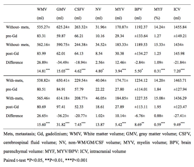 |
104 |
Pitfall of synthetic MRI: Effect of gadolinium on the estimation of brain tissue volumes and myelin based on rapid simultaneous relaxometry
Tomoko Maekawa, Akifumi Hagiwara, Masaaki Hori, Koji Kamagata, Christina Andica, Takuya Haruyama, Kanako Sato, Saori Koshino, Ryusuke Irie, Akihiko Wada, Shigeki Aoki
We investigated the effect of gadolinium on the automatic tissue and myelin volumetry using synthetic MRI. 17 patients with metastases and 19 patients without metastasis were retrospectively analyzed before and after administration of gadolinium. After gadolinium administration, white matter volume (ml), non-white matter/gray matter/cerebrospinal fluid volume (ml), myelin volume (ml), and myelin volume/brain parenchymal volume (%) were significantly increased, whereas gray matter volume (ml), cerebrospinal fluid volume (ml), brain parenchymal volume (ml), and intracranial volume (ml) were significantly decreased regardless of metastasis. The gadolinium had significant effects on the automatic calculation of tissue and myelin volumes estimated by synthetic MRI.
|
|
3776.
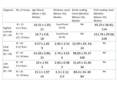 |
105 |
Neuro-anatomical changes in the early blind, late blind during critical developmental time: Voxel Based Morphometry study
Ankeeta Ankeeta, Senthil Kumaran, Rohit Saxena, N Jagannathan
In the early blind individuals, the white matter plasticity during the critical developmental period may compensate for the impairment resulting from the de-afferentation of the visual information that may explain for the early blind subjects show fewer regions with white matter impairments relative to the late blind
|
|
3777.
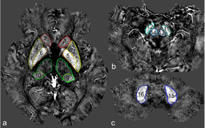 |
106 |
Increased Deposition of Iron in Deep Cerebral Gray Matter Structures in Hemodialysis Patients: A Longitudinal, Susceptibility-Weighted Image Mapping Study
Chao Chai, Saifeng Liu, Huiying Wang, Jinping Li, Tianyi Qian, E. Mark Haacke, Shuang Xia, Wen Shen
The aim of this study was to explore the changes in abnormal iron deposition in hemodialysis patients by longitudinal follow-up MR exam with susceptibility-weighted image mapping (SWIM). SWIM was reconstructed from the magnitude and phase data of SWI to quantify the susceptibility of deep gray matter nuclei in patients with a baseline examination and follow-up examination, and in healthy controls. The results suggest that iron deposition in the gray matter nuclei in patients increased over time, and this process may be a risk factor for neurocognitive dysfunction. The independent risk factors for abnormal iron deposition included the follow-up interval, creatinine, and abnormal calcium-phosphorus metabolism.
|
|
3778.
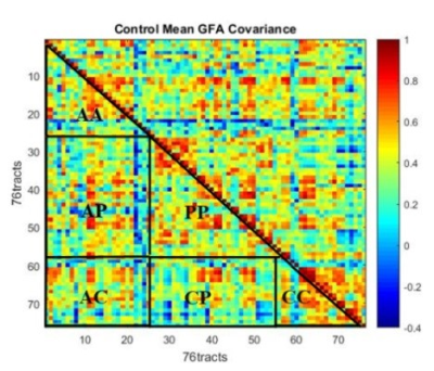 |
107 |
Assessment of white matter de-differentiation in mild cognitive impairment using tract covariance matrix derived from diffusion spectrum imaging
Tao-Han Hung, Yung-Chin Hsu, Yu-Ling Chang, Isaac Wen-Yih Tseng
In this study, we compared the degree of white matter de-differentiation between the patients with mild cognitive impairment (MCI) and normal-aging population using diffusion spectrum imaging (DSI) and whole brain tract-based automatic analysis (TBAA). The degree of de-differentiation, defined as the global level of generalized fractional anisotropy (GFA) covariance between each white matter tract, was found to be significantly higher in the MCI group, as revealed by two-sample T test of partial correlation matrices of the two subject groups. Further analysis showed that covariance was higher among projection tracts.
|
|
3779.
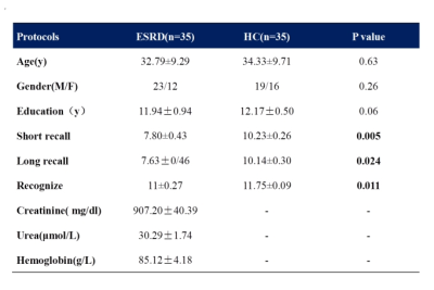 |
108 |
Altered Hippocampal Structure and Functional Connectivity Associated with Memory Impairment in Patients with End-stage Renal Disease
Xueying Ma, Dun Ding, Peng Li, Pan Zhang, Ming Zhang
Memory impairment was common among ESRD patients, but the physiopathologic mechanisms were largely unclear. Hippocampus and prefrontal cortex played an important role in the human memory. Here we analyzed the structurre and functional connectivity of hippocampus and its correlation with memory scores (AVLT-H). We found that the hippocampal volume and FC between the rostral/caudal hippocampi and ventrolateral prefrontal cortex, dorsolateral prefrontal cortex as well as middle temporal gyrus. Correlations were found between hippocampal changes and memory impairment. Our study indicated that the hippocampus should be taken into consideration in the further mechanism study of neurol damage in ESRD patients.
|
|
3780.
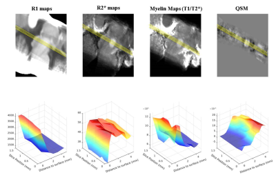 |
109 |
In-vivo Quantitative Structural Imaging of the Superior Colliculus at 9.4T
Joana Loureiro, Marc Himmelbach, Thomas Ethofer, Rolf Pohmann, Pascal Martin, Jonas Bause, Klaus Scheffler, Gisela Hagberg
We investigated the possibility to observe the anatomical details of the superior colliculus (SC), a layered structure located on the tectum of the midbrain, by in vivo MRI at 9.4T. Through image analysis in native space, several brain structures of the mid brain could be identified. The signal variation of all imaging modalities (T1, R2* and QSM) along and across the superior colliculus consistently highlighted the deep white layer VII, adjacent to the periaqueductal grey; the myelinated fibres in the superficial optic layer (layer III) and an iron-rich layer attributed to the intermediate grey layer (IV).
|
|
3781.
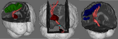 |
110 |
White matter fibre tract changes in patients treated with MR imaging-guided focused ultrasound are associated with lesion location and clinical outcome
Christian Thaler, Qiyuan Tian, Max Wintermark, Pejman Ghanouni, Casey Halpern, Jamie Henderson, Raag Airan, Michael Zeineh, Kim Butts Pauly, Jennifer McNab
DTI is sensitive to tract-specific FA changes and thereby can be used to inform on therapeutic mechanisms and optimal targeting strategies for MRI-guided high intensity–focused ultrasound. Here, we used probablilistic tractography to investigate white matter fibre tract changes in essential tremor patients treated with MRIgHIFU. On the treated side, a significant decrease in FA was detected in the tracts projecting between the thalamus and motor cortex and between the dentate nucleus and the thalamus (i.e. the dentato-rubro-thalamic tract). These decreases in FA were correlated with the degree of tract-lesion overlap. Additionally, the post-treatment FA decrease in the tracts projecting between the thalamus and the motor cortex were positively correlated with clinical improvement. Interestingly, tract changes were also observed in the medial lemniscus of the contralesional side, possibly indicating global changes in brain connectivity.
|
|
3782.
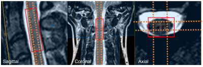 |
111 |
Associations between tissue sodium concentration, age and cross-sectional area in the healthy spinal cord
Bhavana Solanky, Ferran Prados, Marios Yiannakas, Vanessa Bassan, Baris Kanber, Sebastien Ourselin, Olga Ciccarelli, Claudia Gandini Wheeler-Kingshott
Tissue sodium concentration has recently come into the spotlight for a number of neurological conditions, given its potential role in neurodegeneration and due to the advances in MRI technology. This has led to many studies in the brain, but there is a shortage of studies characterising sodium in the spinal cord. Here we use 23Na-MRS to measure sodium in the healthy spinal cord and look at the association between tissue sodium concentrations with age and spinal cord cross sectional area.
|
|
3783.
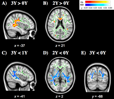 |
112 |
White matter microstructural maturation in adolescent American Football athletes is affected by history of sport-participation and concussion
Yukai Zou, Ikbeom Jang, Nicole Vike, Diana Svaldi, Larry Leverenz, Eric Nauman, Thomas Talavage, Joseph Rispoli
Concussion is a biomechanically induced brain injury that causes mental health concern for adolescent collision-sport athletes, whose brains are still developing and maturing. These athletes normally continue participating in collision events, regardless of history of concussion and risk for future concussion. Using diffusion tensor imaging, we investigated 93 asymptomatic adolescent athletes who participate in high school American Football. Per developmental expectations, we typically observed increasing fractional anisotropy or decreasing mean diffusivity with increasing years of high school experience, but prior history of concussion reversed this trend in a number of brain regions.
|
|
3784.
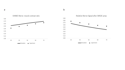 |
113 |
Development and Evaluation of a contrast optimized 3D SHINKEI MR Neurography on patients with Brachial plexus injuries at 1.5T
Prashant Nair, Rajagopal Kadavigere, Yogesh Mariappan, Samir Paruthikunnan, Narayana Rolla, Indrajit Saha
The objective of this work is to optimize the 3D nerve-SHeath signal increased with INKed rest-tissue RARE Imaging (SHINKEI) sequence for the best nerve-muscle T2 contrast in 1.5T and evaluate the clinical feasibility of this optimized sequence in 24 patients with brachial plexus injuries. Based on the evaluation by two experienced radiologists and the comparison to surgical observations, the optimized SHINKEI sequence performed better than fat suppressed TSE (STIR TSE) and Diffusion Weighted Imaging with Background Suppression (DWIBS) sequences that are currently widely used in the clinical protocols
|
|
3785.
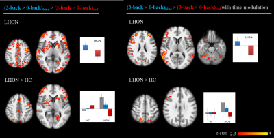 |
114 |
Melanopsin retinal ganglion cells in LHON patients: an fMRI study of brain activations under monochromatic light stimulation
Stefania Evangelisti, Claudia Testa, Chiara La Morgia, Gilles Vandewalle, Claudio Bianchini, David Manners, Paola Fantazzini, Michele Carbonelli, Alfredo Sadun, Caterina Tonon, Valerio Carelli, Raffaele Lodi
We combined light stimulation and fMRI to investigate the contribution of melanopsin-expressing retinal ganglion cells (mRGCs) to visual and non-visual processes in Leber Hereditary Optic Neuropathy (LHON) paradigm of retinal degeneration . Monochromatic visual simulation showed a stronger effect in LHON visual cortex for blue vs red light with relatively long stimuli, supporting the hypothesis of a role for melanopsin in visual processes. When light was combined with a working memory task, blue light modulation of cognitive brain response was maintained in LHON; indeed the effect was stronger than in healthy subjects, probably because of the higher mRGCs/RGCs ratio in LHON retinas.
|
|
3786.
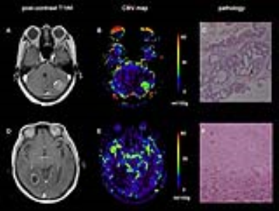 |
115 |
Quantitative Dynamic Susceptibility Contrasted Perfusion in Differential Diagnosis of Recurrence and Radionecrosis for Brain Metastases after Stereotactic Radiotherapy
Did Not Present
Bao Wang, Tianyi Qian, Bin Zhao, Yingchao Liu, Na Sun
Stereotactic radiotherapy (SRS) has become a widely used treatment for patients with brain metastases (BM). However, differential radionecorsis (RN) and tumor recurrence (TR) after SRS for BM by MR images is still challenging. In this study, a quantitative dynamic susceptibility contrasted perfusion weighted imaging (DSC-PWI) technique was applied to solve this problem. The results showed that quantitative DSC-PWI had higher diagnostic efficiency and smaller inter-observer differences than traditional DSC-PWI.
|
|
3787.
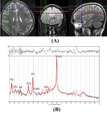 |
116 |
Metabolic alterations in DLPFC in patients with Subclinical Hypothyroidism : An In-vivo 1H MRS study
Did Not Present
Subash Khushu, Mukesh Saini, Poonam Rana, Shilpi modi, Tarun Sekhri
Altered cognitive performance is well known in both hypothyroidism and hyperthyroidism but little is known about deficits in brain functions in Subclinical hypothyroidism (SCH). Aim of the present study was to investigate the metabolic changes in Dorso-Lateral Pre-Frontal Cortex (DLPFC) of SCH patients using 1H MRS. 19 freshly diagnosed SCH patients and 24 age matched healthy controls were recruited and subjected to MRS. Our result show significantly increased concentrations of GSH (p<0.009) and creatine (p<0.017) in the DLPFC and diminished cognitive performance in SCH compared to controls. The increase in the level of GSH in SCH compared to controls may be indicative of an initial compensatory, neuro-protective response due to oxidative stress.
|
|
3788.
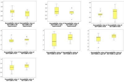 |
117 |
Aberrant Brain Iron Deposition in Gray Matter Nuclei and Hippocampus in Alzheimer Disease with Type 2 Diabetes Mellitus: A Quantitative Susceptibility Mapping Study
Xiaoxin Li, Yanwei Miao, Liang Han, Junyi Dong, Qingwei Song
In this study, brain iron contents in basal nuclei, thalamus and hippocampus of AD+T2DM patients and AD-T2DM patients was quantified with susceptibility values by QSM, and further to explore the correlation between brain iron deposition and cognition level and clinical factors. From the study results, we conclude that the susceptibility values were generally decreased due to aberrant iron deposition in gray matter nuclei in AD+T2DM patients relative to AD-T2DM patients. The susceptibility values of right GP has a better correlation with cognition, which administers to monitor the development of cognitive impairment.
|
|
3789.
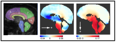 |
118 |
Motion of the cerebellum in patients with Chiari malformation compared to healthy subjects: A quantitative study using spiral cine DENSE MRI
John Oshinski, Soroush Pahlavian, Jordan Schuster , Xiaodong Zhong, Rouzbeh Amini, Francis Loth, Daniel Barrow
The goal of this study was to acquire mid-sagittal cine DENSE images to quantify displacement and strain over the cardiac cycle in patients with Chiari malformation and age-matched controls. The major finding of this study was that both tissue displacement and tissue strain in the parenchyma of the cerebellum was significantly greater in patients with Chiari malformation than healthy controls.
|
|
3790.
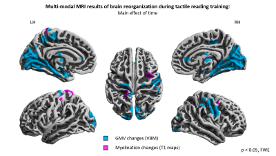 |
119 |
Multi-modal MRI investigation of brain plasticity during prolonged Braille learning in sighted subjects – voxel-based morphometry and quantitative T1 mapping approach
Bartosz Kossowski, Jacek Matuszewski, Lukasz Bola, Anna Banaszkiewicz, Malgorzata Paplinska, Michal Szczepanik, Marcin Szwed, Katarzyna Jednoróg, Artur Marchewka
In order to study structural brain reorganization, multi-modal MRI combining voxel-based morphometry and quantitative T1 mapping was used in longitudinal design on sighted subjects who underwent tactile Braille reading course. Results show that methods are complimentary to each other. Combined approach like that gives insight into different aspects of tissue property changes introducing new opportunities for studying brain plasticity.
|
|
3791.
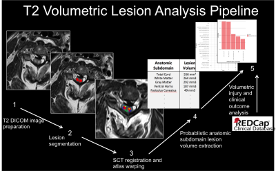 |
120 |
Atlas-based volumetric assessment of T2 abnormality in acute spinal cord injury predicts motor outcomes: A transforming research and clinical knowledge in spinal cord injury (TRACK-SCI) pilot study
David McCoy, Russell Huie, Sara Dupont, William Whetstone, Sanjay Dhall, Rachel Tsolinas, Xuan Duong-Fernandez, Leigh Thomas, Vineeta Singh, Lisa Pascual, Jared Narvid, Nikolaos Kyritsis, Geoff Manley, Adam Ferguson, Michael Beattie, Jacqueline Bresnahan, Jason Talbott
MR evaluation of intrinsic cord signal abnormality relies on gross morphologic imaging measures such as T2-hyperintense lesion length and subjective patterns of T2 signal abnormality. In the current study, we register T2w images and manually segmented lesions from acute SCI patients to a spinal cord (SC) anatomical template in order to calculate volumes of damaged tissue in 22 probabilistic anatomical subdomains of the SC. We identify specific anatomic subdomains in the SC which serve as MR biomarkers of motor impairment and indirectly support neuro-protective strategies targeting ventral horn and lateral column white matter tissue for maximizing motor function after SCI.
|
|
| Back |
| The International Society for Magnetic Resonance in Medicine is accredited by the Accreditation Council for Continuing Medical Education to provide continuing medical education for physicians. |
