|
Electronic Poster Session
Interventional MRI |
Tuesday, 19 June 2018
Electronic PosterInterventional MRI
4031 -4054 Thermometry & MR-HIFU
4151 -4174 MR-Guided Interventions (Not Thermo nor HIFU) |
| |
Thermometry & MR-HIFU
Electronic Poster
Interventional MRI
Tuesday, 19 June 2018
| Exhibition Hall |
13:45 - 14:45 |
| |
|
Computer # |
 |
4031.
 |
1 |
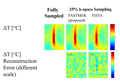
 Fast MRI by Exploiting a Reference Scan (FASTMER) for MR Thermometry Fast MRI by Exploiting a Reference Scan (FASTMER) for MR Thermometry
Efrat Shimron, Haim Azhari
A Compressed Sensing approach for shortening acquisition time in Magnetic Resonance guided Focused Ultrasound (MRgFUS) thermometry is presented. The approach is based on the recently developed Fast MRI by Exploiting a Reference scan (FASTMER) method. The suggested method embeds into the CS optimization problem a regularization term related to a-priori knowledge about the similarity between pre-heating and post-heating acquired data. The results obtained from an in-vitro Focused Ultrasound (FUS) experiment demonstrate that the proposed method provides efficient and accurate temperature mapping from substantially subsampled k-space data. The method is suitable for data acquired with flexible undersampling schemes.
|
|
4032.
 |
2 |
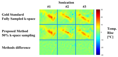
Combining Compressed Sensing and Parallel Imaging for Accelerating Focused Ultrasound MR Thermometry
Efrat Shimron, Haim Azhari
A novel method for accelerated multi-coil Magnetic Resonance guided High Intensity Focused Ultrasound (MRgHIFU) thermometry is presented. This method integrates the two powerful approaches of parallel imaging and Compressed Sensing (CS), and reconstructs temperature rise from sub-sampled k-space data. The proposed reconstruction process utilizes the sparsity of the differences between baseline and post-heating data and operates in a calibrationless manner. The method was validated through a retrospective study of data from a 8-coils in-vivo human prostate treatment and a 2-coils animal experiment.
|
|
4033.
 |
3 |
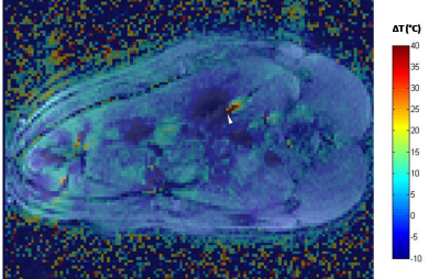
In vivo feasibility of PRF thermometry in the swine pancreas during laser ablation
Céline Giraudeau, Paola Saccomandi, Federico Davrieux, Giuseppe Quero, Emiliano Schena, Michele Diana, Francesco Maria Di Matteo, Guido Costamagna, Jacques Marescaux
The purpose of this preliminary work was to investigate the feasibility to get accurate PRF thermometryimages in the in vivo swine pancreas during laser ablation. We show that PRF thermometry images and consistent temperatures curves can be obtained during the procedure, and these results are confirmed by temperature sensor measurements and histological examination. The presence of a susceptibility artifact (presence of gas bubbles) is discussed.
|
|
4034.
 |
4 |
Three-dimensional assessment of MRI-guided percutaneous liver ablation
Elena Kaye, Kathleen Jedruszczuk , Jeremy Durack, Majid Maybody, Stephen Solomon
Here we present a 3D registration-based technique for assessment of ablative margins following MRI-guided thermal ablation. The method was developed and evaluated in a retrospective study of 26 MRI-guided cryo, laser or microwave ablations of various liver metastases.
|
|
4035.
 |
5 |
Improved MR thermometry during Microwave ablation by correcting for sporadic electromagnetic interference
Aiming Lu, Krzysztof Gorny, Christopher Favazza, Joel Felmlee, David Woodrum
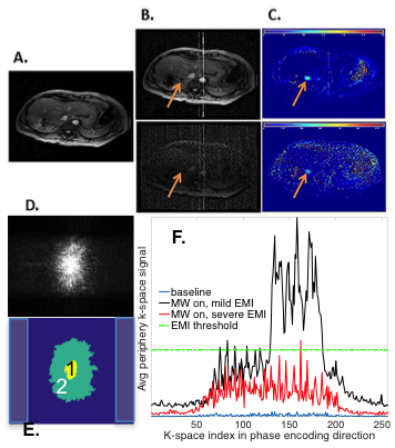
Image-guided microwave ablation (MWA) is a minimally invasive treatment for localized diseases. MRI is advantageous for localizing the lesions, guiding microwave antennae placement, and assessing the extent of ablation. Monitoring of MWA in real-time using MR thermometry has also been demonstrated to be feasible, but can suffer from the image quality degradation due to electromagnetic interference (EMI) from the microwave generator (MWG). A novel approach to correct for EMI-contaminated images is presented here by utilizing the uncontaminated k-space data from neighboring frames. Significantly improved temperature and thermal dose maps have been obtained in our clinical patient studies.
|
|
4036.
 |
6 |
Measurement of Focal Laser Ablation Zones in Prostate Cancer using High Resolution Spectroscopic Imaging of the Water Resonance
Shiyang Wang, Xiaobing Fan, Ambereen Yousuf, Scott Eggener, Gregory Karczmar, Aytekin Oto
MRI-guided Focal laser ablation (FLA) is used to treat prostate cancer. Contrast enhanced imaging can assess the ablated region, but cannot be used during ablation due to effects of high energy ultrasound on MRI contrast agent stability. Our results show that high resolution MR spectroscopic imaging of the water resonance detects changes in T2* and water resonance peak amplitude that clearly delineate the ablation zone without contrast agent injection. Ablation zones measured by MR spectroscopic imaging are consistent with contrast enhanced images. This suggests that MR spectroscopic imaging can guide FLA during ablation to dynamically optimize treatment and improve outcomes.
|
|
4037.
 |
7 |
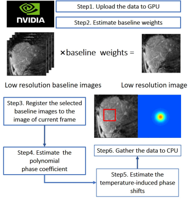
Ultrafast Temperature Estimation from Undersampled K-Space for MR Guided Microwave Ablation
Did Not Present
Ke Wang, Zijing Dong, Fuyixue Wang, Bingyao Chen, Jiafei Yang, Xing Wei, Kui Ying
Improved fast k-space temperature estimation using Golden Angle (GA) radial can effectively accelerate the computation process and reduce the motion artifacts. However, existing MR temperature imaging methods are time-consuming and the limited performance prevent their clinical applications. This work proposed a ultrafast method to largely accelerate temperature estimation process. Using a continuous fast k-space temperature estimation, together with improved algorithm design, GPU acceleration and more efficient computation, we achieve temperature estimation with 2 seconds temporal resolution.
|
|
4038.
 |
8 |
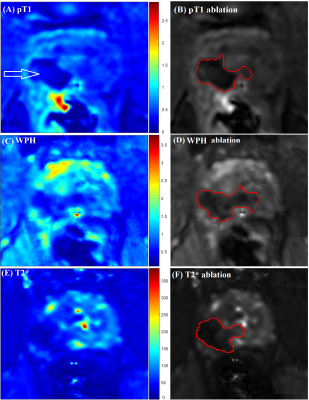
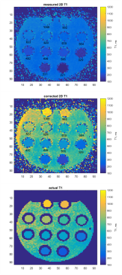
Bloch simulation-based correction for 2D VFA T1 mapping for fat MR thermometry
Ieva Braskute, Roel Deckers, Max Viergever, Chrit Moonen, Lambertus Bartels
For MR-HIFU thermal therapies, specifically in the breast, MR temperature mapping both in aqueous and fatty tissues is desired, as it would improve treatment monitoring compared to MR thermometry in aqueous tissues only. Fat thermometry based on T1-mapping with 2D variable flip angle method offers high temporal resolution; however, 2D T1 mapping suffers from systematic errors caused by non-rectangular 2D slice profile. We propose a Bloch simulation-based 2D T1 correction technique to improve the accuracy of 2D T1 measurements. We demonstrate the proposed correction on simulated and experimental data.
|
|
4039.
 |
9 |
Early clinical feasibility study of body mounted and motorized needle guidance tool in abdominal MRI-guided cryoablation
Nobuhiko Hata, Brian Ninni, Franklin King, Takahisa Kato, Junichi Tokuda, Kemal Tuncali
This study investigates the feasibility of directing cryotherapy probes using a body-mounted and motorized needle guidance device in abdominal MRI-guided cryoablation. Utilizing a motorized needle alignment device offers an alternative solution to the trial-and-error method of probe placement, as it allows one to directly orient cryotherapy probes, efficiently. This study found the method to be feasible for use in cryoablation in 3T with placement accuracy of 8.8mm. Further investigation is warranted to improve the efficiency of probe placement.
|
|
4040.
 |
10 |
Design of Focused Radio Frequency Heating Array Coils for Non-Invasive Hyperthermia with Ultra-High Field MRI
Joshua de Bever, Mihir Pendse, Brian Rutt
This abstract details the implementation of an automated pipeline for designing high channel count RF coils for the purpose of non-invasive Focused RF hyperthermia generated from Ultra-high field MRI parallel transmit coils. The pipeline integrates multiple tools including Sim4Life for EM-FDTD simulations in the Virtual Population physiologically realistic body models, Advanced Design Systems circuit simulator, and Matlab for custom algorithm execution. This work leverages GPU acceleration which has reduced simulation times for an 84 channel RF coil from 77 days on a CPU to 6 hours on three compute nodes equipped with 14 affordable GPUs.
|
|
4041.
 |
11 |
The effect of k-space weighted image contrast (KWIC) and ultrasound focus size on the accuracy of proton resonance frequency (PRF) thermometry
Bryant Svedin, Christopher Dillon, Dennis Parker
Stack of stars acquisitions with k-space weighted image contrast (KWIC) reconstruction provide an effectively high temporal resolution with high spatial resolution. The effective temporal resolution of KWIC reconstructions depend on the size of the object of interest. This abstract investigates the relationship between ultrasound focus size, the size of the KWIC window, and the accuracy/precision of PRF temperature measurements.
|
|
4042.
 |
12 |
Blood-brain barrier disruption and delivery of irinotecan in a rat model using a clinical transcranial MRI-guided focused ultrasound system
Nathan McDannold, Yongzhi Zhang, Chanikarn Power, Natalia Vykhodtseva
The feasibility of controlled blood-brain barrier (BBB) disruption in rats was demonstrated with a low-frequency clinical transcranial MRI-guided focused ultrasound device that operates at 230 kHz (ExAblate Neuro, InSightec) combined with microbubbles. Thirty-six targets were sonicated in one hemisphere in each experiment under closed-loop control based on real-time recordings of acoustic emissions. Disruption was confirmed in maps of R1 relaxation following Gadavist administration. After three weekly BBB disruptions covering an entire hemisphere, we always produced BBB disruption with only minor vascular side effects. We also delivered irinotecan chemotherapy across the BBB without apparent neurotoxicity.
|
|
4043.
 |
13 |
Synergistic effect of MR-HIFU hyperthermia mediated drug delivery followed by ablation
Video Permission Withheld
Edwin Heijman, Nicole Hijnen, Esther Kneepkens, Mariska Smet, Sander Langereis, Holger Grüll
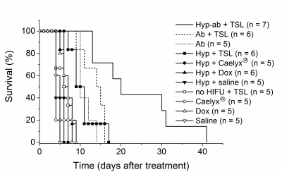
Four Magnetic Resonance-guided High Intensity Focused Ultrasound (MR-HIFU) thermal therapy strategies (no HIFU, hyperthermia, ablation and hyperthermia followed by ablation) in combination with temperature sensitive liposomes (TSLs), co-encapsulating doxorubicin (dox) and ProHance®, were investigated in rhabdomyosarcoma rat tumor model. All HIFU heating strategies combined with TSLs resulted in cellular uptake of dox deep into the interstitial space and significant increase of intratumoral drug concentrations. The combination of hyperthermia-triggered TSLs followed by ablation showed the best therapeutic outcome compared to other strategies due to direct induction of thermal necrosis in the tumor core and efficient drug delivery to the tumor rim.
|
|
4044.
 |
14 |
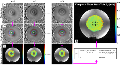
Shear wave elastography using multi-point acoustic radiation force imaging
Lorne Hofstetter, Henrik Odéen, Bradley Bolster, Jr., Douglas Christensen, Allison Payne, Dennis Parker
Characterizing the mechanical properties of tissue during focused ultrasound ablative therapies could provide useful information for pretreatment planning and treatment endpoint assessment. In this work we present a 3D MR acquisition capable of measuring propagating shear waves from a spatially distributed collection of focused ultrasound generated acoustic radiation force impulses. This new multi-point shear wave elastography technique is demonstrated and compared to conventional MR elastography.
|
|
4045.
 |
15 |
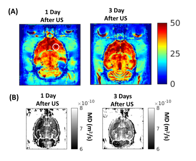
Longitudinal multi-parametric MRI follow-up of ultrasound induced blood-brain barrier disruption in rats
Allegra Conti, Françoise Geffroy, Hermes Kamimura, Fawzi Boumezbeur, Sébastien Mériaux, Benoit Larrat
Focused Ultrasound in conjunction with microbubbles allows the reversible and safe opening of the BBB in a narrow range of Acoustic Pressure (AP). Here we wanted to investigate what happens to the brain tissues at short- and long-terms in case of an excessive AP is reached in vivo. Passive Cavitation Detection has been used to register inertial cavitation during the opening. At 1,3,7,14 and 28 days after the BBB opening, T1,2-weighted images, T2*-maps, DWI, MRS acquisitions and histological analysis have been performed. P-gp and myelin expression, cells deaths, and astrocytes/microglial activations have been also imaged postmortem within 28 days.
|
|
4046.
 |
16 |
Experimental demonstration of a HIFU self-scanning treatment in moving tissue using hybrid ultrasound and MR guidance
Orane Lorton, Laura Gui-Lévy, Pauline Guillemin, Nadia Möri, Philippe Cattin, Sylvain Terraz, Christoph Becker, Rares Salomir
MRI-controlled tumor treatment by High Intensity Focused Ultrasound (HIFU) is challenging in the abdominal organs because of the breathing motion. We experimentally demonstrated a self-scanning method for motion compensation by passively scanning the tissue passing through the static focal point. Ex vivo turkey samples were subjected to a breathing-like non-periodic motion and the HIFU power was modulated using the velocity information from landmark tracking on simultaneous ultrasound images. MR imaging provided targeting and on-line thermometry. Temperature map were computed using the reference-less PRFS method. A dramatic improvement of the isotherms uniformity score was achieved for rectilinear volumetric ablation.
|
|
4047.
 |
17 |
Feasibility of MR guided High Intensity Focused Ultrasound (MRgHIFU) for treating recurrent gynecological tumours: a pilot study
Sharon Giles, Ian Rivens, Katja De Paepe, Veronica Morgan, Georgios Imseeh, Gail ter Haar, Alexandra Taylor, Nandita deSouza
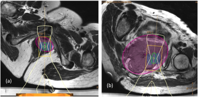
MR guided high intensity focused ultrasound (MRgHIFU) treatment plans were generated for 16 patients with recurrent gynecological tumors. Treatment volumes (i) ignoring risk to non-target regions (TVunconstrained), (ii) considering risk, assuming that patients were optimally prepared (TVoptimal), and (iii) assuming no preparation was possible (TVno-prep), were compared with planning target volume (PTV). 9/16 patients (56%) with tumor volumes ≤53 ml were considered feasible to treat safely if optimally prepared (TVoptimal≥50% PTV). Main limiting factors were transducer focal range, bone in the beam path, and risk to bowel. However, even without preparation, 4/16 patients (25%) remained feasible to treat (TVno-prep ≥50% PTV).
|
|
4048.
 |
18 |
Feasibility of MR guided High Intensity Focused Ultrasound (MRgHIFU) for treating recurrent gynecological tumours: comparing T2W imaging and diffusion weighted imaging (DWI) for treatment planning
Sharon Giles, Jessica Winfield, Ian Rivens, Katja De Paepe, Veronica Morgan, Georgios Imseeh, Gail ter Haar, Alexandra Taylor, Nandita deSouza
MR guided high intensity focused ultrasound (MRgHIFU) treatment plans were generated for 16 patients with recurrent gynecological tumors. Gross tumor volumes (GTV) defined on diffusion-weighted imaging using an echo-planar and turbo-spin echo technique (EPI-DWI, TSE-DWI) were approximately 20% smaller than GTV defined on T2W imaging, but did not result in sequence-dependent differences in planning target volume (PTV). PTVs were more easily defined on DWI. However, there were clinically relevant discrepancies (>4 mm) between T2-defined and DWI-defined PTV locations, worst in the phase-encode direction using EPI-DWI, that need to be accounted for if incorporating DWI into MRgHIFU treatment planning.
|
|
4049.
 |
19 |
Shear stiffness determination using MR-acoustic radiation force imaging
Bradley Bolster, Jr., Henrik Odéen, Hailey McLean, Dennis Parker, Allison Payne
Magnetic resonance acoustic radiation force imaging (MR-ARFI) has been used pre-clinically to detect tissue displacement changes during magnetic resonance guided focused ultrasound treatments. Although displacement is a quantitative metric, shear stiffness is typically used to clinically describe disease states. This work develops and evaluates a model-based technique to determine tissue shear stiffness from MR-ARFI displacement measurements. The MR-ARFI derived values are compared to conventional MR elastography shear stiffness values in a phantom study.
|
|
4050.
 |
20 |
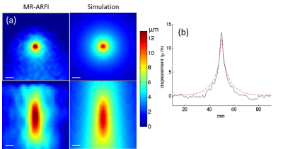
Optical tracking-guided MR-ARFI for targeting focused ultrasound neuromodulation
Sumeeth Jonathan, M Phipps, Vandiver Chaplin, Aparna Singh, Pai-Feng Yang, Allen Newton, John Gore, Li Chen, Charles Caskey, William Grissom
Magnetic resonance-acoustic radiation force imaging (MR-ARFI) pulse sequences permit localization and targeting during focused ultrasound (FUS) therapy. MR-ARFI uses motion-encoding gradients (MEGs) to visualize the tissue displacement caused by the acoustic beam’s radiation force. However, a priori knowledge of the acoustic beam’s position and orientation in space is critical for MR-ARFI so that the MEGs can be placed in the proper orientation. We used an optical tracking system to inform the geometry of MR-ARFI acquisitions. The proposed methods will be used to guide ongoing experiments that use MR-ARFI to produce acoustic beam maps for targeting ultrasound neuromodulation in real-time.
|
|
4051.
 |
21 |
Model Predictive control of MRI-guided HIFU for Hyperthermia
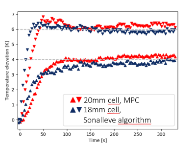
Lukas Sebeke, Xi Luo, Bram de Jager, Edwin Heijman, WMPH Heemels, Holger Grüll
Hyperthermia has been shown in clinical trials to strongly enhance therapeutic efficacy of radio- and chemotherapy. The main challenge in hyperthermia is to achieve spatially homogenous temperatures between about 41-43 oC for ca. one hour. Magnetic Resonance Imaging-guided High Intensity Focussed Ultrasound (MR-HIFU) enables heating of tissues with high spatial accuracy. We developed a new regulatory algorithm based on Model Predictive Control (MPC) for stable MR-HIFU-hyperthermia, which is designed to find effective heating patterns based on predictions of the temperature evolution in tissue. A comparison with the currently available controller is presented, detailing advantages, shortcomings and opportunities.
|
|
4052.
 |
22 |
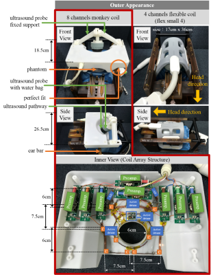
Development of an 8-channel head array for MRI guided monkey ultrasound stimulation
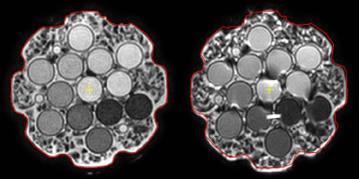
Jo Lee, Xing Yang, Qiaoyan Chen, Changjun Tie, Xiaojing Long, Nan Li, Xiaoliang Zhang, Hairong Zheng, Ye Li
For flexible operating the ultrasound probe in a monkey model, all three instruments, stereotaxic instrument, ultrasound probe and monkey RF coil, must match to each other. In this study, an 8-channel monkey coil was custom-designed to fit the specific stereotaxic instrument, ultrasound probe, and the rhesus monkey’s head in order to get a high quality images and efficient ultrasound operation. Compared to the commercial small flexible coil array, the custom-designed monkey coil provides higher SNR in all three orientations, higher spatial resolution in vivo, and better parallel imaging capability.
|
|
4053.
 |
23 |
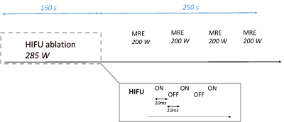
Monitoring High Intensity Focused Ultrasound (HIFU) ablations in real time using interventional MR Elastography (MRE)
Jonathan Vappou, Paolo Cabras, Kisoo Kim, Pramod Rao, Afshin Gangi, Elodie Breton
This study introduces the use of interventional Magnetic Resonance Elastography (iMRE) for monitoring HIFU ablations, using the acoustic radiation force as a means for generating the shear waves necessary for MRE directly from the HIFU focus. This method allows for monitoring tissue elasticity and temperature in real time during the ablation. Its feasibility is illustrated in vivo on porcine muscle. Tissue was found to stiffen significantly (+125%) when the temperature increased, and changes in tissue stiffness were found to be irreversible. These findings suggest that tissue stiffness may be an interesting biomarker, complementary to thermal dose, to monitor HIFU ablations.
|
|
4054.
 |
24 |
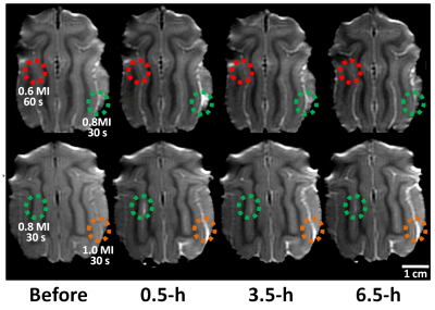
Longitudinal Observation of Focused Ultrasound Induced Blood-Brain Barrier Opening of Cat Brain from 7-T Contrast-Enhanced MRI
Xianfeng Feng#, Tingting He#, Xiao Yu, Chih-Hung Tsai, Wen-Yen Chai, Chao-Ting Wang, Wei Xiong, Bin Xu, Yifeng Fan, Hao-Li Liu*, Hsin-Yi Lai*
Blood-brain barrier (BBB) has long been impeding the application of many therapeutic agents in treating diseases in central nervous system. Results showed that microbubble-mediated focused ultrasound can open BBB in cats noninvasively and locally. The size and duration of BBB opening can be accurately monitored by 7T MRI longitudinally. The combination of two techniques has the potential to be further applied for further clinical practice in the future.
|
|
MR-Guided Interventions (Not Thermo nor HIFU)
Electronic Poster
Interventional MRI
Tuesday, 19 June 2018
| Exhibition Hall |
14:45 - 15:45 |
| |
|
Computer # |
|
4151.
 |
1 |
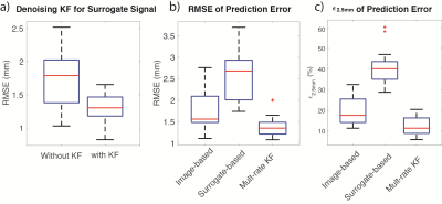
Motion Prediction using a Multi-Rate Kalman Filter with Golden Angle Radial Acquisition for Real-Time MRI-Guided Interventions
Xinzhou Li, Samantha Mikaiel, Holden Wu
Real-time MRI can provide high soft-tissue contrast without ionizing radiation for interventional procedure guidance. To achieve accurate and low-latency tracking of target tissues for decision support and feedback control, this work proposes a motion prediction framework based on a multi-rate Kalman filter and real-time golden-angle radial MRI. The proposed framework leverages the unique sampling pattern of golden-angle radial acquisition to combine image-based with surrogate-based motion tracking. Initial results demonstrate that the proposed framework can achieve significantly reduced error in motion prediction and provide low-latency feedback for real-time MRI guided interventions.
|
|
4152.
 |
2 |
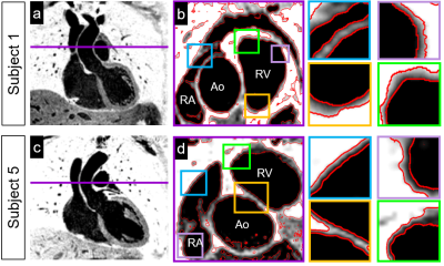
Simultaneous 3D Whole-Heart Bright-Blood Visualization of the Coronary Sinus and Heart Anatomy and Black-Blood PSIR Quantification of Atrial Wall Thickness for Non-Contrast Enhanced Interventional Planning
Giulia Ginami, Karina Lopez, Radhouene Neji, Camila Munoz, Sebastien Roujol, Peter Mountney, Reza Razavi, Rene Botnar, Claudia Prieto
Atrial wall thickness quantification has the potential of providing important clinical information when planning electrophysiological interventions. Imprecise delivery of thermal energy during catheter ablation can prevent the success of the procedure. Furthermore, pre-interventional knowledge of subject-specific variations in the anatomy of the coronary sinus (CS) is crucial for adequate catheterization. Here, we propose a free-breathing 3D whole-heart phase-sensitive inversion recovery sequence suitable for non-contrast enhanced interventional planning, offering simultaneous visualization of the atrial walls and CS anatomy. The sequence is integrated in a framework with image-based navigation and non-rigid respiratory motion correction for 100% scan efficiency and improved image sharpness.
|
|
4153.
 |
3 |
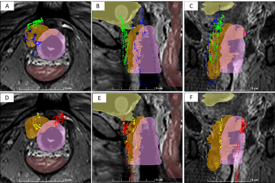
Active-tracked versus Passive-tracked MRI-guided Cervical-cancer Brachytherapy catheter placement in 11 patients: Improved accuracy and reduced procedure time
Jose De Arcos, Ehud Schmidt, Wei Wang, Junichi Tokuda, Kamal Vij, Charles Dumoulin, Ravi Seethmaraju, Robert Cormack, Akila Viswanathan
We performed MRI-guided placement of high dose rate (HDR) brachytherapy catheters in 11 cervical-cancer patients within a 3.0 T MRI scanner. We compared placing MR-tracked metallic stylets to passively-tracked conventional stylets. Comparisons were performed during three procedure stages: coarse stylet navigation to the approximate region of the tumor; fine-tuned navigation to the clinician’s desired (final) location; stylet pull-back (withdrawal) from the body, which provided catheter trajectories for Radiation Treatment Planning. Active-tracking’s main benefits; (I) catheters placed much closer to the clinician’s intended location, including via complex manipulations requiring complete withdrawal and repositioning, (II) placement durations similar to transrectal-ultrasound guided HDR procedures.
|
|
4154.
 |
4 |
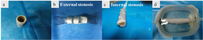
Noninvasive assessment of great vessel stents using a susceptibility-based imaging method
Caiyun Shi, Shi Su, Xin Liu, Dong Liang, Haifeng Wang, Jim Ji, Guoxi Xie
Previous studies have demonstrated that a susceptibility-based positive contrast MR method exhibits excellent efficacy for visualizing MR compatible metal devices by taking advantage of their high magnetic susceptibility. However, the method was not evaluated in the assessment of stent restenosis. The purpose of this study is to assess whether the susceptibility-based positive method can be used to assess the stent restenosis, with the comparison of two typical MR positive contrast techniques, i.e., SUMO and GRASP. The experimental results showed that the susceptibility-based method not only provides better localization of the stent than SUMO and GRASP but also has capabiltiy to assess the stent restenosis.
|
|
4155.
 |
5 |
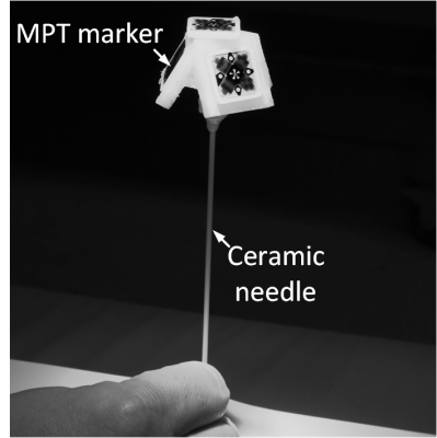
Percutaneous MR-guided Interventions Using an Optical Moiré Phase Tracking System
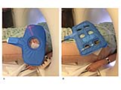
Urte Kägebein, Frank Godenschweger, Frank Wacker, Oliver Speck, Bennet Hensen
The development of an appropriate guidance support is essential to simplify and shorten MRI guided percutaneous interventions. We present a new tracking system and sequence enabling intuitive, interactive and precise instrument navigation with the aid of an optical Moiré Phase tracking system from the inside of the MRI scanner. The system was evaluated regarding targeting error and skin to target time by experienced interventional radiologist and novice users, revealing a precise (0.99mm±0.47mm), fast (155s±62s) and simple real-time needle guidance.
|
|
4156.
 |
6 |
Feasibility of MRI image based synthetic CT generation in radiotherapy using deep convolutional neural network
Yafen Li, Wen Li, Yaoqin Xie, Jun Xia
Generating electronic density information for MRI images is crucial for MRI-based dose calculation in a MRI-only workflow of radiotherapy. To address this problem, we proposed a deep convolutional neural network plus with an auto-context model to predict synthetic CT from MRI images of routine-sequence. The highly accuracy of generated synthetic CT results shows that the proposed method is effective and robust.
|
|
4157.
 |
7 |
Transcatheter intra-arterial perfusion (TRIP)-MRI for the evaluation of immediate Irreversible Electroporation effects in the VX2 rabbit liver tumor model.
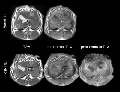
Matteo Figini, Kang Zhou, Liang Pan, Junjie Shangguan, Chong Sun, Bin Wang, Na Shang, Quanhong Ma, Daniele Procissi, Andrew Larson, Zhuoli Zhang
Six rabbits with liver tumors underwent anatomical and perfusion MRI immediately after irreversible electroporation (IRE) treatment of the tumors, using a catheter inserted in the left hepatic artery to deliver the contrast agent (TRIP-MRI). All the treated regions showed low perfusion as measured by the Area under the tissue response Curve. In some cases, a rim or a few spots with higher AUC were present, and were presumably associated with the penumbra of reversible electroporation that is known to be present in IRE-treated tissues. TRIP-MRI could be a valuable non-invasive tool to assess IRE effect and to immediately plan retreatment.
|
|
4158.
 |
8 |
Using 2D image slices for obtaining the guidewire coupling modes of a PTx transmit array
Felipe Godinez, Arian Beqiri, Jose Teixeira, Joseph Hajnal, Shaihan Malik
A multislice method for measuring relative coupling between elements of a parallel transmit (PTx) array and conductive structures such as guidewires at multiple internal locations. Fast 2D image slices are used to measure the relative coupling along the guidewire inside a large phantom. This is useful during conditions where the coupling modes can only be measured from within the object volume and where current sensors cannot be placed. Such conditions exist in large phantoms and can exist in humans.
|
|
4159.
 |
9 |
Intraarterial chemotherapy of glioblastoma following osmotic blood-brain barrier opening under real-time MRI guidance
Chengyan Chu, Monica Pearl, Yanrong Chen, Anna Jablonska, Xiaolei Song, Miroslaw Janowski, Piotr Walczak
Blood-brain barrier (BBB) prevents effective chemotherapy of brain tumors. Intra-arterial (IA) injection of hyperosmotic mannitol has been attempted for many years to permeabilize the BBB, however due to high variability, this procedure never became a routine clinical practice. We have previously shown that real-time MRI may circumvent that obstacle in large animal model. However, for drug screening purposes the mouse model if preferred. Here, we have shown that real-time, interventional MRI is also instrumental to precisely open BBB in rodents, and more importantly combining it with subsequent delivery of chemotherapeutic drug melphalan provides therapeutic benefit warranting consideration of clinical application.
|
|
4160.
 |
10 |
Comparison of MR-guided joint injection with a fluoroscopic approach for pediatric MR arthrography
Video Permission Withheld
Sphoorti Shellikeri, Anne Marie Cahill, Victor Ho-Fung, Randolph Setser, Michael Acord, Seth Vatsky, Fernando Escobar, Abhay Srinivasan
Magnetic resonance imaging (MRI) provides excellent soft-tissue contrast and multiplanar capability in joint imaging, but often requires direct joint injection of gadolinium to provide adequate depiction of cartilage and ligaments. MRI-guided joint interventions are not commonly performed as routine practice. In MR arthrography, the contrast injections are usually performed under fluoroscopy and is followed by a diagnostic MRI to evaluate the abnormalities in the rotator cuffs. This work describes our experience with combined interventional and diagnostic MR shoulder arthrography, and evaluates the efficacy of MR-guided injection, comparing technical success, diagnostic quality, and procedure times to fluoroscopic-guided arthrography.
|
|
4161.
 |
11 |
Application of SSFSE T1-weighted imaging for MRI-guided bone biopsy
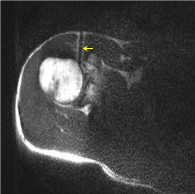
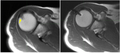
Elena Kaye, Daniel Litwiller, Maggie Fung, Stephen Solomon, Majid Maybody
MRI guided biopsy plays an important role in the diagnosis of bone lesions. Although T1w FSE offers exceptional signal-to-noise ratio and resolution, its acquisition time is relatively long. In this study, we evaluate a single-shot FSE with centric partial Fourier encoding, variable refocusing flip angle, and an inversion recovery preparation pulse (vrfSSFSE-IR) for MRI-guided bone biopsy application with the goal to reduce the overall procedure time.
|
|
4162.
 |
12 |
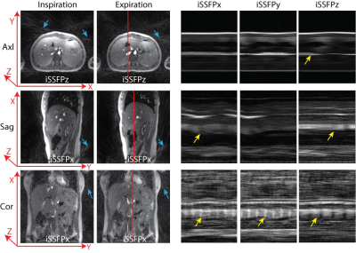
Real-Time Golden Angle Radial iSSFP: Impact of the Gradient Spoiler Direction on Motion and Flow Effects
Samantha Mikaiel, Thomas Martin, Xiaodong Zhong, Kyunghyun Sung, Holden Wu
Real-time visualization is crucial to the success of MRI-guided minimally invasive cancer interventions. We have developed golden-angle (GA) ordered radial integrated-SSFP (iSSFP), which can suppress banding artifacts associated with bSSFP while maintaining similar contrast. However, the addition of the gradient spoiler in iSSFP removes the flow and motion compensation along the axis of the gradient. In this work, we analyze pelvic and abdominal iSSFP scans acquired in the axial, sagittal and coronal planes to investigate the effects of having the gradient along different directions with respect to motion/flow. GA radial iSSFP can potentially improve tissue contrast for real-time MRI-guided interventions, after careful consideration for the imaging plane, gradient spoiler direction, and motion/flow direction.
|
|
4163.
 |
13 |
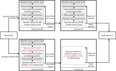
Accuracy and Time Efficiency of Real-Time MRI-Guided Remote-Controlled Targeted Needle Placement During Motion Using Hydrostatic Actuators
Samantha Mikaiel, James Simonelli, Xinzhou Li, Yu-Hsiu Lee, Yong Seok Lee, Kyunghyun Sung, David Lu, Tsu-Chin Tsao, Holden Wu
In this work, we investigate the accuracy and time efficiency for real-time MRI-guided targeted needle placement using a rolling-diaphragm hydrostatic actuator system during motion. We show that the actuator-assisted approach was able to guide the needle to targets with greater accuracy and in less time than the conventional step-and-shoot strategy for both static and dynamic conditions using a programmable motion phantom. The new actuator system can potentially enable physicians to remotely perform real-time MRI-guided interventions during motion.
|
|
4164.
 |
14 |
Non-contrast MRA Guidance in Pediatric Endovascular Interventions
Lingyun Chen, Onno Wink, Amber Pokorney, Ryan Robison, Robyn Augustyn, Marrit Thorkelson, Richard Towbin
The goal of this research project is to investigate the feasibility of using non-contrast 3D MRA imaging in the planning and guidance of catheter-based pediatric vascular disease treatment. It may result in a reduction in patient radiation exposure, contrast (Gadolinium and Iodine) usage and procedure time as compared to traditional 2D fluoroscopy guidance.
|
|
4165.
 |
15 |
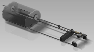
A Phantom for Evaluation of Motion Quantification Methods in MR-Guided Radiotherapy
Video Permission Withheld
Lukas Leiner, Gernot Echner, Matthias Borutta, Reiner Umathum, Arthur Magill, Steffen Seeber, Sebastian Flassbeck, Florian Friedrich, Anna Fischer, Nicolas Behl, Mark Ladd, Florian Maier
MR-guided radiotherapy enables real-time tracking of targets during therapy application, with the aim of improving targeting accuracy and, hence, treatment efficiency. To evaluate MR-based motion quantification techniques, a phantom that can generate reproducible, known motion is an extremely valuable tool. A modular motion phantom was designed and implemented that provides precise and reproducible linear motion, including accurate position information. The phantom was found to be MR-compatible. An initial evaluation of real-time bSSFP imaging was performed. The developed device can be used as research tool for MR-guided therapy and as a clinical quality assessment tool to improve treatment of patients.
|
|
4166.
 |
16 |
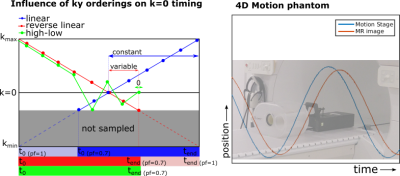
Imaging latencies for Cartesian and Golden Angle 2D MRI in real-time MR-guided radiotherapy
Pim Borman, Rob Tijssen, Clemens Bos, Chrit Moonen, Bas Raaymakers, Markus Glitzner
For tumor tracking it is vital to minimize the latency between the moment of anatomic change and its appearance on the MR image. Apart from the temporal footprint of the acquisition, the readout trajectory itself influences the latency. We explore how the latency is minimized by shifting the collection of the center of k-space to the end of the acquisition. For Cartesian sequences this is achieved by changing the profile order, whereas in golden angle acquisitions a smaller window or k-t filter is able to achieve this.
|
|
4167.
 |
17 |
A Three-dimensional Template Matching Technique in Target Tracking Technique of MRgHIFU for liver
Tomiki Morita, Etsuko Kumamoto, Daisuke Kokuryo, Kagayaki Kuroda
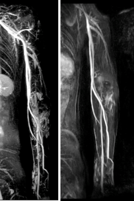
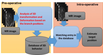
MRgHIFU treatment for liver requires a tracking technique to “lock on” to the focal spot at the target tissue region during respiratory-induced motion. We proposed three-dimensional focus tracking technique using template matching method. In this method, by creating a deformity / mutation model reconstructed from multi-slice volume data and extracting multiple blood vessel branch points, the focal spot can be estimated from the relative positional relationship. In this study, we optimized template size of template matching for extracting branching points in preoperative. Experimental results demonstrated to improve accuracy of blood vessel extraction.
|
|
4168.
 |
18 |
Robust quantitative diffusion weighted MR on an MR-Linac system
Tim Schakel, Rob Tijssen, Hans Hoogduin, Marielle Philippens
Quantitative diffusion weighted imaging is demonstrated on a clinical MR-Linac system. ADC values were measured using a standardized diffusion phantom using two different diffusion sequences, single shot EPI and single shot TSE: SPLICE. In general, accurate and repeatable ADC measurements could be acquired using both sequences. EPI showed more susceptibility and eddy current related image distortions, whereas SPLICE was geometrically more robust. Large deviations in ADC start to occur when moving further away from the isocenter, suggesting the need for gradient non-linearity correction of ADC values.
|
|
4169.
 |
19 |
Real-time SNR enhancement in surgical region for intraoperative MRI combining a stationary and a small freely-moving RF coils
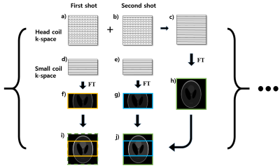
Jun-Hee Kim, Won-Joon Do, Jong-Woo Lim, Sang-Chul Lee, Sung-Hong Park
This study introduces a new technique to enhance SNR in a surgical region for real-time intraoperative MRI that combines a static big and an endoscopic small RF coils. Segmented imaging using the static RF coil and fast imaging using the small RF coil were performed simultaneously with a reduced FOV. The images from the static and freely-moving RF coils were updated in multiple shots and in every shot, respectively. This technique not only reduces computing time, but also improves SNR in surgical region without intensity variation, which would be beneficial in intraoperative MRI.
|
|
4170.
 |
20 |
3D specific absorption rate estimation from focused ultrasound sonications using the Green’s function heat kernel
Nick Freeman, Henrik Odéen, Dennis Parker
In-vivo determination of tissue thermal and acoustic properties, such as the specific absorption rate (SAR) for focus ultrasound, are important for accurate thermal modeling in treatment planning, monitoring, and control using, e.g., Pennes bioheat transfer equation. In this work we derive and present a numerical method using the Green’s function and MR thermometry data as input to estimate SAR non-invasively. The method is as accurate but substantially faster (on the order of seconds, compared to minutes) compared to two other SAR determination methods. Simulation and phantom experiments in a tissue mimicking gel phantom are performed.
|
|
4171.
 |
21 |
Assessment of Immediate Response to Irreversible Electroporation for Targeted Ablation of Liver Tissues Using Dynamic Contrast Enhancement in a Rabbit Model
Junjie Shangguan, Matteo Figini, Chong Sun, Liang Pan, Bin Wang, Quanhong Ma, Kang Zhou, Na Shang, Zhuoli Zhang
Irreversible electroporation (IRE) may be visualized by MRI immediately post-procedure to monitor and assess tissue response peri-operatively. Dynamic contrast-enhanced MRI (DCE-MRI) allows measurement of tissue perfusion. We will demonstrate that DCE-MRI allows early visualization of IRE ablated tissue margins for prediction of the ablated region and quantification of tissue response. 6 rabbits underwent IRE ablation of the liver and pre-IRE and post-IRE DCE-MRI. A decrease in post-IRE apparent diffusion coefficients in the ablated region compared with baseline indicates tissue damage. Post-IRE AUC is decreased compared with baseline, suggesting decreased but still present blood perfusion in the ablated region post-IRE.
|
|
4172.
 |
22 |
Dedicated 3-Channel Surface Coil for Interventional Magnetic Resonance Imaging at 3T - First Evaluation
Video Permission Withheld
Lena Sonnow, Wesley Gilson, Arne Hengerer, Clifford Weiss, Himanshu Bhat, Frank Wacker, Jan Fritz
We developed a dedicated interventional MRI coil with 3 receiver channels and a wide central aperture for interventional access, which we benchmarked against a 4-channel system surface coil. We assessed signal-to-noise-ratios (SNR), geometry (g)-factors and instrument artifact size in a phantom and 100 subjects. The interventional MRI prototype coil produced similar SNR values and g-factors than the 4-channel system coil. There was no significant difference in artifacts, which were suitable for accurate and safe procedures. The central aperture of the interventional MRI prototype coil improved access and the coil performed error-free during a large variety of interventional MRI procedures.
|
|
4173.
 |
23 |
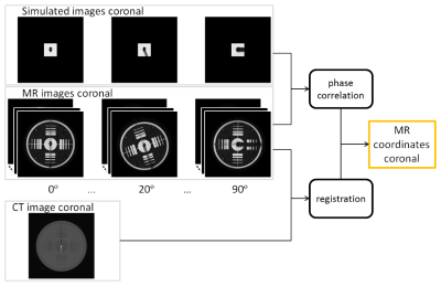
Validation study of high framerate source localization for MR-guided HDR brachytherapy
Ellis Beld, Marinus A. Moerland, Max A. Viergever, Jan J.W. Lagendijk, Peter R. Seevinck
For the development of MR-guided high-dose-rate (HDR) brachytherapy, the accuracy and precision of an MR-based HDR source localization method were investigated in a phantom study, by a comparison with CT. MR images were acquired for various angulations of the source with respect to B0, and a 3D CT scan was acquired. The MR source positions were compared to the CT source position. The results demonstrated a high, subvoxel accuracy (0.4-0.6 mm) and a high precision (≤0.1 mm) at high temporal resolutions (0.15-1.2 s per slice). This proved that our proposed MR-based source localization method is valuable for MR-guided HDR brachytherapy.
|
|
4174.
 |
24 |
Platform for investigating spatial and intensity parameters for phase-sensitive MRI focal localization with steerable single element focused ultrasound transducers through skull
Spencer Brinker, Frank Preiswerk, Nathan McDannold
A platform is designed and constructed for investigating Focused Ultrasound (FUS) spatial and intensity focal parameters during phase-sensitive MRI focal localization. Recently, there has been a large interest to use single element transducers to deliver therapy to the brain. Spatial accuracy and intensity safety limits need further investigation. The platform developed in this project uses 3D Slicer software to incorporate MRI and neuronavigation camera coordinates for guiding an automated 3D hydrophone mapping system. A 272 kHz single element FUS transducer, ex vivo skull, and gel phantom are used to demonstrate components of the integrated MRI and navigation based hydrophone mapping system.
|
|
| Back |
| The International Society for Magnetic Resonance in Medicine is accredited by the Accreditation Council for Continuing Medical Education to provide continuing medical education for physicians. |
