|
Electronic Poster Session
Acquisition, Reconstruction & Analysis |
Tuesday, 19 June 2018
Electronic PosterAcquisition, Reconstruction & Analysis
4079 -4102 Motion Correction in the Brain
4103 -4126 Motion & Artefact Correction
4127 -4150 Encoding & Beyond
4199 -4222 System Imperfections & Artifacts: Characterization & Correction
4223 -4246 Pulse Sequences & Beyond
4247 -4270 MR Fingerprinting |
| |
Motion Correction in the Brain
Electronic Poster
Acquisition, Reconstruction & Analysis
Tuesday, 19 June 2018
| Exhibition Hall |
13:45 - 14:45 |
| |
|
Computer # |
|
4079.
 |
49 |
 Comprehensive head motion modelling and correction using simultaneously acquired MR and PET data Comprehensive head motion modelling and correction using simultaneously acquired MR and PET data
Francesco Sforazzini, Jakub Baran, Alexandra Carey, Nadim Shah, Gary Egan, Zhaolin Chen
Head motion is one of the major issues in neuroimaging. With the introduction of MR-PET scanners, motion parameters can now be estimated from two independent modalities acquired simultaneously. In this work, we propose a new data-driven method that combines MR image registration and PET data driven approach to model head motion during the complete course of MR-PET examination. Without changing the MR-PET acquisition protocol, the proposed method provides motion estimates with a temporal resolution of ~2 secs. Results on a phantom dataset show that the proposed method can significantly reduce motion artefact in brain PET images and improve image sharpness compared with the MR based methods.
|
|
4080.
 |
50 |
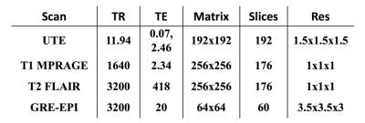
Fast and Robust: Prospective Motion Correction Combined with Compressed Sensing for 3D Time-of-Flight MRA
Video Permission Withheld
Christoph Forman, Michaela Schmidt, Peter Speier, Martin Schramm, Steffen Schroeter, Daniel Splitthoff, Tobias Kober
We present the combination of prospective motion correction and compressed sensing for 3D Time-of-Flight MRA. This enables fast TOF imaging in high isotropic resolution, while preserving image quality and diagnostic value even in the presence of motion. The method was evaluated in five healthy volunteers and image quality was compared to a conventional TOF MRA. Prospective motion compensation successfully enabled robust diagnostic image quality in the highly accelerated scan. The promising results as well as the full integration of the proposed method in a standard clinical scanner enable a comprehensive evaluation in patients in the near future.
|
|
4081.
 |
51 |
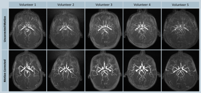
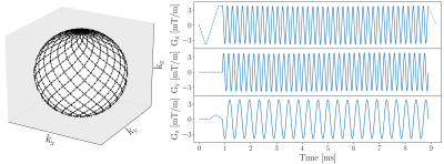
Fast and Accurate 3D Rigid-Body Motion Information From Spherical Lissajous Navigators at Small K-Space Radii
Richard Buschbeck, N. Jon Shah
A new navigator concept is presented which enables the measurement of accurate rigid-body motion information from spherical navigators at extremely small k-space radii. This is achieved by using a 3D Lissajous navigator trajectory and a rotation estimation algorithm known from computer vision. The new method provides motion information with sufficient accuracy and precision for many motion correction techniques and significantly outperforms current spherical navigator methods in terms of acquisition and computation time.
|
|
4082.
 |
52 |
Slice-wise motion tracking during simultaneous EEG-fMRI
Malte Laustsen, Mads Andersen, Patrick Lehmann, Rong Xue, Kristoffer Madsen, Lars Hanson
Slice-wise motion tracking during combined electroencephalography (EEG) and echo planar imaging (EPI) is developed. Using gradient-induced noise on the EEG for tracking, no interleaved navigator modules or additional hardware is needed. The motion parameters are determined after a calibration and training scan. The method is explored in a phantom and in vivo.
|
|
4083.
 |
53 |
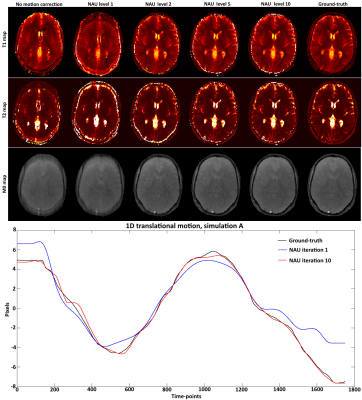
Zero-Dimensional Self Navigated Autofocus for Motion Corrected Magnetic Resonance Fingerprinting
Gastao Cruz, René Botnar, Claudia Prieto
Magnetic Resonance Fingerprinting (MRF) provides simultaneous multi-parametric maps from a continuous transient state acquisition of many time-point images. Motion occurring during the MRF acquisition can create artefacts in the consequent T1/T2 maps. Here we propose to derive an intermediate 1D motion model from the acquired MRF data itself via self-navigation of the k-space central point and further refine the motion estimates using an autofocus algorithm for MRF motion correction. The proposed approach was evaluated in simulations.
|
|
4084.
 |
54 |
Segmented 3D fat navigators for faster brain motion tracking at 7T
Video Permission Withheld
Frédéric Gretsch, Daniel Gallichan
Fat Navigators have previously demonstrated their utility for high-accuracy motion tracking. We investigate a segmented 3D version of the navigators which allows significant acquisition time reduction while maintaining high motion accuracy.
|
|
4085.
 |
55 |
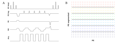
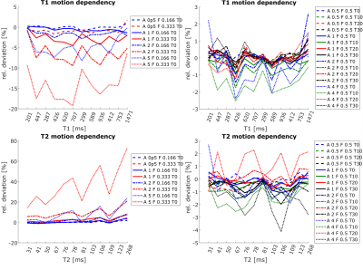
Evaluating the influence of motion on FISP-MRF
Gregor Körzdörfer, Peter Speier, Steffen Schröter, Martin Schramm, Josef Pfeuffer, Bernhard Hensel, Mathias Nittka
Magnetic Resonance Fingerprinting (MRF) is an MR technique that generates parameter maps by matching pseudo randomly generated MR signals to a precalculated dictionary. MRF sequences in general consist of a long series of closely spaced excitations. An assumption underlying MRF is that a partially motion-corrupted signal is not able to significantly alter results. Corrupted signal segments are supposed to have no respective counterpart in the dictionary and therefore do not affect the pattern match. This assumption is evaluated in this study. Controlled motion was added to phantom and in vivo MRF experiments, and the results were related to realistic patient movement recorded by a 3D camera system.
|
|
4086.
 |
56 |
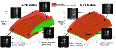
Evaluating Motion Artifact Correction of the Linearized Geometric Solution in bSSFP MRI
Michael Hoff, Qing-San Xiang
The linearized geometric solution (LGS) is known for demodulating balanced steady state free precession (bSSFP) MRI signal of its dependence on magnetic field inhomogeneity, but has also recently shown signs of mitigating motion artifact. Here the extent of motion artifact correction by the LGS is explored via simulations of variable motion schedules, image noise, and T1/T2 ratio. It corrects motion artifact to a greater extent than a complex image averaging solution, except in scenarios of low signal-to-noise ratio (SNR). This study motivates clinical applications of bSSFP in imaging fluid structures that normally suffer from motion and/or field inhomogeneity artifacts.
|
|
4087.
 |
57 |
Revealing sub-voxel motions of brain tissue using phase-based amplified MRI (aMRI)
Video Permission Withheld
Itamar Terem, Wendy Ni, Maged Goubran, Greg Zaharchuk, Kristen Yeom, Michael Moseley, Mehmet Kurt, Samantha Holdsworth
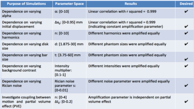
Problem:Amplified Magnetic Resonance Imaging (aMRI) was recently introduced as a new brain motion detection and visualization method. In this work we strive to improve aMRI by incorporating a phase-based motion amplification algorithm. Methods:Phase-based aMRI was developed, validate using digital phantom simulations and compared with EVM-based aMRI in healthy volunteers at 3T. Data were also acquired on a patient with Chiari I malformation, and displacement maps were produced using free form deformation (FFD) of the aMRI output. Results:Phantom simulations showed that phase-based aMRI has a linear dependence of amplified displacement on true displacement. Amplification was independent of temporal frequency, phantom size, Rician noise, and partial volume effect, but had a slight dependence on phantom shape. Phase-based aMRI supported larger amplification factors than EVM-based aMRI, and was less sensitive to noise and artifacts. Abnormal biomechanics were seen on FFD maps of the Chiari I malformation patient.Conclusion: Phase-based aMRI can be used for quantitative analysis of minute changes in brain motion, and may reveal subtle physiological variations of the brain due to pathology. Preliminary data shows the potential of phase-based aMRI to assess abnormal biomechanics in Chiari I malformation.
|
|
4088.
 |
58 |
A Comparison of Prospective versus Retrospective Motion Correction for Reliable T1-Weighted Neuroimaging
Video Permission Withheld
Steven Kecskemeti, Abigail Freeman, Andrew Alexander
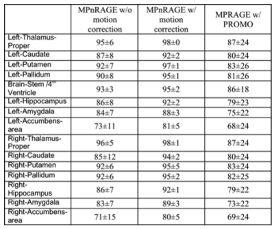
Prospective motion corrected MPRAGE T1-weighted images were compared against retrospectively motion corrected MPnRAGE T1-weighted images. 14 young children were scanned without sedation and without the head being stabilized with pads. Dice-overlap of automatic tissue segementation masks (FAST and FLIRT) were used as a measure of reliability. MPnRAGE with motion correction showed exceptional regional label consistency (>=80% Dice overlaps for all 16 regions and >90% in 12 of the regions). Conversely, MPRAGE-PROMO demonstrated lower performance than even MPnRAGE without motion correction.
|
|
4089.
 |
59 |
Combined prospective motion correction and retrospective B0 correction for EPI using optical tracking and predictive B0 maps.
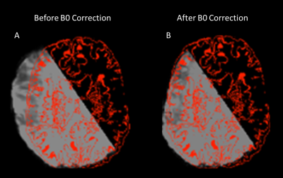
James Smith, Olivier Mougin, Kingkarn Aphiwatthanasumet, Matthew Clemence, Andrew Peters, Richard Bowtell, Paul Glover, Penny Gowland
We demonstrate a combination of prospective motion correction and retrospective B0 unwarping using predictive field maps to improve the registration of EPI images. Prospective motion correction is performed using an optical tracking camera and custom scanner code. Field maps were generated using a multilinear fit of subject orientation using data gathered at known orientations. Results show a higher correlation when registering EPI data to a high-resolution anatomical, post-correction.
|
|
4090.
 |
60 |
Influence of tracking system latency on prospective motion correction
Patrick Hucker, Bruno Riemenschneider, Stefan Kroboth, Maxim Zaitsev
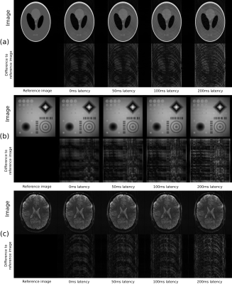
Tracking latency in prospective motion correction (PMC) with external tracking systems is commonly assumed to influence image quality, however no studies attempted to quantify this effect. This work presents a method to visualize latency-induced artifacts in simulated MR images. At first a MR sequence with PMC is simulated using vendor tools and recorded or synthetic motion with varying latency is added. Thereafter the effective encoding trajectory based on the exported gradient/ADC events is calculated. The resulting trajectory is used to simulate MR data with the inverse FFT, visualizing artifacts due to the tracking latency.
|
|
4091.
 |
61 |
A dynamic MR-signal model to capture 3D motion-fields at ultra-high frame-rate
Niek Huttinga, Cornelis van den Berg, Peter Luijten, Alessandro Sbrizzi
We present a framework that has the potential to capture non-rigid 3D motion at 50 Hz, hereby drastically accelerating state-of-the-art techniques. Our model directly and explicitly relates the motion-field to the k-space data and is independent of the spatial resolution, allowing for extremely high under-sampling. We illustrate proof-of-principle validations of our method through a simulation test and whole-brain 3D in-vivo measured data. Results show that the 3D motion-fields can be reconstructed from extremely under-sampled k-space data consisting of as little as 64 points, enabling 3D motion estimation at unprecedented frame-rates.
|
|
4092.
 |
62 |
Contact-Free Respiration Motion Monitoring Using a Markerless Structured Optical System in MRI
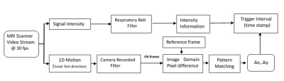
Chen Zhang, Jin Liu, Jiarui Cai, Haikun Qi, Jinnan Wang, Chun Yuan, Huijun Chen
Respiration motion artifacts remain a major problem in MRI, particularly at higher imaging resolution and field strength. In this study, we investigated the feasibility of using a MR-compatible in-bore camera system to perform contactless monitoring of respiratory information during MRI of human subjects. A good match to the respiratory data is apparent in terms of the timing of trough, and ICC between trigger intervals from the respiratory belt data and video-derived signals is 0.97, meaning that this optical system can monitor and correct in end-expiratory, in addition to simultaneously detecting random motion based on our previous work.
|
|
4093.
 |
63 |
Wasserstein GAN for Motion Artifact Reduction of MR images
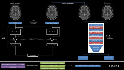
Sandro Braun, Pascal Ceccaldi, Xiao Chen, Benjamin Odry, Boris Mailhe, Mariappan Nadar
Subject motion is a common artifact in MR acquisition that can severely degrade image quality. We take advantage of the recent advances in deep generative network to compensate motion and generate images of increased quality, measured by evaluating changes in MSSIM and normalized L2 distance (NRMSE). We trained an image to image network to predict motion compensated magnitude images given motion-corrupted input images, coupled with an adversarial network to help refine those predicted images. For the discriminator loss, we use the Wasserstein objective. The results suggest clear improvements on MSSIM and NRMSE metrics for the majority of cases.
|
|
4094.
 |
64 |
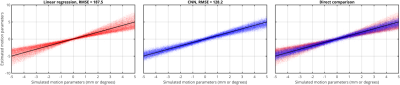
Using Machine Learning to accelerate fat-based head motion-navigators – a preliminary simulation study
Daniel Gallichan
We hypothesise that machine learning approaches could be applied to speed up motion-correction navigators – potentially obviating the need for image reconstruction and co-registration. In this preliminary 2D simulation study we investigate the ability of a simple 5-layer convolutional neural network to predict motion-parameters based on difference images between two head poses. Our results indicate that the CNN is able to outperform linear regression over the range of parameters tested, supporting our aim to develop this concept in more detailed future work.
|
|
4095.
 |
65 |
Deep learning-based real time MRI
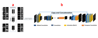
Vahid Ghodrati, Jiaxin Shao, Ziwu Zhou, Yu Gao, Fadil Abbas Ali, Fei Han, Peng Hu
In this work, we use the dilated U-net to reconstruct the dynamic free breathing cine images from regular under sampled raw data. Also, we consider different under sampling rate to determine maximum achievable rate. Moreover, we modify the acquisition procedure of sequence to show the possibility of prospective dynamic imaging. The proposed method is capable of reconstructing high quality real time 2D cardiac images with up to 6X acceleration with excellent image quality and minimal latency of only 6ms on a typical workstation.
|
|
4096.
 |
66 |
Accelerating cardiac dynamic imaging with video prediction using deep predictive coding networks
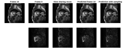
Xue Feng, Craig Meyer
Deep predictive coding networks based on stacked recurrent convolutional neural network have shown great success in video prediction since they can learn to recognize and analyze the motion patterns of each element from previous frames. In this study we adopted this model to predict future frames in cardiac cine images and used a k-space substitution method to improve the prediction accuracy. It showed promises in accelerating cardiac dynamic imaging.
|
|
4097.
 |
67 |
Machine learning algorithms for detection of motion artifacts: a general approach
Alessandro Sciarra, Hendrik Mattern, Oliver Speck
Despite all the developments to overcome MRI motion artifacts, there are still open questions. When do we need to repeat a scan? Is the image quality sufficient for segmentation or to make a diagnosis? Is the motion correction working properly? Independent of the type of image acquired (structural, diffusion, functional, etc.), machine learning algorithms can detect automatically motion artifacts and provide feedback in real time. In this work different machine learning algorithms have been tested to detect motion artifacts in synthetic and in vivo data.
|
|
4098.
 |
68 |
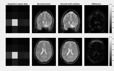
Motion correction in MRI using deep learning
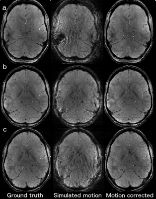
Patricia Johnson, Maria Drangova
Subject motion in brain MRI remains an unsolved problem. We propose a machine learning approach for motion correction of brain images. Our initial objective is to train a neural network to perform a motion corrected image reconstruction on image data with simulated motion artefacts. Training pairs were generated using an open source MRI data set; a unique motion profile was applied to each 2D image. A deep neural network was developed and trained with over 3000 image pairs. The images predicted by the network, from motion-corrupted k-space, have improved image quality compared to the motion corrupted images.
|
|
4099.
 |
69 |
Ultra-High-Resolution Dental MR Imaging Using an Ultra-Short-TE Sequence with Prospective Motion Correction
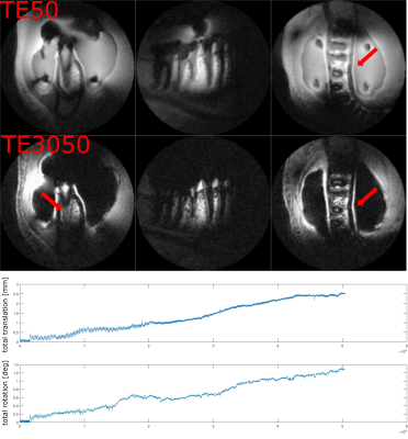
Xiang Gao, Philipp Amrein, Johannes Fischer, Davide Piccini, Patrick Hucker, Ute Ludwig, Jan-Bernd Hövener, Maxim Zaitsev
High-resolution dental MR Imaging with clear boundaries is of interest for clinical applications e.g. for tooth implant planning. Due to the fast signal decay of the dental tissues, 3D ultra-short-TE (3D-UTE) sequences are employed, which combine rapid center-out half-projection readouts with a phyllotaxis ordering on the surface of k-space sphere. Even though 3D-UTE is relatively robust against subject motion, motion artifacts still limit the achievable image quality. In this work, we implement a 3D-UTE sequence with a camera-based prospective motion correction and apply it in an in vivo experiment to achieve motion-free dental imaging with an isotropic resolution of 0.35mm.
|
|
4100.
 |
70 |
Motion robust Magnetic Resonance Fingerprinting using Echo-Planar Imaging and intensity based motion correction
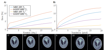
Mario Wenning, Benedikt Rieger, Lothar Schad, Sebastian Weingärtner
Quantitative magnetic resonance imaging (MRI) enables the quantification of biomarkers and the detection of tissue changes. Recently, various magnetic resonance fingerprinting (MRF) sequences were proposed to enable simultaneous quantification of T1 and T2/T2*relaxation times. In this study, we demonstrate motion sensitivity of the original MRF and MRF-EPI. Intensity-based image registration is used in six healthy subjects to eliminate motion artifacts in MRF-EPI. The effectiveness is demonstrated by a significant improvement in the Dice coefficient. As the algorithm is independent of patient and slice, it facilitates motion robust acquisition of parameters maps with MRF ready for clinical use.
|
|
4101.
 |
71 |
Separation And Quantification Of Head Motion Modes By Pilot Tone Measurements
Video Permission Withheld
Peter Speier, Bacher Mario, Jan Bollenbeck, Matthias Fenchel, Tobias Kober
Pilot Tone Navigation, a new highly integrated electromagnetic navigation method, was applied to characterize head motion in a guided volunteer experiment. Nodding ("yes") and shaking ("no") motion could be separated and quantified by optimizing linear signal combinations from 20 receive channels on fluoroscopically acquired images. These initial results suggest that head motion can be continuously monitored via the regular MRI receive chain without interaction with the acquisition using a single Pilot Tone generator.
|
|
4102.
 |
72 |
Novel and Efficient Generation of Diffeomorphic Motion Phantom
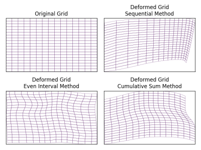
Xia Zhu, Dipanjan Sengupta, Theodore Willke, Andrew Beers, Jayashree Kalpathy-Cramer
Dealing with motion artifacts is a fundamental preprocessing step in magnetic resonance imaging (MRI). In this work, we present three novel methods to generate MRI phantoms for diffeomorphic motion which can be used to validate motion correction algorithms. Previous research efforts for diffeomorphic motion generation, employ a brute force approach in which random pixel/voxel displacements are repeatedly generated until a topologically valid displacement is found. Such methods are not only time consuming but also doesn’t guarantee a valid displacement. Our approach algorithmically ensures that the topological properties are always maintained resulting in guaranteed diffeomorphic motion with much faster runtime.
|
|
Motion & Artefact Correction
Electronic Poster
Acquisition, Reconstruction & Analysis
Tuesday, 19 June 2018
| Exhibition Hall |
13:45 - 14:45 |
| |
|
Computer # |
|
4103.
 |
73 |
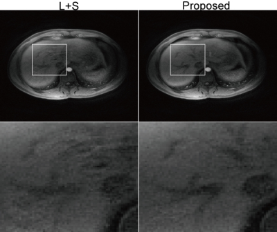
Sliding Motion Corrected Low-rank Plus Sparse Reconstruction for Free-breathing Liver DCE-MRI
Wenyuan Qiu, Dongxiao Li, Fan Liu, Xinyu Jin, Thanh Nguyen, Martin Prince, Yi Wang, Pascal Spincemaille
Liver dynamic contrast enhanced MRI (DCE-MRI) requires high spatiotemporal resolution to clearly visualize enhancement patterns. Image reconstruction quality is often compromised due to unavoidable respiratory motion during the long-time acquisition. This abstract presents a novel sliding motion corrected low-rank plus sparse (SMC-LS) reconstruction algorithm for free-breathing liver DCE-MRI. The sliding motion of the liver along the superior-inferior direction is estimated directly from the under-sampled k-space data. Low-rank and sparse regularization are enforced on the sliding motion corrected image reconstructions. Results demonstrated that SMC-LS can substantially reduce motion blurring and preserve more details in free breathing liver DCE-MRI.
|
|
4104.
 |
74 |
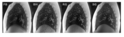
Self-Calibrated Soft Gating for Respiratory Resolved 3D+Time Lung ZTE
Anne Menini, Peng Lai, Graeme McKinnon, Florian Wiesinger
3D + Time lung ZTE reconstruction can provide combined anatomical imaging and motion extraction for RTP and PET/MR-type applications. In this work, we propose a soft-gating method for non-Cartesian imaging, via a solver preconditioner, to improve the sharpness and SNR of 4D ZTE. No a priori knowledge is used for the soft gating weights, as they are self-calibrated based on coil-image bin to bin similarities. The proposed soft gating method provides a SNR gain while maintaining resolution.
|
|
4105.
 |
75 |
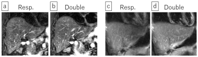
Prospectively Respiratory-Cardiac Double-Triggered Three-Dimensional T2-Weighted Abdominal MRI
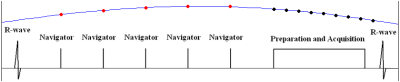
Yuji Iwadate, Atsushi Nozaki, Yoshinobu Nunokawa, Shigeo Okuda, Mitsuharu Miyoshi, Masahiro Jinzaki, Hiroyuki Kabasawa
We developed a prospective respiratory-cardiac double-triggering technique with navigator echo and incorporated it into 3D T2-weighted variable refocusing flip-angle fast spin echo (Cube) for abdominal MRI. The respiratory and cardiac navigator signals synchronized well with the signals from the external devices, and resultant double-triggered Cube images had higher sharpness scores of the left liver lobe than the respiratory-triggered Cube images. The double-triggering technique is feasible and can be applied to other applications where both respiratory and cardiac motions are problematic.
|
|
4106.
 |
76 |
Improved non-rigid motion suppression for free-breathing PROPELLER with adaptively weighted blade combination
Hai Luo, Gaojie Zhu, Xiang Zhou, Meining Chen, Chao Wang, Xia Liu, Wei Bian, Ziyue Wu
PROPELLER has been applied to suppress respiratory motion for free-breathing abdomen imaging but the results are often unsatisfactory with existing weighting mechanisms. In this study, a novel adaptive weighting method is proposed to maximize the respiratory motion suppression without using a fallible reference blade. First, mutual information is used to measure the motion correlation across different blades. Second, principal components analysis is applied to adaptively reject/keep the acquired data by assigning proper weights to all blades. Our experiments show that the proposed method can provide abdominal images with less blurring and less partial volume artifacts compare to the conventional PROPELLER.
|
|
4107.
 |
77 |
Joint Non-rigid Motion and Image MRI SENSE Reconstruction
Javier Royuela-del-Val, Alejandro Godino-Moya, Rosa María Menchón-Lara, Santiago Sanz-Estébanez, Federico Simmross-Wattenberg, Marcos Martín-Fernández, Carlos Alberola-López
In this work a method of the reconstruction of magnetic resonance images in the presence of non-rigid motion is presented. A non-rigid deformation model is introduced in the reconstruction problem in order to obtain 1) a motion-free image and 2) a set of non-rigid transformations that describe the motion of the structures being imaged. The method is able to reconstruct the motion information without reconstructing the images in additional motion states avoiding extra computational costs and resulting in a better posed reconstruction problem. Preliminary results for 2D cardiac cine MRI are shown.
|
|
4108.
 |
78 |
Tracking Respiratory Motion Throughout Arbitrary MRI Sequences via Pilot Tone Navigation
David Rigie, Thomas Vahle, Ryan Brown, Tiejun Zhao, Matthias Fenchel, Peter Speier, Kimberly Jackson, Fernando Boada
In this work, we demonstrate a flexible approach to track respiratory motion throughout arbitrary MRI sequences without requiring any additional patient setup time or sequence modification. A reference RF signal, which has previously been referred to as the “pilot tone” (PT), is tracked throughout MR imaging, and its amplitude modulation provides information about respiratory motion. Specifically, we demonstrate continuous tracking of respiratory motion throughout multi-echo and multi-shot sequences on a human volunteer via PT navigation.
|
|
4109.
 |
79 |
Comparison of three surrogate-based respiratory motion correction methods for 3D high resolution cardiac MRI
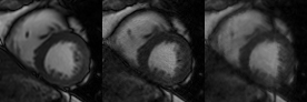
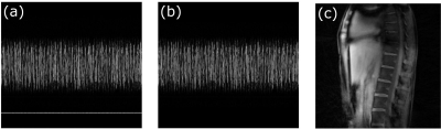
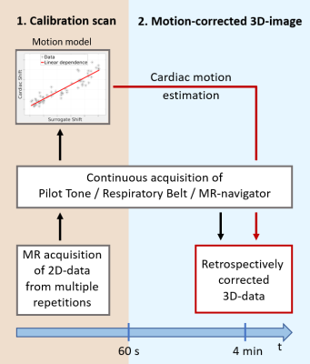
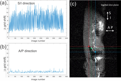
Juliane Ludwig, Peter Speier, Frank Seifert, Tobias Schaeffter, Christoph Kolbitsch
Respiratory motion correction has been proposed to improve scan efficiency and ensure high image quality for 3D cardiac MRI. Nevertheless, these techniques often require dedicated data acquisition and cannot necessarily capture intra-cycle variations of the breathing. Here we compare three different surrogate signals (“pilot tone”, respiratory belt and MR-navigator) for a surrogate-based motion correction approach which provides motion information with high temporal resolution and can be combined with a wide range of different MR acquisition schemes. The temporal stability of these surrogates is assessed and motion correction of a 3D cardiac MR scan is demonstrated.
|
|
4110.
 |
80 |
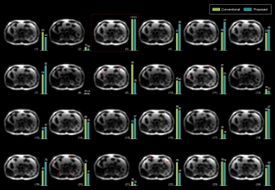
Motion-correction algorithm for free-breathing, fast-heart rate small animal studies on clinical scanners
Matthew Tarasek, Jeannette Roberts, Deirdre Cassidy , Thomas Foo, Desmond Yeo, Randall Carter, Brian Bales
Small animal scanning methods are common for preclinical efficacy evaluation of contrast agents, and naïve animal studies done on clinical scanners are becoming more prevalent. Unfortunately, scanning small animals on large-bore clinical scanners creates several challenges such as motion effects due to organ movement and very fast heart-rates. Here we provide a retrospective motion-correction algorithm for fast-heart-rate/free-breathing small animals based on diaphragm tracking. Results show a ten-fold reduction in baseline contrast region signal uncertainty and significant accuracy improvement of pharmacokinetic time-constant estimation.
|
|
4111.
 |
81 |
Atlas-based breathing motion correction for dynamic lung XeMRI
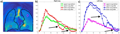
ADAM SZMUL, Bartlomiej Papiez, Ozkan Doganay, Fergus Gleeson, Daniel Bulte, Julia Schnabel, Vicente Grau
In this work we propose a framework to compensate for the breathing motion for dynamic lung XeMRI (DXeV), aligning images from different breathing phases and therefore increasing the accuracy of the ventilation analysis process. We build a lung atlas, to delineate a plausible shape of the lungs in the ventilation images, which is further used to co-register all ventilation volumes in the sequence to the reference lung atlas. After applying the proposed breathing motion correction method, the tidal breathing motion has been largely compensated and all masks of ventilation volumes correspond to each other spatially.
|
|
4112.
 |
82 |
Implementation of a control system for prospective respiratory motion correction during Cardiovascular MR imaging
Stephen Jermy, Ali Alhamud, Ian Burger, Ntobeko Ntusi, Ernesta Meintjes
Navigated free-breathing techniques are commonly used during cardiovascular MR (CMR) acquisitions when breath-holding techniques are not a viable option. Navigated free-breathing techniques with acceptance windows suffer from much longer scan times. A control system using an adaptive Kalman filter was developed to continuously update the slice position throughout the imaging segment during free-breathing CMR, allowing the acquisition to continue throughout the respiratory cycle, regardless of diaphragm position, thereby improving respiratory efficiency and reducing scan times.
|
|
4113.
 |
83 |
Motion Correction based on Z-spectral Consistency for APTw/CESTw MRI Applications in Body Oncology
Jochen Keupp, Elwin de Weerdt
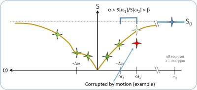
CESTw imaging in body areas with significant physiological motion is strongly hampered because of the inherent motion sensitivity of this MRI technique. In particular, slow physiological motion in the abdomen, e.g. peristaltic motion or bladder filling may lead to Z-spectral inconsistency and artifacts in CESTw images. Here, a method is introduced which analyzes Z-spectral consistency to mask CESTw image areas affected by motion. The technique is successfully demonstrated on a volunteer example in liver CESTw MRI, indicating a future application in body oncology.
|
|
4114.
 |
84 |
Automatic Motion Artifact Detection as Scan-aided Tool in an Autonomous MRI Environment
Wenwen Jiang, Okai Addy, William Overall, Bob Hu, Juan Santos
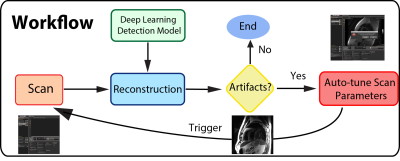
Motion artifacts in MRI can be confused with pathology, and render the scans not diagnosable. Ideally during examination, scan operators should identify these artifacts immediately, and reacquire the scan. But it can be challenging to do so in a clinical time-constraint workflow. Motion artifacts are especially difficult to identify accurately, as they come in a wide variety. Here, we propose an autonomous scan control framework, based on deep learning, to detect motion artifacts immediately after reconstruction. The deep learning model was integrated into a real-time imaging system, and enabled an interactive scanning pipeline.
|
|
4115.
 |
85 |
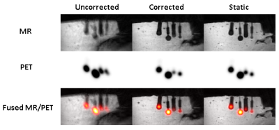
MR-assisted PET reconstruction in the presence of respiratory motion: A phantom study
Cihat Eldeniz, H. Michael Gach, Richard Laforest, Hongyu An
Simultaneous PET/MR provides an unprecedented opportunity for motion correction. We aimed at developing an integrated MR-assisted PET motion correction method which would allow accurate PET quantification in the presence of respiratory motion. In this study, we evaluated the impact of motion correction on lesions of various sizes by using a deformable motion phantom. A static scan was used as the ground truth for PET activity. The combined MR and PET motion correction recovered the activity from 55% to >97% of the static scan activity.
|
|
4116.
 |
86 |
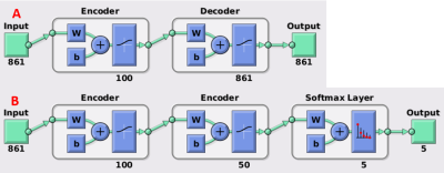
MR/PET motion correction using deep learning
Yasheng Chen, Cihat Eldeniz, Richard Laforest, Hongyu An
In abdominal imaging, the simultaneous acquisition of MR and PET provides a unique opportunity to take advantage of MR (which has high spatial and temporal resolution) to resolve the respiratory motion artefacts in PET. But this motion correction scheme requires MR motion scans during the whole PET session. To improve the imaging efficiency, we use simultaneously acquired MR/PET signal to train a PET re-binning motion correction classifier, which can be deployed to correct the motion in the PET scans without concurrently acquired MR motion detection. We have demonstrated the feasibility of this online motion correction scheme with a phantom study.
|
|
4117.
 |
87 |
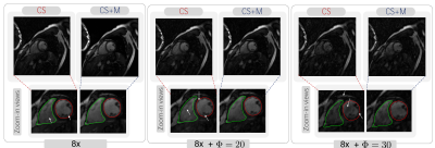
CS+M: A Simultaneous Reconstruction and Motion Estimation Approach for Improving Undersampled MRI Reconstruction.
Angelica I. Aviles-Rivero, Guy B. Williams, Martin J. Graves, Carola-Bibiane Schönlieb
Current research in MRI is based on using CS implications to reconstruct high-quality images from a subset of $$$k$$$-space data acquired in an incoherent manner. In this work, we introduce a mathematical framework for improving undersampled MRI data reconstruction, which we call CS+M, where M stands for motion. The significance here, and unlike existing solutions is that by modeling explicitly and simultaneously the inherent complex motion patterns, given by physiological or involuntary motion, in a CS setting, synergies in a complex variational problem are created. These synergies have clinical potentials in terms of improving image quality while reducing motion artifacts.
|
|
4118.
 |
88 |
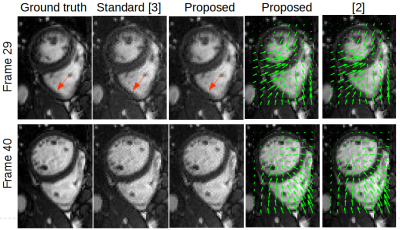
An alternating algorithm for dynamic cardiac MRI reconstruction and motion estimation
Ningning Zhao, Daniel O'Connor, Dan Ruan, Ke Sheng
This work deals with the problem of dynamic MRI reconstruction and motion estimation jointly. Specifically, a multi-scale affine optical flow model is incorporated into the compressed sensing framework. Simulation results demonstrate the efficiency of the proposed algorithm in image reconstruction and motion estimation against the standard CS based method for MRI reconstruction.
|
|
4119.
 |
89 |
3D Motion Estimation and Correction of Motion in Sequential Slices of Kidney Diffusion-Weighted MRI
Sila Kurugol, Bahram Marami, Onur Afacan, Simon Warfield, Ali Gholipour
In this paper we introduced a motion-compensated model estimation technique for renal DW-MRI. The technique has two main components: 1) we adapted an approach based on robust state estimation, which was recently utilized to solve slice-based motion estimation, to track physiological motion (including respiratory motion); 2) we used weighted least squares to estimate diffusion tensor model and calculate diffusion parameters from motion-compensated data. Overall, our method achieved the highest FA values in the medulla, compared to no motion correction and volume to volume registration which resulted in reduced FA values, artifacts, and blurrier FA, MD and AD maps.
|
|
4120.
 |
90 |
Correction of Slice-Specific Nyquist Ghost Artifact for Simultaneous Multi-Slice EPI
Uten Yarach, Oliver Speck
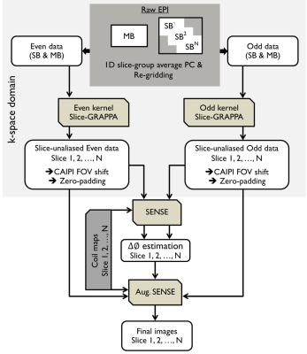
The slice-specific ghosting artifact has been challenging for SMS-EPI. A standard 1D-based phase correction usually leads to increased residual ghosting artifacts in case of subject motion or eddy current variations at different time points. In this study, we propose a dynamic 2D slice-specific phase correction to address the slice-specific phase error. This correction scheme does not require additional time for acquiring 2D phase difference scans. It is a combination between slice-GRAPPA and model-based framework. The result shows that the ghost-to-signal ratios for MB3Ry2 in human brain obtained by the proposed and vendor-provided reconstruction were 1.37% and 2.66%, respectively.
|
|
4121.
 |
91 |
Nyquist Artifact Correction for Multi-band and Multi-shot EPI Using Multiplexed Sensitivity Encoding (MUSE) - A Self-reference 2D Phase Correction Method
Xiaoxi Liu, Di Cui, Edward S. Hui, Queenie Chan, Nan-Kuei Chen, Hing-Chiu Chang
Echo planar imaging (EPI) suffers from Nyquist artifact that is typically corrected by using 1D phase correction with reference scan. In oblique-plane acquisition, 2D phase corrected is warranted to effectively reduce Nyquist artifact. However, Nyquist artifact correction is challenging in the case of multi-band EPI due to the presence of slice-dependent phase error. In this study, we aim to develop a self-reference 2D phase correction method to eliminate the Nyquist artifact in multi-band multi-shot EPI data, which utilizes the recently developed multiplexed sensitivity encoding (MUSE) reconstruction algorithm.
|
|
4122.
 |
92 |
Removal of Pseudo-lesion Artifact in Diffusion-Weighted Echo-Planar Imaging Using Virtual Coil Acquisition and Multiplexed Sensitivity Encoding (MUSE)
Xiaoxi Liu, Edward S. Hui, Hing-Chiu Chang
In routine brain DW-EPI with SENSE, the pseudo-lesion artifact due to residual aliasing of eyeball has been previously reported. We have found that the incidence of pseudo-lesion artifact was over 50% when performing double-oblique imaging on stroke patients. The possible cause is highly likely related to inference between residual Nyquist ghost and unfolding process in SENSE reconstruction. In this study, we propose a self-reference method to effectively remove pseudo-lesion artifact in double-oblique DW-EPI using virtual coil acquisition and multiplexed sensitivity encoding (MUSE). Our proposed method reveals higher image quality, better SNR performance, and lower artifact level than conventional SENSE reconstruction.
|
|
4123.
 |
93 |
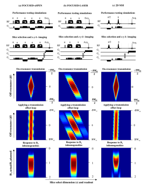
Characterization and suppression of stripe artifact in velocity-selective magnetization-prepared unenhanced MR angiography
Taehoon Shin, Qin Qin
While velocity-selective magnetization-prepared MR angiography (VS-MRA) has shown great promise for diverse arterial territories, artifactual stripes often occur in resultant angiograms. We investigate the formation of the stripe artifact using extended phase graph analysis and show that the stripes contain not only the fundamental frequency determined by the area of VS unipolar gradient but higher-order components with multiples of the fundamental frequency. Based on this finding, we propose and test efficient strategies for suppression of the artifact in a phantom and healthy human subjects.
|
|
4124.
 |
94 |
Metal artifact reduction in MRI-based radiation therapy
Video Permission Withheld
H Michael Gach, Stacie Mackey, Mo Kadbi, Jacqueline Zoberi, Jose Garcia-Ramirez, Yuan (James) Rao, S Murty Goddu, Perry Grigsby, Hiram Gay, Christina Tsien, Jiayi Huang, Jeff Michalski
A large percentage of patients receiving MRI simulations for radiotherapy treatment planning have metal in their bodies. Often the metal is in or near the target or organs at risk. Metal creates susceptibility artifacts that can saturate the tissue signal and distort the tissue geometry. In this case report, examples of the benefits of metal artifact reduction using slice encoding for metal artifact correction (SEMAC) are presented for patients scanned at 1.5 T. Critical regions that were obscured by artifact were restored using SEMAC, thus allowing MRI-based treatment planning.
|
|
4125.
 |
95 |
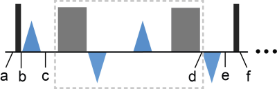
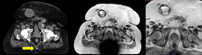
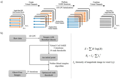
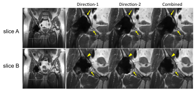
Robust Virtual Coil SAKE with Automatic Rank Thresholding for Removal of EPI Ghost Artifacts
Zhenda Xu, Mengye Lyu, Yilong Liu, Markus Barth, Ed Wu
Ghost artifacts in EPI can arise from phase error between positive- and negative-echo and inter-shot phase error. Virtual coil SAKE (VC-SAKE) can be used to eliminate these phase errors simultaneously, but its performance relies on selection of parameters. In particular, proper rank thresholding is extremely important. In this study, we proposed to automatically determine the optimal rank threshold by minimizing the image entropy, and evaluated its performance with various datasets from different scanner systems.
|
|
4126.
 |
96 |
Clinical Evaluation of Pile-up and Ripple Artifact Suppression Near Metal by Alternating Gradients
Xinwei Shi, Kathryn Stevens, Brian Hargreaves
Multi-Spectral Imaging techniques have been shown to significantly reduce metal-induced artifacts. However, they often suffer from pile-up/ripple artifacts near metal, where metal-induced-off-resonance gradients “cancel” the frequency-encoding gradient. We have previously proposed a method to reduce these artifacts by combining two acquisitions with alternating-sign readout and slice-select gradients. Here we demonstrate that the alternating-gradient method is compatible with robust PCA, which provides 2-fold acceleration beyond parallel imaging and half-Fourier acquisition to compensate for the additional acquisition. Our study shows that the alternating-gradient method can significantly reduce pile-up/ripple artifacts in volunteers with hip and knee arthroplasties, and provides additional diagnostic advantages compared to the standard sequence.
|
|
Encoding & Beyond
Electronic Poster
Acquisition, Reconstruction & Analysis
Tuesday, 19 June 2018
| Exhibition Hall |
13:45 - 14:45 |
| |
|
Computer # |
|
4127.
 |
97 |
fMRI at 7 Tesla with 0.5mm Isotropic Resolution and Full Field of View
Patrick Liebig, Robin Heidemann, Bernhard Hensel, Yuehui Tao, Wei Liu, David Porter
fMRI protocols at ultra-high field typically use pixel sizes below 1mm. With single-shot EPI, this results in a prolonged readout train relative to the T2* decay time, resulting in image blurring and a limit to the true resolution that can be achieved. This effect can be mitigated by using multi-shot EPI to reduce the echo-train length, but this is associated with a reduction in temporal SNR. Previous work demonstrated a less severe reduction in temporal stability with readout-segmented EPI than with interleaved EPI. This study investigates the application of readout-segmented EPI to ultra-high resolution fMRI of the motor cortex.
|
|
4128.
 |
98 |
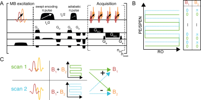
A CAIPIRINHA-based approach to the referenceless reconstruction of high-definition SPEN images with simultaneous multislice
Gilad Liberman, Samuel Cousin, Eddy Solomon, Lucio Frydman
Spatiotemporal Encoding (SPEN) is an ultrafast imaging technique where the low-bandwidth axis is rasterized in a joint spatial/k-domain. SPEN benefits from increased robustness to inhomogeneities, folding-free reconstruction of subsampled data, and an ability to combine multiple interleaved or signal averaging scans in a referenceless fashion. SPEN’s relatively high SAR, however, complicates its volumetric uses. Here we show how this can be solved by merging a controlled aliasing for parallel imaging (CAIPIRINHA) protocol involving phase-cycling of multi-banded excitation pulses in independent scans, so as to enable a referenceless reconstruction of interleaved multislice acquisitions delivering high in-plane definition and excellent inter-slice decoupling.
|
|
4129.
 |
99 |
Constrained Lossy Compression for MR Raw Data Transmission
Matthew Restivo, Adrienne Campbell-Washburn, Peter Kellman, Hui Xue, Rajiv Ramasawmy, Michael Hansen
Computationally intensive image reconstruction algorithms can be made accessible to the diagnostic workflow by streaming data to remote workstations in real-time. Due to bandwidth constraints, data compression is an important tool to ensure that network transmission is not a bottleneck. However, since image quality losses are unacceptable for clinical MRI, it is important to constrain any compression losses below the thermal noise.
Here we propose a framework for online data compression based on constraining SNR loss using a custom-built compression library. Greater than 5-fold data reduction was achieved by accepting a negligible SNR loss.
|
|
4130.
 |
100 |
Anisotropic Field-of-Views in 3D Golden Angle Radial Imaging
Guruprasad Krishnamoorthy, Jouke Smink, Marc Kouwenhoven, Marcel Breeuwer
3D Golden angle Radial sequence (3D-GArad) permits reconstruction with varying temporal / spatial resolution from the same dataset. It is less sensitive to motion, flow artifacts and supports ultra-short echo times. It supports self-gated retrospective motion correction techniques. One of the limitations of the current implementations of 3D-GArad is, it only supports isotropic field-of-view (FOV), which leads to redundant sampling and increased scan time when the imaging volume has anisotropic dimensions. In this work, we have developed a method based on the work by Larson et al., to support anisotropic FOV with different FOV-shapes for 3D-GArad.
|
|
4131.
 |
101 |
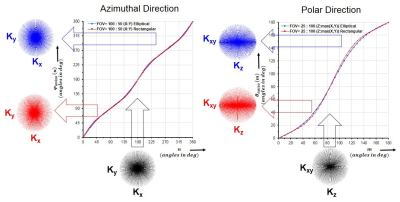
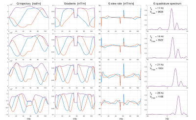
A quadrature filter approach to diffusion weighted imaging with application in pore size estimation
Hans Knutsson, Filip Szczepankiewicz, Cem Yolcu, Magnus Herberthson, Evren Özarslan, Markus Nilsson, Carl-Fredrik Westin
We present a novel approach to gradient waveform design for diffusion weighted MRI. The approach is founded on the temporal frequency domain formulation of the signal attenuation. The objective is to obtain a set of circularly polarized oscillating gradient waveforms that are optimal in terms of b-value, frequency selectivity and in-plane rotational invariance. A new pore size estimation algorithm is presented. We show phantom and in vivo results based on MRI scans using an opimized waveform set.
|
|
4132.
 |
102 |
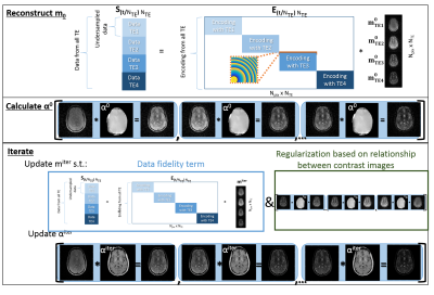
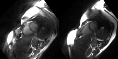
In vivo O-space Turbo Spin Echo Imaging with New Reconstruction Algorithm
Nadine Dispenza, Hemant Tagare, R. Constable, Galiana Gigi
We report the first in-vivo T2 weighted O-space TSE images, and demonstrate that a single TSE dataset can generate T2w images for any of the echo times while still tolerating further undersampling from parallel imaging. In addition to reconstructions based on a refinement of a previously introduced filtering algorithm, we introduce a more general and rigorous reconstruction approach that exploits the geometric relationship between images in the T2w series. The proposed reconstruction algorithm reduces T2 blur and improves contrast agreement to Cartesian TSE images.
|
|
4133.
 |
103 |
Enhancing Spatial-Temporal Resolution in Simultaneous Multi-Slab Echo Volumar Imaging
Kishore Vakamudi, Steen Moeller, Sudhir Ramanna, Akio Yoshimoto, Essa Yacoub, Ricardo Otazo, Alam Syed, Stefan Posse
We develop multi-band echo-volumar-imaging (MB-EVI) by combining multi-slab encoding with EVI to investigate BOLD sensitivity, resting-state connectivity in different frequency bands, and spatial-temporal resolution limits. We also study the effect of compressed sensing (CS) reconstruction on fMRI sensitivity. Mapping of major resting-state networks with 3mm3 voxel size was feasible in multiple frequency bands. High spatial resolution (2mm3) improved delineation of neuroanatomy and enabled sensitive mapping task-based activation. CS with 2.4-fold undersampling showed negligible loss in image quality and moderate region-specific losses in BOLD sensitivity. MB-EVI provides flexibility for maximizing spatial-temporal resolution, volume-coverage and BOLD-sensitivity for mapping task-activation and functional connectivity.
|
|
4134.
 |
104 |
Respiration resolved imaging using continuous steady state multiband excitation with linear frequency sweeps
Laurence Jackson, Anthony Price, Jana Hutter, Lucilio Cordero-Grande, Alison Ho, Paddy Slator, Ana Dos Santo Gomes, Joshua van Amerom, Maria Murgasova, Laura McCabe, Mary Rutherford, Joseph Hajnal
Free-breathing abdominal MRI is challenging due to the unpredictable and complex 3D deformation caused by respiration. We present a novel acquisition where short TR single-band or multiband slice acquisitions are swept smoothly across the anatomy of interest while preserving steady state conditions. This is achieved by adding a frequency offset to successive RF pulses shifting the excited slice by fraction of slice thickness for each acquired k-space. This creates a highly efficient acquisition with high redundancy in respiration. A method is presented to produce 4D respiration-resolved volumes from this data and examples of human placenta and kidney are presented.
|
|
4135.
 |
105 |
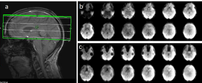
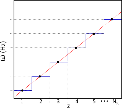
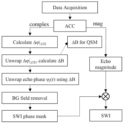
SWI+: A robust artifact-free SWI procedure with improved contrast
Yongquan Ye, Jinguang Zong, Jingyuan Lyu, Weiguo Zhang
A robust image reconstruction and processing strategy to achieve minimal susceptibility artifact for SWI is developed and demonstrated. With a multi-echo GRE data acquisition and a newly developed robust rapid phase unwrapping algorithm, an enhanced version of SWI imaging procedure, namely SWI+, is developed to address the pitfalls in classic SWI, which collects only one echo and simply employs high pass filtering on phase images.
|
|
4136.
 |
106 |
Reducing eddy currents in radial cardiovascular MRI using tiny 3D golden-angle
Alexander Fyrdahl, Karen Holst, Martin Ugander, Andreas Sigfridsson
Three-dimensional double golden-angle radial imaging has shown great potential in free-breathing whole-heart cardiac cine imaging using data binning, and respiratory resolved cardiac imaging. However, the original golden-angle trajectory involves large jumps in k-space, which leads to eddy current artifacts in bSSFP imaging. For successful binned cardiac imaging, two conditions must be fulfilled; first, the trajectory should provide high k-space uniformity, and second, the trajectory should avoid eddy current induced degradation of image uniformity. The purpose of this work was to develop a double golden-angle radial trajectory that fulfills both of these conditions.
|
|
4137.
 |
107 |
Model-based Reconstruction with Automatic Scaling for Real-Time Phase-Contrast Flow MRI with Complementary Sets of Radial Spokes
Zhengguo Tan, Jost Kollmeier, Arun Joseph, Oleksandr Kalentev, Dirk Voit, Klaus-Dietmar Merboldt, Thorsten Hohage, Jens Frahm
Real-time phase-contrast flow MRI is extended from sequential acquisitions of flow-compensated and flow-encoded data with the same set of radial spokes to interleaved acquisitions with different radial spokes oriented by a small Golden angle, thereby improving spatial accuracy and reducing streaking artifacts. To apply model-based reconstructions for this sampling scheme, an automatic scaling of unknowns is proposed, which is capable of balancing partial derivatives and regularizations during the iterative nonlinear inversion.
|
|
4138.
 |
108 |
Hybrid O-Space and FRONSAC Imaging
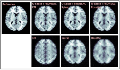
Haifeng Wang, Yuchou Chang, Dong Liang
Nonlinear spatial encoding magnetic (SEM) fields can accelerate data acquisitions and improve the imaging quality. In this work, the O-Space and FRONSAC imaging are combined into a hybrid nonlinear spatial encoding approach with dynamic nonlinear gradients. The preliminary experiment of phase mapping shows that the proposed method can be implemented in the current O-Space system. Simulations based on the preliminary experiment demonstrate that this approach can accelerate data acquisitions and reduce artefacts caused by highly undersampling acquisitions.
|
|
4139.
 |
109 |
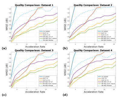
MR Image Reconstruction via Denoising (MR-RED)
Adam Rich, Rizwan Ahmad
In this work, the feasibility of employing denoising to recover MR images from undersampled data is demonstrated. By embedding denoisers into the Laplacian-based regularization functional and solving the resulting optimization problem, state-of-the-art results are achieved. Performance of several denoisers and compressed sensing methods is compared in four cardiac MRI datasets. We show that denoising-based reconstruction can outperform soft-thresholding-based algorithms in terms of normalized MSE.
|
|
4140.
 |
110 |
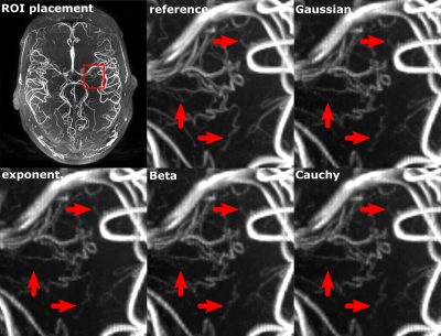
Optimizing Cartesian compressed sensing for ultra-high resolution Time of Flight angiography
Hendrik Mattern, Oliver Speck
Ultra-high resolution Time of Flight (ToF) angiography requires long scan times. Compressed sensing (CS) can reduce the scan time but requires pseudo-random sampling and may blur image details. We present a framework to create Cartesian pseudo-random sampling patterns from any given probability density function and to evaluate quantitatively vessel depiction with CS reconstruction. Results from 0.30 mm and 0.15 mm isotropic ToF data show that moderate acceleration with CS can reduce the scan time considerably while providing vessel depiction similar to the non-accelerated reference.
|
|
4141.
 |
111 |
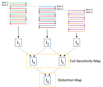
Improved Parallel Echo-Planar Imaging (EPI) with Ghost Removal and Distortion Correction
Yuan Zheng, Yu Ding, Xiaodong Ma, Weiguo Zhang
We have developed a procedure for reconstructing high-quality EPI images with parallel imaging, and presented both in-vitro and in-vivo results. Interleaved EPI and the PLACE technique are used to generate coil sensitivity maps (CSM) and a distortion map. These maps do not suffer from ghosting and match the distortion of the imaging data. The CSM are used in the PEC-SENSE reconstruction to generate images with significantly reduced ghosting artifacts. Geometric distortions are subsequently corrected using the distortion map. This technique can be used to improve the image quality of many EPI applications, such as diffusion imaging, when parallel imaging is involved.
|
|
4142.
 |
112 |
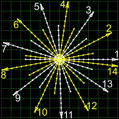
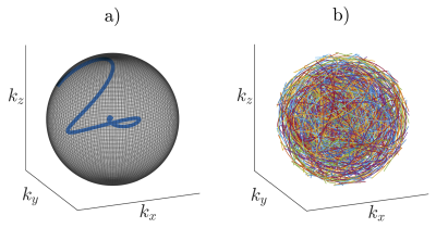
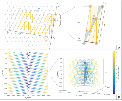
Efficient $$$k$$$-space coverage using a 3D accelerated low-discrepancy trajectory
Tobias Speidel, Volker Rasche
Acquiring three-dimensional (3D) images in MRI is a time consuming process. The overall duration of acquiring a Nyquist sampled 3D dataset can be significantly shortened by enhancing the efficiency of $$$k$$$-space sampling. This can be achieved by accelerating each $$$k$$$-space read-out and by additionally increasing the coverage of $$$k$$$-space for every trajectory interleave. In this work, we present a 3D $$$k$$$-space trajectory that is highly accelerated in terms of $$$k$$$-space velocity that leads to a low-discrepant coverage of suchlike using a considerably reduced number of read-outs.
|
|
4143.
 |
113 |
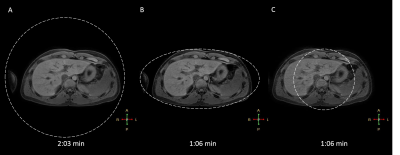
3D Golden Angle Stack-of-Stars with Anisotropic Field-of-View
Joao Tourais, Guruprasad Krishnamoorthy, Marc Kouwenhoven, Jouke Smink, Marcel Breeuwer
Radial sampling techniques are often used in dynamic MRI because they are robust to flow and motion, support short echo times, and have a diffuse aliasing pattern. However, standard implementations of radial imaging do not support in-plane anisotropic FOV, which leads to sampling redundancy when the object being imaged has anisotropic in-plane dimensions (e.g. abdomen, chest, spine, leg, etc.). In this work we demonstrate the feasibility of 3D golden angle stack-of-stars acquisition with an in-plane anisotropic FOV in abdominal acquisitions.
|
|
4144.
 |
114 |
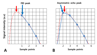
Asymmetric-echo-based Ultrashort Echo-time (ASE-UTE) Imaging
Jinil Park, Jeongtaek Lee, Jang-Yeon Park
The first 3D-UTE sequence used FID acquisition techniques using ramp sampling to minimize TE, but defining the center of k-space precisely was a difficult problem (true-k0). In case of echo acquisition containing echo peaks, it is better to define true-k0 than FID acquisition although true-k0 problem still exists because echo peaks often do not agree with true-k0 due to the echo shifts caused by system imperfections such as gradient delays and eddy currents. Here, we propose an asymmetric-echo-based 3D-UTE (ASE-UTE) sequence where echo peaks are acquired on the ramp.
|
|
4145.
 |
115 |
Simultaneous Multi-slice Ultra-short Echo Time Imaging Using POMP Half Pulses and CAIPI
Christoph Rettenmeier, V. Andrew Stenger
Phase offset multi-planar (POMP) encoding is applied in a simultaneous multi-slice (NSMS = 4) excitation scheme using half sinc pulses. In combination with a gradient echo 2D spiral readout trajectory and controlled aliasing in parallel imaging (CAIPI) via a model-based reconstruction a fast ultra-short echo time (UTE) sequence is developed. Sequence details and performance tests on short T2* phantoms are presented.
|
|
4146.
 |
116 |
Improvement of 2D-multi-slice, multi-shot Cartesian T2-weighted TSE using golden angle shot reordering
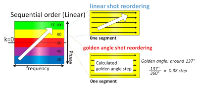
Yuki Furukawa, Takashige Yoshida, Masami Yoneyama, Kouhei Yuda, Mariko Okura, Nobuo Kawauchi
2D multi-slice, multi-shot Cartesian TSE sequence is frequently used for routine MRI but it is sensitive to motion. We evaluated the feasibility of golden angle shot reordering for 2D-multi-shot TSE with 60 patients. Golden angle shot reordering reduced effectively ghost artifacts by randomly filling in each segment of Cartesian k-space. We demonstrated that golden angle shot reordering intrinsically has a motion robustness without penalty for image quality and scan time. This technique might be useful for all anatomies in routine clinical examination..
|
|
4147.
 |
117 |
APIR4EMC: Autocalibrated Parallel Imaging Reconstruction for Extended Multi-Contrast Imaging
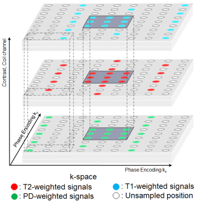
Chaoping Zhang, Alexandra Cristobal-Huerta, Juan Antonio Hernandez-Tamames, Stefan Klein, Dirk Poot
Multi-contrast images of the same region are routinely acquired in clinical MRI. This abstract proposes a fast reconstruction method for multi-contrast imaging: Autocalibrated Parallel Imaging Reconstruction for Extended Multi-Contrast (APIR4EMC). Unlike conventional parallel imaging (GRAPPA) which reconstructs the image for individual contrast, APIR4EMC additionally includes the inter-contrast signals in the prediction of unsampled positions in the k-space of each contrast and simultaneously reconstructs images for all contrasts. Experiments show significant improvement of APIR4EMC compared to conventional parallel imaging in terms of artifacts and SNR.
|
|
4148.
 |
118 |
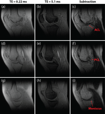
Wind Out-In Dual-Echo Yarn-Ball with Application to Knee Imaging
Robert Stobbe, Christian Beaulieu
3D centre-out Yarn-Ball k-space acquisition is implemented in a wind-out/wind-in dual-echo format for the first time. As with standard 3D-Radial acquisition this technique facilitates short (first ‘echo’) TE with utility for imaging tissues with short T2*. However, the advantage of centre-out Yarn-Ball is much greater k-space sampling efficiency than 3D-Radial. Smooth variation from wind-out to wind-in minimizes potential errors in k-space trajectory evolution as a result of eddy-currents. Spoiled-steady-state dual-echo 3D Yarn-Ball images with 0.7x0.7x0.7 mm3 voxels were acquired from the knee of a healthy volunteer in 6 minutes, and the difference image shows ligament and meniscus conspicuity.
|
|
4149.
 |
119 |
A rapid 3D spiral readout with uniform sampling density and smooth T2* weighting
Maria Engel, Lars Kasper, Christoph Barmet, Thomas Schmid, Klaas Pruessmann
A stack of disks trajectory for fast 3D acquisitions with long readouts is introduced and compared to interleaved stack of spiral and Yarn-ball trajectories. Isotropic 1.6 mm whole-brain coverage is achieved in less than a second.
|
|
4150.
 |
120 |
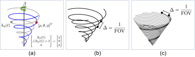
Redesigned Cones Trajectory based on Discretization of Cones Coordinate
Kwang Eun Jang, Dwight Nishimura, Shreyas Vasanawala
We propose a new non-Cartesian sampling scheme based on a 3D cones k-space trajectory. The design framework uses a cones coordinate system that uniquely represents a point in 3D k-space with a rotation of a spiral that resides on a conic surface. The new trajectory is obtained by discretizing the proposed coordinate system. Incorporation of a variable-density spiral improves the sampling efficiency. Simulations and phantom studies demonstrate the improved efficiency of the new scheme compared to the conventional cones design.
|
|
System Imperfections & Artifacts: Characterization & Correction
Electronic Poster
Acquisition, Reconstruction & Analysis
Tuesday, 19 June 2018
| Exhibition Hall |
14:45 - 15:45 |
| |
|
Computer # |
|
4199.
 |
49 |
Variability of B1+ and B0 fields in the human brain at 7T.
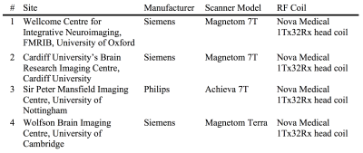
William Clarke, Olivier Mougin, Ian Driver, Catarina Rua, Susan Francis, Richard Bowtell, Richard Wise, Adrian Carpenter, Keith Muir, Stuart Clare
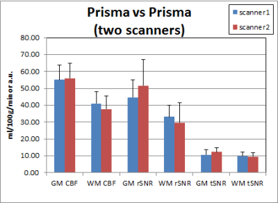
B1+ and B0 whole brain field-maps were collected from three human subjects, on four different 7 tesla scanners. Three different scanner systems from two different vendors were used; all acquisitions used the same RF coil. The data was used to assess the inter-subject and inter-scanner variability, and to calculate typical field values over a range of different brain regions. Systematic difference in B1+ calibration and B0 shimming between scanners were found, necessitating user validated B1+ calibration and B0 shimming for quantitative multi-site studies.
|
|
4200.
 |
50 |
Comparison of ASL quantification results across 3T scanners with different hardware/software – insight for multisite ASL studies
Yu Fen Chen, James Higgins, Todd Parrish
Combination of data across different scanner platforms is a major concern for multisite studies. The quantitative nature of arterial spin labeling (ASL) is thought to be free from such concerns, but our results on data collected on the same scanner before and after hardware upgrade demonstrate that ASL is also subject to this confound. Normalization to a reference region greatly reduces variation even for non-quantitative data, and is a recommended approach for removing scanner-related variations.
|
|
4201.
 |
51 |
Initial Investigation of Multi-Probe Magnetic Field Monitoring on a Whole-Body Human System Operating at 10.5 Tesla: Application to Spin Echo Diffusion Weighted EPI
Video Permission Withheld
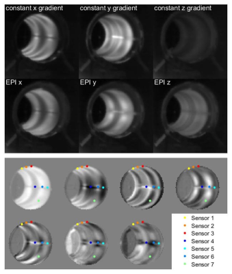
Ruoyun Ma, Edwards Auerbach, Kamil Ugurbil, Pierre-François Van de Moortele
DW-EPI is subjected to image distortion induced by eddy currents. Magnetic field camera has been shown to provide corrected reconstruction based on accurate measurement of actual k-space trajectories. In this study, initial effort was made to implement this approach in the context of a whole body human system operating at 10.5T where susceptibility challenges are greater. After characterization of the probe behavior and careful design of the probe signal acquisition scheme, field evolution during the EPI readout was mapped. Image reconstruction corrected with the measured encoding information demonstrated a significant reduction of image distortion in DW-EPI.
|
|
4202.
 |
52 |
Motion Encoding Gradient Nonlinearity Correction for Magnetic Resonance Elastography
Joshua Trzasko, Arvin Arani, Philip Araoz, Matt Bernstein, Richard Ehman, John Huston III, Yunhong Shu, Ek Tsoon Tan
Due to engineering limitations, the spatial encoding gradient fields produced by an MRI scanner are never perfectly linear. Gradient non-ideality is typically associated with image geometric distortion, but it also imparts spatially varying bias in the signal generated during gradient-based motion encoding. In this work, we theoretically investigate how motion encoding gradient nonlinearity affects MR elastography and develop a corrective strategy applicable to any MRE protocol.
|
|
4203.
 |
53 |
Image Distortion Correction via Gradient Nonlinearity Encoding at 9.4T MRI with Radial Ultra short TE (UTE) Sequence
Shanshan shan, Mingyan Li, Haiwei Chen, Feng Liu, Stuart Crozier
Radial-encoding sequences can be more sensitive to gradient trajectory deviations caused by gradient nonlinearity (GNL) than Cartesian encoding. In this work, we developed a method to alleviate image distortion caused by GNL specifically for radial-encoding. Experimental results acquired from a 9.4 T pre-clinical MRI scanner with using Ultra-short TE sequence were used to validate the proposed method.
|
|
4204.
 |
54 |
Distortion measurements empirically validate gradient model impact on diffusion weighting
Dariya Malyarenko, Enzo Barberi, Yuxi Pang, Julien Sénégas, Ajit Devaraj, Johannes Peeters, Teodor Stanescu, Thomas Chenevert
Distortion fields induced by gradient nonlinearity were measured for two channels of two MRI systems to validate vendor-provided gradient design models intended for correcting spatially nonuniform diffusion weighting. Excellent agreement was observed between the model and the empiric displacement measurements recast into first two diagonal elements of system nonlinearity tensor for both systems. The measurements were sensitive enough to detect sub-millimeter differences between nominally symmetric right-left and anterior-posterior gradients. Unexplained minor deviation from spatially symmetric design was detected for right-left gradient of both systems by diffusion and distortion measurement.
|
|
4205.
 |
55 |
Concomitant Field Compensation Improves Quality of Clinical Fast Spin Echo Images Acquired on an Asymmetric MRI Gradient System
Shengzhen Tao, John Huston III, Erin Gray, Joshua Trzasko, Myung-Ho In, Yunhong Shu, Matt Bernstein
The fast-spin-echo (FSE) acquisitions are the workhorse for routine MRI, but can be affected by concomitant field (CF)-induced phase errors. Recently, a compact 3T (C3T) MRI platform equipped with an asymmetric, high-performance gradient was developed. The asymmetric design of this gradient induces zeroth/first-order CFs (in addition to the second-order CF on conventional whole-body gradients), which can degrade image quality of FSE acquisitions. We have previously developed real-time gradient pre-emphasis and frequency shifting techniques to compensate for these additional CFs. Here, we demonstrate that these compensations significantly improve image quality of FSE using a clinical T2w-FSE protocol and a preliminary dataset.
|
|
4206.
 |
56 |
Fast eddy current characterization for the development of in-bore devices
Video Permission Withheld
David Brunner, Betram Wilm, Simon Gross, Benjamin Dietrich, Christoph Barmet, Klaas Pruessmann
Devices operating in the magnet bore, such as coil arrays, receivers or shim inserts become ever more sophisticated as well as the employed image acquisition schemes. However, quantitative testing for their influence on the gradient switching performance is often difficult and time consuming. In this work we present a methodology for measuring, evaluating and predicting eddy current effects fast and quantitatively, employing a dynamic magnetic field camera and a framework of linear time invariant system characterization. For demonstration, standard components and several frequently applied RF coils are assessed.
|
|
4207.
 |
57 |
Extending the Simple Method for Adaptive Gradient-Delay Compensation in Radial MRI
Sebastian Rosenzweig, Hans Christian Martin Holme, Robin Wilke, Martin Uecker
Lately, radial k-space trajectories have become very popular especially for fast MRI. However, radial sampling is prone to streaking artifacts caused by gradient delays. Here, we propose two extensions for a simple but powerful method that compensates gradient delays by estimating the corresponding sample shift using cross-correlation of opposed spokes. First, we show that the opposed spokes don't have to be acquired in calibration scans but can be taken directly from the actual measurements. Second, we show that it is also possible to generate synthetic spoke-pairs for gradient delay estimation.
|
|
4208.
 |
58 |
Measuring Eddy Currents Induced by Switching Gradient/Shim Currents
Paul Chang, Sahar Nassirpour, Anke Henning
In this work, we measured eddy currents for a very high order B0 shim system. The eddy currents were measured using two methods: a low-resolution B0 mapping sequence and a NMR field camera. The high temporal resolution of the field camera allowed the eddy currents to be corrected using a digital pre-emphasis setup.
|
|
4209.
 |
59 |
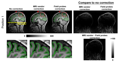
Using field monitoring to map and correct image distortions due to gradient nonlinearities
Ying-Hua Chu, Yi-Cheng Hsu, Fa-Hsuan Lin, Maxim Zaitsev
We demonstrate how a field probe array can be used to measure gradient nonlinearities. Brain imaging results show that the distortion was most prominent at image voxels away from the iso-center. Compared to MRI vendor’s solution, our measurements and reconstruction could further reduce the image distortion by 35%.
|
|
4210.
 |
60 |
Improved thermal modelling and prediction of gradient response using sensor placement guided by infrared photography
Jennifer Nussbaum, Bertram Wilm, Benjamin Dietrich, Klaas Pruessmann
In MRI, the dynamics of gradient fields can be predicted by assuming that the system is linear and time-invariant. However, time-invariance can be violated by thermal effects. To address this shortcoming, it has been proposed to expand the GIRF model by thermal variation parametrized with the help of temperature sensors. It has remained open how many relevant thermal degrees of freedom the system actually had and where the matching number of sensors should be placed to capture them. In the present work, we address these questions using infrared photography and principal component analysis of gradient heating. Response modelling is additionally enhanced by accelerating GIRF measurements and including cross-terms. The improvements are shown in the prediction of gradient waveforms for image reconstruction and in the effects on the images.
|
|
4211.
 |
61 |
Reduction of artefacts in Bloch-Siegert based $$$B_1^+$$$-Mapping in CPMG-Sequences with imperfect refocussing pulses
Video Permission Withheld
Volker Sturm, Thomas Kampf, Lukas Buschle, Ke Zhang, Peter Jakob, Mirko Pham, Christian Ziener, Martin Bendszus, Sabine Heiland, Felix Kurz
The Bloch-Siegert-effect imprints the rf-coil’s transmission field in the phase of the acquired signal. Consequently the measured amplitude information can be used to determine other MR-Parameter. A previously published sequence based on a CPMG spin echo train uses both information pathways and allows simultaneous acquisition of $$$T_2$$$- and $$$B_1^+$$$-maps. However, the formerly proposed method produces potentially erroneous $$$B_1^+$$$-map in inhomogeneous $$$B_0$$$-fields.
This work presents a method to reduce those errors by combining signal from different echoes. This allows a robust measurement of $$$B_1^+$$$-maps in inhomogeneous $$$B_0$$$-fields as present in high-field MRI with simultaneous $$$T_2$$$ quantification.
|
|
4212.
 |
62 |
Mapping B0 inhomogeneities through a self-contained analysis of single-shot 2D MRI data
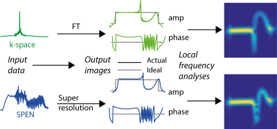
Gilad Liberman, Samuel Cousin, Lucio Frydman
Ultrafast MRI is susceptible to main field distortions ΔB0 that affect these images’ quality and faithfulness –particularly along the low-bandwidth dimension. This work shows that these distortions can be mapped from the spatial images themselves, without requiring additional information. This information becomes available from a time-frequency analysis of the signals, after freeing them from phase-wrapping complications. This hypothesis is explained, and results are demonstrated using both simulations and single-shot human brain images collected by spatiotemporal encoding (SPEN) techniques. The method opens a route to enhancing SPEN’s and EPI’s robustness to field inhomogeneities.
|
|
4213.
 |
63 |
Accurate Slice-Selective MRI near Metallic Implants with FOCUSED-xSPEN
Gil Farkash, Gilad Liberman, Lucio Frydman
Metals frequently cause dramatic spatial inhomogeneities in the static field B0. This causes severe localization errors, including slice z-displacements and slice-thickness variations. Clinically used techniques often avoid such errors by adding a phase encoding loop in z, prolonging examination times. This study examines a novel Fully refOCUSED cross-term SPatio-temporal ENcoding (FOCUSED-xSPEN) approach, as a slice selection technique for 2D MRI in the presence of metal implants. The method relies on xSPEN’s proven immunity to in-plane distortions caused by B0 heterogeneities, to design a new excitation scheme that delivers faithful 2D slices near implants with reduced scan times.
|
|
4214.
 |
64 |
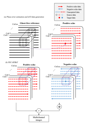
EPI Nyquist ghost removal by incorporating iterative k-space based phase error estimation and correction with SPIRiT (PEC-SPIRiT)
Yilong Liu, Mengye Lyu, Victor Xie, Ed Wu
EPI acquisition suffers from Nyquist ghost due to inconsistency between opposite readout directions. As proposed in our previous work, such inconsistency induced phase error can be removed by incorporating the Phase Error Correction with SENSE (PEC-SENSE). In this study, we employ a k-space based phase error estimation, and remove the EPI Nyquist ghost by incorporating phase error correction with SPIRiT (PEC-SPIRiT). Here, a FLASH scan is used for calibration. The proposed method was evaluated using both phantom and in vivo studies, demonstrating its robustness against distortion mismatch and at high accelerations.
|
|
4215.
 |
65 |
Undersampled Spiral Magnetic Resonance Fingerprinting with Water and Fat Blurring Correction
Teresa Nolte, Daniel Truhn, Nicolas Gross-Weege, Mariya Doneva, Peter Koken, Aaldert Elevelt, Volkmar Schulz
In the presence of aqueous and fatty tissues, Magnetic Resonance Fingerprinting (MRF) acquisitions with spiral readout suffer from blurring artifacts. We propose to correct undersampled spiral MRF data by combining MRF with a Dixon acquisition and a subsequent conjugate phase reconstruction correction. With the proposed method, the blurring artifacts are removed from the MRF data. T1 and T2 parameter maps with improved homogeneity are obtained.
|
|
4216.
 |
66 |
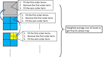
An efficient eddy current correction method for fat quantification by bipolar readout sequence
Chao Zou, Chuanli Cheng, Yangzi Qiao, Hao Peng, Changjun Tie, Qian Wan, Xin Liu, Hairong Zheng, Min Pan
Multi-echo GRE sequence with bipolar readout gradients can reduce the achievable echo spacing compared to unipolar readout gradients for fat fraction (FF) quantification, and has higher acquisition efficiency. The eddy current induced phase (EC-phase) in bipolar sequence corrupts the phase consistency between the echoes, making it one of the confounding factors for the accurate fat quantification. In this study, we proposed an accurate EC-phase estimation method in bipolar acquisition sequence for FF quantification. The proposed method is validated through comparing the FF quantification from the EC-phase corrected bipolar data to the well-established unipolar acquisition through phantom experiments.
|
|
4217.
 |
67 |
Fat Shift Correction in Bipolar Multi-Echo Dixon Imaging using Water-Fat Separation in k-Space
Holger Eggers
Fat is shifted relative to water in opposite directions in the odd and even single-echo images obtained with bipolar multi-echo sequences. Considering this effect in a water-fat separation permits suppressing associated artifacts, but has required regularization so far to cope with a locally ill-conditioned inverse problem in k-space. In this work, an alternative approach to limiting noise amplification in the water-fat separation is proposed and explored, which promises to avoid the loss of image sharpness observed with regularization.
|
|
4218.
 |
68 |
Single echo radial random alternating TE acquisition for B0 field inhomogeneity robust fat suppression
Dongyeob Han, Taehwa Hong, Dong-Hyun Kim
A single echo method which provides B0 field inhomogeneity robust fat suppression was proposed using a radial acquisition with random alternating echo time. To validate this study, simulation and in vivo experiments were performed. This technique would be useful in fat suppressed applications (e.g., MSK, liver, ...) which are required fast scan and robustness of B0 field inhomogeneity.
|
|
4219.
 |
69 |
Accelerated Imaging of Metallic Implants Using a 3D Convolutional Neural Network
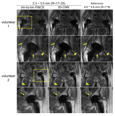
Xinwei Shi, Kathryn Stevens, Brian Hargreaves
Multi-Spectral Imaging (MSI) methods, such as SEMAC and MAVRIC-SL, resolve metal-induced field perturbations by applying additional encoding in the spectral dimension, at the cost of increased scan time. In this work, we introduce a 3D-CNN-based reconstruction to accelerate MSI utilizing spatial-spectral features of aliasing artifacts. We demonstrate in in vivo experiments that the proposed method can accelerate MAVRIC-SL acquisitions by a factor of 3 when used alone, and 17-25 when combined with parallel imaging and half-Fourier acquisition. The 3D-CNN showed significant improvement in image quality compared with parallel image and compressed sensing (PI&CS), with negligible additional computation time.
|
|
4220.
 |
70 |
bin-SENSE: Accelerated MRI Near Metal With No Additional Hardware
Philip Lee, Xinwei Shi, Daehyun Yoon, Evan Levine, Brian Hargreaves
MAVRIC-SL suppresses artifacts induced by field inhomogeneities induced by metal, but requires increased scan time due to the acquisition of a fourth spectral dimension. We accelerate MAVRIC-SL acquisitions limited to the body coil by exploiting fieldmap information implicitly provided by the MAVRIC-SL acquisition. SENSE is applied to undo the coherent aliasing, using the spectral profiles as the spatial weights required for matrix inversion. A region growing method is applied to estimate the fieldmap and spectral profiles from uniformly undersampled data. We demonstrate 1.3x retrospective acceleration at a minimal g-factor penalty with no additional hardware requirement.
|
|
4221.
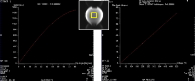 |
71 |
A fast quality assessment and follow-up of transmit and receive coil paths
Franck Mauconduit, Chantal Ginisty, Séverine Desmidt, Séverine Roger, Lionel Allirol, Lucie Hertz-Pannier, Alexandre Vignaud
To operate a MRI system within the best conditions of reproducibility, Quality Assurance (QA) is mandatory to prevent defects and to detect abnormalities in subparts of the system. When using third-party or home-made coils, it is particularly important to guaranty stability of transmit and receive coil paths. For this purpose, we implemented a fast and user-friendly integrated assessment and follow-up to prevent or quickly detect failures.
|
|
4222.
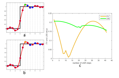 |
72 |
Interlaced Total Variation for Gibbs Artifact Reduction in the Presence of Zero Padding
Kaixuan Zhao, Zhongbiao Xu, Feng Huang, Yingjie Mei, Yanqiu Feng
The Gibbs artifact can be effectively eliminated by searching the local sub-voxel shifts (LSS) that minimize total variations, but it is only applicable to images reconstructed without zero padding. This work extends the LSS method to Gibbs artifact reduction in the presence of zero padding by introducing an interlaced total variation (iTV). The simulation and in vivo experimental results demonstrate that the proposed method can effectively remove Gibbs artifact in the presence of zero padding.
|
|
Pulse Sequences & Beyond
Electronic Poster
Acquisition, Reconstruction & Analysis
Tuesday, 19 June 2018
| Exhibition Hall |
14:45 - 15:45 |
| |
|
Computer # |
|
4223.
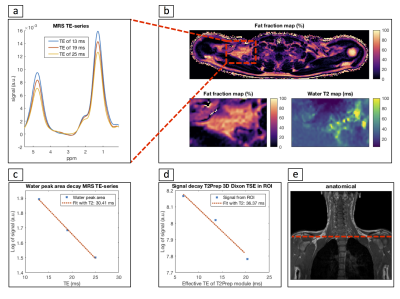 |
73 |
In-vivo water T2 mapping in tissues containing water and fat using a T2-prepared 3D Dixon TSE sequence and a pre-calibrated fat spectrum model
Dominik Weidlich, Maximilian Diefenbach, Sarah Schlaeger, Andreas Hock, Stefan Ruschke, Dimitrios Karampinos
Water T2 is seen as a potential biomarker for brown adipose tissue presence and as a marker of disease activity in patients with neuromuscular disorders. Measuring water T2 in tissues containing water and fat is challenging due to the need to separate the effects of water and fat during T2 quantification. The present work proposes an adiabatic T2 prepared 3D TSE Dixon sequence in combination with a novel DIXON water-fat decomposition method and demonstrates in vivo its feasibility to measure water T2 of brown adipose tissue in the supraclavicular fossa.
|
|
4224.
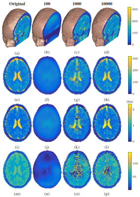 |
74 |
3D CSI-Electrical Properties Tomography
Reijer Leijsen, Wyger Brink, Cornelis van den Berg, Andrew Webb, Rob Remis
The goal of Contrast Source Inversion – Electrical PropertiesTomography is to iteratively reconstruct the conductivity and permittivity of tissue from $$$B_1^+$$$ field data. Up to now, this method has only been implemented in two dimensions. Here we demonstrate a proof-of-principle of a three-dimensional (3D) extension, thereby potentially turning CSI-EPT into a volumetric EPT technique. We present the basic 3D CSI-EPT equations, illustrate the performance of 3D CSI-EPT for realistic 3D body models, and show that accurate reconstructions are obtained even at tissue boundaries. The influence of the magnitude of the electric field on the reconstructions is also discussed.
|
|
4225.
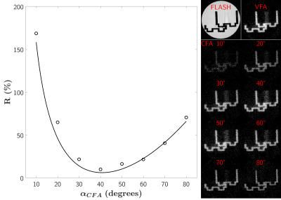 |
75 |
Inversion-Recovery UTE multispoke sequence: comparison of two excitation schemes
Lucas Soustelle, Julien Lamy, François Rousseau, Jean-Paul Armspach, Paulo Loureiro de Sousa
Inversion-Recovery UTE sequences have been used to highlight and quantify short-T2 structures. Covering a 3D volume remains time-consuming, and a trade-off often needs to be found between k-space undersampling and acceptable appearing streaking artifacts. As such, acquiring several radial spokes within a single repetition time represents a supplementary sequence acceleration possibility. In this work, we propose to quantify the short-T2 signal difference between a constant and an optimized variable flip angle strategy in a multispoke acquisition module, and within a long-T2 suppression condition in vitro.
|
|
4226.
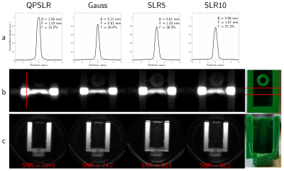 |
76 |
On the use of frequency modulated pulses in sat-UTE
Lucas Soustelle, Ericky Caldas de A. Araujo, François Rousseau, Jean-Paul Armspach, Pierre G. Carlier, Paulo Loureiro de Sousa
Conventional slice selection in 2D-UTE sequences is challenging as eddy currents and gradient non-idealities make difficult to achieve an appropriate slice selection and a minimum TE. The sat-UTE sequence proposes a simplification that separates slice selection from excitation, ensuring an easily implementable 2D-UTE sequence. The selection part was originally proposed with a short gaussian pulse, constraining slice selectivity to a gaussian profile and demanding exceedingly high RF power for ensuring efficient saturation. In this work, we propose an alternative implementation using low-peak-amplitude and selective quadratic phase pulses for slice saturation, providing an efficient and sharp short-T2 slice selection in sat-UTE.
|
|
4227.
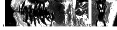 |
77 |
Fast High-Resolution Endodontic MRI
Video Permission Withheld
Philipp Madörin, Matthias Weigel, Thomas Connert, Sebastian Kühl, Oliver Bieri
The aim of this study was to explore standard MRI hardware and sequences as an alternative radiation-free modality to Cone Beam Computer Tomography for imaging the dental root canals rapidly and with high-resolution for the planning and guidance of the bur to the root canal orifice in combination with an intraoral surface scan.
|
|
4228.
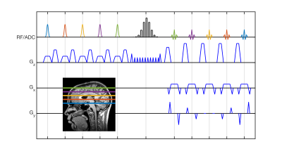 |
78 |
Accelerated Spin Echo Imaging using Simultaneous Spin-Echo Refocusing in conjunction with a PINS Refocusing Pulse
Francesco Padormo, Rebecca Feldman, Priti Balchandani
We proposed a novel alternative to the Turbo Spin Echo. It combines ideas from the Simultaneous Spin-Echo Refocusing method with PINS pulse design to result in a low SAR acquisition.
|
|
4229.
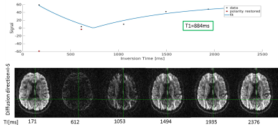 |
79 |
Rapid multi-inversion SMS-EPI integrated with gradient-echo, spin-echo and diffusion-weighted EPI acquisitions
Daniel Park, Thomas Witzel, Ilana Leppert, Yi-Fen Yen, Qiuyun Fan, Christine Tardif, Jonathan Polimeni
Recently inversion-recovery techniques have been combined with EPI in an efficient scheme in which a non-selective inversion is merged with a slice reordering scheme, providing rapid quantitative T1 maps. Here we merge this approach with several recent EPI technologies, including SMS-EPI, and several image contrasts including T2-, T2*, and diffusion weighted acquisitions. Not only does this allow the efficient combination of multiple contrasts but it opens the door to a rapid, all-EPI examination.
|
|
4230.
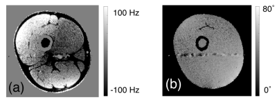 |
80 |
A Gradient Spoiled Spin-Echo Sequence for Simultaneously Measuring B1+, B0, T1, T2, T2*, and Velocity of a Two-Dimensional Slice
Nicholas Dwork, Adam Kerr, John Pauly
A single sequence is presented that quantifies several parameters simultaneously of a two-dimensional slice.
|
|
4231.
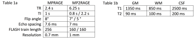 |
81 |
Evaluating RF and gradient spoiling in MPRAGE and MP2RAGE using novel, efficient algorithms for 3D extended phase graph and ideal-spoiling models
M. Dylan Tisdall
To optimize spoiling efficiency, we evaluate several design choices for RF- and gradient-spoiling in MPRAGE-type sequences. Optimization is performed using two efficient algorithms, one for the ideal-spoiling condition, and one for a 3D extended phase graph model that realistically captures spoiling effects. We used novel implementations of both algorithms, with publicly available source, that attempt to maximize computational efficiency. Our results suggest possible avenues for improving spoiling in MPRAGE-type sequences.
|
|
4232.
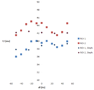 |
82 |
Description of the off-resonance dependency in slice-selective FISP MRF
Gregor Körzdörfer, Bartosz Guzek, Yun Jiang, Josef Pfeuffer, Bernhard Hensel, Peter Speier, Mathias Nittka
In Magnetic Resonance Fingerprinting (MRF), quantitative parameter maps are generated by matching pseudo randomly generated MR signals with a precalculated dictionary. A common implementation of MRF is based on an unbalanced SSFP (FISP) sequence in combination with a slice-selective excitation. In this work, we demonstrate its sensitivity to off-resonances in specific configurations, investigate the underlying spin physics and derive according mitigation approaches.
|
|
4233.
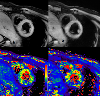 |
83 |
mGRE and TFE based cardiac SWI at 3.0 T
Did Not Present
Xiaoqi Wang
In this work, a 3.0 T multiple echo sequence was developed for cardiac scans, and a feasibility study was carried to assess whether susceptibility-weighted imaging (SWI) provides image contrast for the detection of myocardium disease. Currently collected images indicates that cardiac SWI at 3.0 T provides a prominent option for detecting myocardial abnormality, while further optimization to obtain sensitivity and accuracy is needed.
|
|
4234.
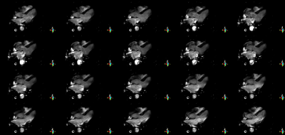 |
84 |
Imaging cardiac fat by chemical shift encoded MRI application in CINE format at 3 T
Did Not Present
Xiaoqi Wang
Ectopic fat deposition impacts cardiac health, and non-invase quantification is required. In this work, a chemical shift encoded MRI is designed for cardiac applications. Images of Proton density fat fraction (PDFF) water (W) and fat(F), and T2* were generated for each frame of cardiac motion, formed CINE movies for cardiac cycles. The novel sequence was flexible in clinical applications with short acquisition time and made sensitive findings in this research.
|
|
4235.
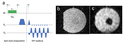 |
85 |
AC magnetic sensing using the SIRS effect combined with bSSFP at ultra-low field
Bragi Sveinsson, Neha Koonjoo, Bo Zhu, Thomas Witzel, Matthew Rosen
Direct detection of neuronal currents has long been a goal within MRI, with the aim of improving upon the spatial and temporal resolution of BOLD fMRI. So far, good results have been shown in phantoms but detection in vivo has proven difficult. A promising current detection technique is Stimulus-Induced Rotary Saturation (SIRS), but the BOLD signal can contaminate SIRS measurements, possibly explaining inconclusive in vivo results so far. A new sequence was developed and tested in an ultra-low-field (ULF) regime (6.5 mT) where paramagnetic effects such as BOLD are reduced and is more suited for SIRS measurements in vivo.
|
|
4236.
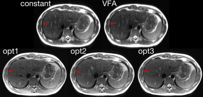 |
86 |
Reducing Blurring while Controlling Contrast in Abdominal Imaging with Variable Flip Angle Single Shot Fast Spin Echo
LI Zhao, Manuel Taso, Daniel Litwiller, David Alsop
Single-shot fast spin echo is used clinically to provide fast, motion-robust abdominal images. However, signal decay along the echo train results in blurred images. Our previous work on optimal variable flip angle design demonstrated improved SNR on brain imaging. Here, we extend this framework to show simultaneous T2 blurring and contrast control in abdominal imaging with single-shot fast spin echo.
|
|
4237.
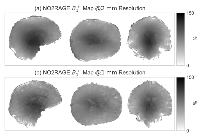 |
87 |
Fast Measurement of B1+ using the NO2RAGE pulse sequence
Riccardo Metere, Tobias Lenich, Kerrin Pine, Harald Möller
The knowledge of the spatial distribution of the transmit field of the coil, B1+, is essential in a number of MRI applications, particularly at higher field strength. SA2RAGE is a recently developed technique for B1+ mapping, derived from the MP2RAGE scheme. The sensitivity to B1+ is obtained from a preparation pulse and optimized delays between block gradient acquisitions. Here, we propose a much faster (but otherwise comparable) method, referred to as NO2RAGE, where the B1+ sensitivity is obtained by omitting the preparation, nullifying the delays, and adjusting the other sequence parameters. The technique compares favorably against other in vivo methods.
|
|
4238.
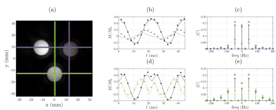 |
88 |
Efficient imaging of Rabi modulated trajectories using balanced Steady-State Driven Trajectory (bSSDT) imaging
James Korte, Bahman Tahayori, Peter Farrell, Yasmin Blunck, Leigh Johnston
We propose an efficient technique to image localised steady-state trajectories, termed balanced Steady-State Driven Trajectory (bSSDT) imaging and implement the protocol to investigate the properties of Rabi modulated steady-state trajectories. In bSSDT imaging, a sequence of points on the voxel-wise steady-state trajectory is acquired. The resultant 4-D data offers a potentially rich source of information about the underlying tissue properties. The proposed imaging technique is a pseudo-continuous excitation version of balanced steady-state free precession (bSSFP) imaging, with relaxation of the magnetisation to the equilibrium steady-state in bSSFP replaced by control of the magnetisation to a steady-state trajectory in bSSDT.
|
|
4239.
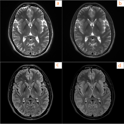 |
89 |
Fast BLADE with variable flip angle
Yuyu Wang, Le Zhang, Muhammed Labeeb, Dehe Weng
An effective approach with variable flip angle to speed up BLADE acquisition is proposed for general MR imaging. It improved the efficiency of BLADE by enlarging the width of one blade. The image quality is comparable with that of conventional BLADE method.
|
|
4240.
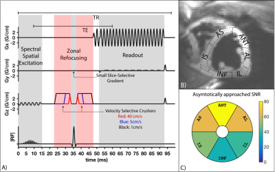 |
90 |
Velocity Sensitivity of Inner-Volume Cardiac Echo Planar Imaging
Ahsan Javed, Terrence Jao, Krishna Nayak
Inner volume echo planar imaging (IV-EPI) is feasible and useful for cardiac diffusion tensor imaging and arterial spin labelling . IV-EPI inherently provides velocity-selective saturation caused by the crusher gradients around the 180º refocusing-pulse. In this study, we characterize this velocity-dependent effect on myocardial signal in systolic and diastolic cardiac phases. We find that a simple velocity distribution model fits the data well, and that the data from our experiments allows us to make recommendation for designing gradients to avoid myocardial signal loss for diastolic and systolic IV-EPI imaging.
|
|
4241.
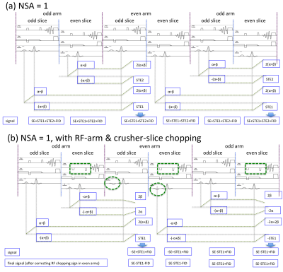 |
91 |
Improving Image Quality of A Spiral T1 Spin-Echo Sequence
Zhiqiang Li, Melvyn Ooi, Dinghui Wang, James Pipe
Cartesian SE is widely used for T1 imaging, but is slow and often susceptible to flow artifacts. A spiral T1SE technique has been proposed for rapid T1 imaging with minimal flow artifacts. However, inconsistent through-plane signal has been observed with certain protocols. In this work we investigate the root causes and propose a combined RF/gradient chopping scheme to minimize this inconsistency, and improve the T1 contrast. The improvement was demonstrated with phantom and volunteer data.
|
|
4242.
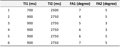 |
92 |
Optimized parameters for MP2RAGE images on 7-T MRI
Uksu Choi, Hirokazu Kawaguchi, Yuichiro Matsuoka, Tobias Kober, Ikuhiro Kida
The MP2RAGE sequence at ultra-high magnetic fields can be advantageous for investigation of microstructural changes because of reduced B1 inhomogeneity and images with different contrasts (uniform (UNI), T1, and fluid and white matter suppression (FLAWS)). In this study, we used several evaluative methods to optimize the inversion times and flip angles for UNI, T1, and FLAWS images of the MP2RAGE sequence at 7 T. We found the optimized inversion times with flip angles for UNI and T1 images, but different parameters for FLAWS images. The results suggested that different parameters of MP2RAGE at 7 T should be applied for different contrast types.
|
|
4243.
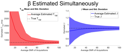 |
93 |
B1- and Fat-Corrected T1 Mapping Using Chemical-Shift Encoded MRI
Nathan Roberts, Timothy Colgan, Xiaoke Wang, Diego Hernando, Scott Reeder
Spatially varying B1 inhomogeneities and fat content are well known to be confounders of quantitative T1 mapping that use multi flip angle techniques. Separate B1 calibration maps can be acquired to correct flip angle errors cause by B1 inhomogeneities, but this requires an additional acquisition. In this work we consider an alternative approach that acquires variable flip angles and variable repetition times, and simultaneously estimates B1 inhomogeneity, T1, fat-fraction and R2*. The feasibility and noise performance of this approach are evaluated using theoretical Cramer-Rao Lower Bound analysis and simulations. Our results demonstrate that this approach is feasible, but suffers from relatively poor noise performance at typical acquisition parameters.
|
|
4244.
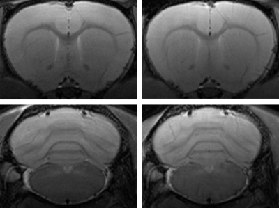 |
94 |
T2 measurement in SE-WMRI fast spin echo imaging
Did Not Present
Edzer Wu, Po-Wei Cheng, Tzi-Dar Chiueh, Jyh-Horng Chen
To investigate the contrast in SE-WMRI spin echo imaging, we measured five tubes of SPIO solution with various concentrations using standard and SE-WMRI Multi-Slice-Multi-Echo (MSME) sequence. T2 relaxation times were then calculated based on the 16 acquired images per scan. Results show that with a 1.4-fold acceleration, the T2 relaxation time of SE-WMRI MSME remains the same within the error bar as standard imaging.
|
|
4245.
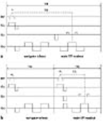 |
95 |
On the Analysis of EPI Phase Correction with Small Tip Angle Excitation to reduce minimum required TE: Application to Whole-Brain Submillimetre-Resolution fMRI at 3T
Seong Dae Yun, N. Jon Shah
EPI has been widely used for fMRI due to its relatively fast imaging speed. However, as one of its drawbacks, ghost artefacts need to be corrected. In the community, an approach that utilises three non-phase encoded navigator echoes is commonly used for the correction. Although this scheme is quite effective, as the matrix size increases for high-resolution imaging the navigator echoes constitute a significant contribution in increasing the minimum required TE. To overcome this issue, this work employs an alternative navigator echo scheme. Here, its performance was analysed and whole-brain submillimetre-resolution fMRI (0.75 × 0.75 mm2) was demonstrated at 3T.
|
|
4246.
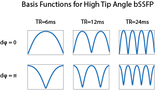 |
96 |
Generation of Arbitrary Spectral Profiles using Orthonormal Basis Combinations of bSSFP MRI
Michael Mendoza, Nicholas McKibben, Neal Bangerter
We present a technique for generating an arbitrary spectral profile by using multiple-acquisition bSSFP. Multiple phase-cycled bSSFP images with increasing TRs were acquired and Gram-Schmidt orthogonalization was applied to spectral basis functions to generate an orthonormal basis. This generated orthonormal basis was used to approximate an arbitrary spectral profile by using linear combinations of the calculated basis functions. A variety of spectral functions were simulated and used as a template to approximate spectral profiles in water and fat phantoms.
|
|
MR Fingerprinting
Electronic Poster
Acquisition, Reconstruction & Analysis
Tuesday, 19 June 2018
| Exhibition Hall |
14:45 - 15:45 |
| |
|
Computer # |
|
4247.
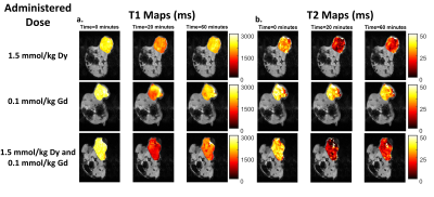 |
97 |
Simultaneous Observation of Two Paramagnetic MRI Contrast Agents via Dynamic T1 and T2 Mapping with Magnetic Resonance Fingerprinting: Initial In Vivo Results
Christian Anderson, Yuning Gu, Mette Johansen, He Hu, Yun Jiang, Michael Kavran, Bernadette Erokwu, Susann Brady-Kalnay, Nicole Steinmetz, Xin Yu, Mark Griswold, Chris Flask
In this work simultaneous T1 and T2 mapping via MR Fingerprinting was used to observe the distribution of dysprosium and gadolinium MRI contrast agents in a mouse tumor model. Changes from baseline in T1 and T2 were observed using each agent alone and an increased effect was seen when agents were used in combination. Additionally, a decrease in the dysprosium dose lead to a reduction in the level of T1 and T2 enhancement observed. These results indicate that MRF might be able to be used to track multiple MRI contrast agents in vivo.
|
|
4248.
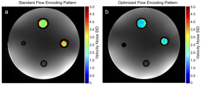 |
98 |
Optimization of the Flow Encoding Pattern under Consideration of Spatio-Temporal Velocity Gradients in flow-MR Fingerprinting
Sebastian Flassbeck, Simon Schmidt, Mark Ladd, Sebastian Schmitter
It has been shown that the quantification of blood flow velocities through the use of bipolar gradients is possible within the MRF framework, called flow-MRF. In this work, we investigate four different flow encoding patterns for flow-MRF and analyze their impact on flow quantification. Finally we present an optimized pattern.
|
|
4249.
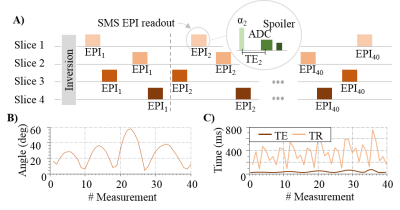 |
99 |
Time efficient whole-brain coverage in Magnetic Resonance Fingerprinting using echo-planar imaging, slice-interleaving and simultaneous multi-slice imaging
Benedikt Rieger, Mehmet Akçakaya, Lothar Schad, Sebastian Weingärtner
Magnetic resonance fingerprinting (MRF) is a promising method for simultaneous multi-parameter quantification. However, spatial coverage is limited in the original method due to the rapid image readout and the balanced sequence design. The aim of this study is to enable time-efficient volumetric coverage in Cartesian MRF-EPI for whole-brain quantification in clinically acceptable scan times. A slice-interleaved acquisition is shown to offer quantification precision comparable to single-slice measurements in a fraction of the measurement time. By combining this with simultaneous multi-slice imaging, whole-brain coverage with 40 slices was achieved in one minute, resulting in high quality T1 and T2* maps.
|
|
4250.
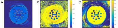 |
100 |
From phantoms to patients: Paving the way for MRF in clinical practice
Marcel Gratz, Jasmin Jäger, Mathias Nittka, Stephan Kannengiesser, Josef Pfeuffer, Gregor Koerzdoerfer, Rainer Kirsch, Florian Meise, Harald Quick, Vikas Gulani, Mark Griswold, Michael Forsting, Lale Umutlu
This study puts the focus on estimating the stability of MRF data in phantoms, volunteers and patients and investigates the options to integrate MRF into routine clinical workflows. The phantom and in-vivo scans yielded consistent and reproducible results throughout different scan days. Thus, MRF enables a fast and reliable generation of quantitative data in vivo and, along with appropriate reference values, has the potential to improve diagnostics in neuroimaging.
|
|
4251.
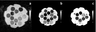 |
101 |
Doped Polyvinylpyrrolidone Solutions for Tunable T1, T2, and ADC in Multiparametric Phantoms
Michael Boss, Yun Jiang, Stephen Russek, Mark Griswold
A multiparametric phantom for quantitative MRI and magnetic resonance fingerprinting was developed. Solutions of PVP were used to tune the apparent diffusion coefficient (ADC), and then doped with Mn to control T1 and T2. The addition of Mn did not affect the ADC, but did significantly shorten T1 and T2, demonstrating the potential of independently tuning relaxation times and ADC.
|
|
4252.
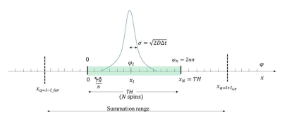 |
102 |
Diffusion Propagator Formalism in Bloch Equation Simulation of Magnetic Resonance Fingerprinting
Shota Hodono, Yun Jiang, Naren Nallapareddy, Vikas Gulani, Mark Griswold
By considering spin diffusion as a random walk process and introducing a Gaussian probability function to the Bloch equation, a diffusion propagator was developed, which allows diffusion to be accounted for in magnetic resonance fingerprinting (MRF) simulations. In this study, signal intensities generated by Bloch simulations with the diffusion propagator were compared with theoretical values for the diffusion-weighted spin echo sequence. Additionally, the diffusion propagator approach was applied to the MRF fast imaging with steady-state precession sequence and the resulting signal evolution was qualitatively compared with that generated by the Extended Phase Graphs method.
|
|
4253.
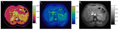 |
103 |
Normative T1 and T2 relaxation times and measurement repeatability of abdomen organs at 3T using 2D MR Fingerprinting
Sanjay Sridaran, Ananya Panda, Yong Chen, Jesse Hamilton, Shivani Pahwa, Katherine Wright, Yun Jiang, Joshua Batesole, Mark Griswold, Nicole Seiberlich, Vikas Gulani
T1 and T2 mapping of abdominal organs provides valuable information for characterization of abdominal pathologies, but is limited by technical difficulties. Previous work has demonstrated the utility of MR Fingerprinting (MRF) for abdominal relaxometry. However, there are still limited normative data for different abdominal organs. In this study, 2D MRF was applied to 22 subjects to establish normative values for abdominal organs at 3T and assess repeatability. Our results demonstrated a good agreement with the literature and repeatability of the measurement. The values obtained in this study can also serve as a reference for future clinical studies.
|
|
4254.
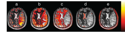 |
104 |
Dictionary free anatomical segmentation of Magnetic Resonance Fingerprinting brain data with Independent Component Analysis
Rasim Boyacioglu, Dan Ma, Debra McGivney, Louisa Onyewadume, Ozden Kilinc, Chaitra Badve, Vikas Gulani, Mark Griswold
Magnetic Resonance Fingerprinting signal evolutions are sensitized to certain tissue properties during data acquisition. The matching step can be suboptimal due to dictionary limitations or tissue related constraints (e.g. partial volume, magnetization exchange). Here, we propose to apply Independent Component Analysis (ICA) to 4D MRF data after image reconstruction without explicit dictionary matching for tissue characterization, lifting the requirement for a relaxation model. ICA of whole brain MRF data segments the brain into multiple components with single tissue types such as gray matter, white matter and CSF for healthy subjects and also tumor in the case of glioblastoma patients.
|
|
4255.
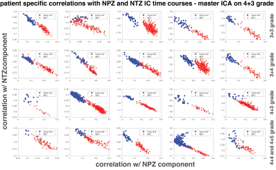 |
105 |
Prostate cancer tissue characterization with spatio-temporal analysis of Magnetic Resonance Fingerprinting data
Rasim Boyacioglu, Ananya Panda, Yun Jiang, Debra McGivney, Vikas Gulani, Mark Griswold
Multiple tissue properties (T1, T2, diffusion, etc.) and system parameters can be acquired simultaneously with Magnetic Resonance Fingerprinting (MRF). The dictionary formation and matching steps may be confounded by the lack of certain tissue properties (e.g. magnetization exchange) in the dictionary and thus, the differentiation of tissues of interest may be suboptimal even though variable MRF signal evolutions set them apart. Here, as a dictionary-free alternative, spatio-temporal analysis of MRF time series is proposed for tissue characterization. Independent Component Analysis (ICA) based correlation analysis of prostate MRF data can distinguish between healthy and cancer tissue without explicit dictionary matching.
|
|
4256.
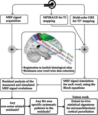 |
106 |
Magnetic Resonance Fingerprinting (MRF) Can Reveal Microstructural Variations in the Brain Gray Matter
Shahrzad Moeiniyan Bagheri, Viktor Vegh, David Reutens
The importance of creating accurate anatomical maps of the human brain has motivated the development of in vivo, observer-independent and reproducible mapping methods that account for inter-individual variation. However, a) whole brain coverage and b) a multi-modal approach that accounts for the combined effects of all microscale tissue properties has yet to be developed. To bridge this gap, we propose a statistical feature-based residual analysis framework that makes use of unique tissue-specific MRF signals after taking account of T1 and T2* effects. In four cortical areas of six participants, this approach showed consistent similarity measurements between regions, indicating that MRF signals contain information about micro-level tissue properties.
|
|
4257.
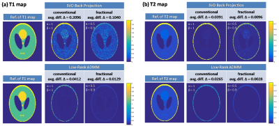 |
107 |
MRF-FrM: A Preliminary Study on Improving Magnetic Resonance Fingerprinting Using Fractional-order Models
Haifeng Wang, Leslie Ying, Xin Liu, Hairong Zheng, Dong Liang
Magnetic resonance fingerprinting (MRF) is an exceptional promise for simultaneous quantification of T1 and T2 maps, based on the traditional Bloch equation formalism of MR and numerous readouts. In this work, a Fractional-order model of the Bloch equations is applied to create the dictionary of the T1 and T2 maps used in MRF. The simulations show that the proposed method can improve the evaluation accuracy of the T1 and T2 maps comparing with the conventional MRF methods with the traditional first-order model of the Bloch simulation.
|
|
4258.
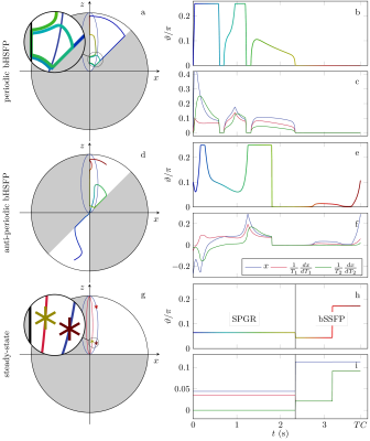 |
108 |
Hybrid-State Free Precession for Measuring Magnetic Resonance Relaxation Times
Jakob Assländer, Daniel Sodickson, Riccardo Lattanzi, Martijn Cloos
This work analyzes the spin physics in steady-state free precession sequences modified to smoothly vary sequence parameters, as suggested in MR Fingerprinting. We arrive at the conclusion that a transient state develops only in one direction, while the magnetization in the other two dimensions transitions adiabatically between steady states. We provide solutions of the Bloch Equations for this hybrid state and demonstrate the superior T1- and T2-encoding performance of the hybrid state compared to the steady state.
|
|
4259.
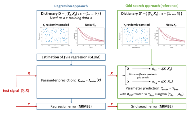 |
109 |
DICTIONARY-FREE MR FINGERPRINTING PARAMETER ESTIMATION VIA INVERSE REGRESSION
Fabien Boux, Florence Forbes, Julyan Arbel, Emmanuel Barbier
MR Fingerprint requires an exhaustive search in a dictionary, which even for moderately sized problems, becomes costly and possibly intractable. In this work, we propose an alternative approach: instead of an exhaustive search for every signal, we use the dictionary to learn the functional relationship between signals and parameters. This allows the direct estimation of parameters without the need of searching through the dictionary. The comparison between a standard grid search and the proposed approach suggest that MR Fingerprinting could benefit from a regression approach to limit dictionary size and fasten computation time.
|
|
4260.
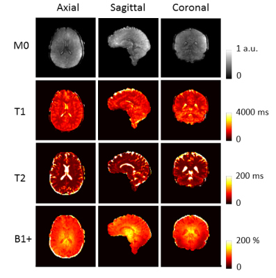 |
110 |
Fast parameter mapping at 7T with SSFP MR Fingerprinting using 3D radial trajectories
Guido Buonincontri, Pedro Gómez, Matteo Cencini, Giada Fallo, Laura Biagi, Rolf Schulte, Michela Tosetti
When using ultra-high field MRI scanners (UHF, B0>= 7T), quantitative imaging is challenging due to B0 and B1+ non-uniformities. Magnetic resonance fingerprinting (MRF) represents a great opportunity for quantitative imaging at UHF as it can estimate these effects at the same time of the parameters of interest. Here, we demonstrate in vivo at 7T a novel 3D MRF approach based on a three-dimensional radial k-space acquisition, estimating M0, T1, T2 and B1+ simultaneously in 5 minutes.
|
|
4261.
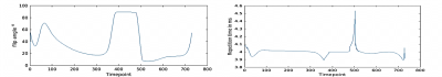 |
111 |
MORE-MRF: Towards Motion Resolved Cardiac Multi-Parametric Mapping with Magnetic Resonance Fingerprinting
Olivier Jaubert, Gastao Cruz, Torben Schneider, Rene Botnar, Daniel Rueckert, Claudia Prieto
Magnetic Resonance Fingerprinting (MRF) estimates multi-parametric maps simultaneously with short acquisition times. Cardiac MRF has been recently proposed to quantify myocardial T1 and T2 maps for 2D single-phase cardiac imaging. This ECG-triggered breath–hold approach provides a single-phase cardiac image and uses a non-continuous (segmented) acquisition. Here we sought to investigate myocardial MRF at multiple cardiac phases from a continuous acquisition, which could simultaneously provide cardiac function and quantitative tissue characterisation. The proposed approach was evaluated in simulations and healthy subjects, considering eight cardiac phases per cycle.
|
|
4262.
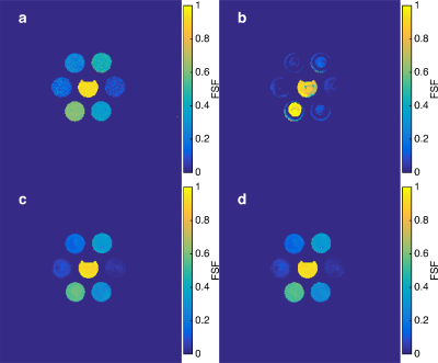 |
112 |
Unbalanced Steady-State Free Precession MR Fingerprinting with Simultaneous Fat Signal Fraction, T1, T2 and B0 Estimation
Jason Ostenson, Bruce Damon, E. Brian Welch
Unbalanced steady-state free precession MR fingerprinting (uSSFP-MRF) may be used to quantify fat signal as well as water T1 and T2, but is subject to off-resonance (B0) and blurring. We propose a variable echo time uSSFP and post-processing method that estimates fat signal fraction, water T1 and T2, and B0 within a single sequence. The method is tested in phantoms and in vivo. Preliminary results indicate the proposed method provides sharp fat fraction maps that account for chemical-shift and B0 effects that normally lead to blurring, as well as generate water-only T1 and T2 maps.
|
|
4263.
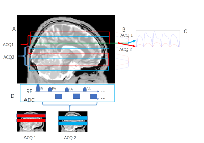 |
113 |
Multiband Multi-slab Magnetic Resonance Fingerprinting Using Overlapping Slabs and Temporal-Sharing Reconstruction
Di Cui, Hing-Chiu Chang, Queenie Chan, Xiaoxi Liu, Edward Hui
Magnetic Resonance Fingerprinting (MRF) is a novel and rapid technique to quantify multiple MR parameters, and single-slab MRF was later developed for better slice coverage. In this study, we proposed a new method to extend single-slab MRF to multiband multi-slab acquisition. The slab boundary artifacts due to imperfect slab profile in multi-slab acquisition is overcome by a overlapping-slab acquisition strategy, and MRF reconstruction from the multiband multi-slab data can be obtained using our previously proposed temporal sharing reconstruction method.
|
|
4264.
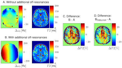 |
114 |
Influence of Off-resonance on FISP Magnetic Resonance Fingerprinting (FISP-MRF)
Bartosz Guzek, Gregor Körzdörfer, Mathias Nittka, Josef Pfeuffer
FISP-MRF is a robust method to acquire T1 and T2 maps. The unbalanced gradient in slice-selection direction should reduce the sensitivity of FISP-MRF on off-resonances. This work investigated the dependency of T1 and T2 values on field inhomogeneities in simulations and measurements on phantoms and in vivo by applying different spatial magnetic field offsets. Depending on the sequence implementation, the measured T1 and T2 values were altered in different ways by the off-resonances. Outcomes showed that the actual implementation of the dephasing moment strongly affects the results. Using a constant instead of variable TR further reduced the sensitivity to off-resonances.
|
|
4265.
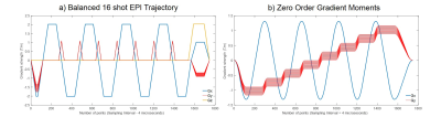 |
115 |
Balanced multi-shot EPI for accelerated Cartesian MR Fingerprinting: An alternative to spiral MR Fingerprinting
Arnold Julian Vinoj Benjamin, Pedro Gómez, Mohammad Golbabaee, Zaid Mahbub, Tim Sprenger, Marion Menzel, Michael Davies, Ian Marshall
This study shows the practical implementation of an accelerated Cartesian Magnetic Resonance Fingerprinting (MRF) scheme using a multi-shot Echo Planar Imaging (EPI) readout. Its performance is compared with conventional spiral MRF and the fast convergence of accelerated iterative reconstructions for this method is shown.
|
|
4266.
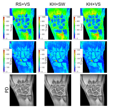 |
116 |
Acceleration of the Cartesian acquisition of MR fingerprinting using the keyhole acquisition and view-sharing reconstruction
Ryoichi Sasaki, Yasuhiko Terada
MR fingerprinting (MRF) is a promising approach that enables quantification of multiple tissue parameters at a single scan. MRF uses spiral sampling which requires a high fidelity of gradient hardware. Meanwhile, MRF with the Cartesian sampling is feasible for conventional scanners with the moderate hardware fidelity, but it comes at the cost of the elongation of the acquisition time. Here we propose an acceleration method for the Cartesian MRF acquisition that enables the 8-fold acceleration. The quantitative errors and undersampling artifacts in the T1 and T2 maps are alleviated by using the keyhole acquisition and view-sharing reconstruction.
|
|
4267.
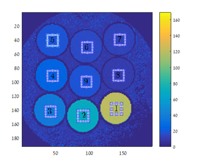 |
117 |
Bloch-Torrey Simulation to Quantify Diffusion Effects on Multi-Parametric Sequences with Application for MR Fingerprinting
Inbar Seroussi, Nir Sochen, Noam Ben-Eliezer, Ofer Pasternak
Quantifying the effect of self-diffusion on multi-parametric sequences, such as those used for Magnetic Resonance Fingerprinting (MRF) is important to increase the accuracy of dictionary based parameter estimation. To quantify diffusion, we propose a signal simulation approach, which replaces the Bloch equation with the Bloch-Torrey equation, and accounts for protocol and scan dependent parameters. We apply this framework on a Multi Spin Echo (MSE) protocol and quantify the diffusion encoding introduced by the spoiler gradients in this sequence. We further show that increasing the spoiler strength would allow detecting diffusion by including the diffusion effect in the dictionary.
|
|
4268.
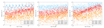 |
118 |
High-dimensional and accurate dictionary fitting with B-spline interpolation and gradient-based optimization
Willem van Valenberg, Stefan Klein, Frans Vos, Lucas van Vliet, Dirk Poot
Dictionary matching in MR fingerprinting acquisitions quantifying many parameters, such as T1 T2, T2’, B0, B1+, and PD, becomes prohibitively expensive due to the dense grid in each dimension of the dictionary that is required. In this work we propose B-spline interpolation of the dictionary to reduce the dictionary size and to enable efficient nonlinear least-squares fitting by gradient-based optimization methods. The experiments show that the method substantially reduces fitting error, especially when the SNR is high and dictionary resolution is the limiting factor for improving the precision of the quantitative parameters.
|
|
4269.
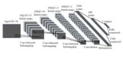 |
119 |
Accelerated Magnetic Resonance Fingerprinting Using Convolutional Neural Network
Ying Liao, Qiang Zhang, Di Cui, Edward Hui, Huijun Chen
The purpose of this work is to accelerate the acquisition of Magnetic Resonance Fingerprinting (MRF) using Convolutional Neural Network (CNN). Compared with traditional MRF reconstruction using 1000 time points, our CNN model shows better reconstruction fidelity in T2 and similar reconstruction fidelity in T1 using 300 time points. Our study suggests that CNN-based method may be an effective tool in the acceleration of MRF reconstruction.
|
|
4270.
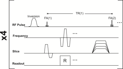 |
120 |
Simultaneous Diffusion, PD, T1, and T2 Mapping with Optimized MR Fingerprinting EPI
Ouri Cohen, Matthew Rosen
MR Fingerprinting (MRF) is a recent technique for simultaneous acquisition of multi-parametric tissue maps. Beyond the proton density (PD), T1 and T2 maps routinely obtained with conventional MRF sequences, quantitative diffusion maps are of great clinical interest but are challenging to acquire due to long scan times and poor resolution. In previous work we’ve demonstrated an optimized MRF EPI sequence that allows short scan times. Here we characterize its diffusion sensitivity and demonstrate its potential for simultaneous mapping of PD, T1, T2 and diffusion.
|
|
| Back |
| The International Society for Magnetic Resonance in Medicine is accredited by the Accreditation Council for Continuing Medical Education to provide continuing medical education for physicians. |
