|
Electronic Poster Session
Cardiovascular |
Wednesday, 20 June 2018
Electronic PosterCardiovascular
4748 -4771 Novel Techniques & Methods
4772 -4795 Cardiovascular Image Processing
4867 -4890 Myocardial Tissue Characterization
4891 -4914 Motion & Beyond |
| |
Novel Techniques & Methods
Electronic Poster
Cardiovascular
Wednesday, 20 June 2018
| Exhibition Hall |
13:45 - 14:45 |
| |
|
Computer # |
|
4748.
 |
1 |
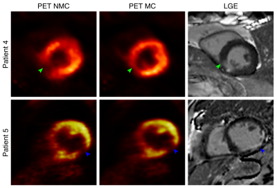
 Respiratory motion-corrected simultaneous myocardial viability PET and coronary MR angiography: initial clinical validation Respiratory motion-corrected simultaneous myocardial viability PET and coronary MR angiography: initial clinical validation
Camila Munoz, Karl Kunze, Radhouene Neji, Christoph Rischpler, René Botnar, Stephan Nekolla, Claudia Prieto
Cardiac PET-MR imaging has shown promising results for the comprehensive assessment of coronary artery disease. Here we present an initial clinical validation of a recently demonstrated respiratory motion-corrected PET-MR framework in patients with chronic total occlusion. Simultaneous visualization of the coronary lumen by Coronary MR Angiography (CMRA) and myocardial viability by 18F-FDG PET was compared against X-ray angiography and LGE-MRI. We demonstrate that the proposed framework produces diagnostic images in both modalities in a short and time-efficient examination of ~12 minutes. Motion correction improved visible length and sharpness of the coronary arteries by CMRA and delineation of the myocardium and noise reduction by 18F-FDG PET, resulting in good agreement with X-ray angiography and LGE-MRI.
|
|
4749.
 |
2 |
Non-invasive MR-based blood oximetry via multi-parametric, non-linear estimation: initial multi-center experience in adult and pediatric patients
Juliet Varghese, Lajja Desai, Rizwan Ahmad, Lee Potter, Ning Jin, Cynthia Rigsby, Paul Tannous, Aimee Armstrong, Michael Markl, Kan Hor, Subha Raman, Orlando Simonetti
Non-invasive estimation of blood oxygen (O2) saturation by magnetic resonance (MR) imaging would be useful in evaluating shunt severity in congenital heart disease, and oxygen delivery and consumption energetics in heart failure and pulmonary hypertension. A T2-based, non-linear, multi-parameter method has been developed to non-invasively determine O2 saturation in the heart and great vessels. In this multi-center study, the feasibility and accuracy of the technique is evaluated against gold-standard catheterization measurements in the cardiac chambers and vessels in a preliminary cohort of adult and pediatric patients with cardiovascular disease.
|
|
4750.
 |
3 |
Imaging inflammation following MI using hyperpolarized pyruvate and 3D Spectral-Spatial EPI
Jack Miller, Andrew Lewis, Carolyn Carr, Oliver Rider, Stefan Neubauer, Damian Tyler
Current clinical imaging techniques offer only limited assessment of innate immune cell driven inflammation, which is an emerging therapeutic target in myocardial infarction. However, macrophages have a defined metabolic phenotype and are highly glycolytic when activated following injury. Here we show that hyperpolarized [1-$$$^{13}$$$C]pyruvate imaging specifically detects this phenotype, which is altered by pharmacological blockade that modulates monocyte/macrophage inflammatory function both in vitro and in vivo. We conclude that cardiac hyperpolarized [1-$$$^{13}$$$C]lactate several days post insult reflects immunology, and not necessarily ischaemia.
|
|
4751.
 |
4 |
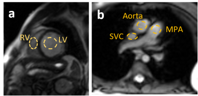
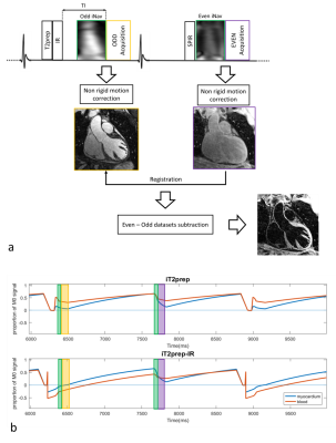
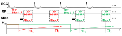
Simultaneous 3D whole-heart bright-blood and black blood imaging for cardiovascular anatomy and wall assessment with interleaved T2prep-IR
Giorgia Milotta, Giulia Ginami, Radhouene Neji, Claudia Prieto, René Botnar
Visualization of cardiovascular anatomy is important for diagnosis, risk stratification and planning of interventional procedures both in patients with congenital and non-congenital heart disease. This study proposes a novel free-breathing 3D whole-heart flow-independent approach for bright-blood visualization of coronary lumen and cardiac structures and for black-blood delineation of aortic, atrial, and coronary artery walls. The use of image-based navigation and non-rigid respiratory motion correction allows for 100% scan efficiency, predictable scan time and improved image sharpness.
|
|
4752.
 |
5 |
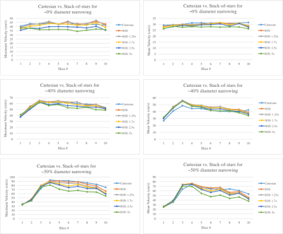
Accelerated Coronary 4D-Flow MRI: towards noninvasive functional assessment of stable coronary artery disease
Zixin Deng, Michael Loecher, Anthony Christodoulou, Christopher Nguyen, Zhengwei Zhou, Jaime Shaw, Yibin Xie, Xiaoming Bi, Zhaoyang Fan, Daniel Ennis, Debiao Li
In patients with suspected coronary artery disease (CAD), invasive catheterization is commonly used to determine the need for coronary revascularization. However, recent studies have shown that >50% of patients who undergo invasive catheterization have non-significant coronary lesions, hence the procedure was unnecessary. Recent work using 4D-Flow and the Navier-Stokes equations has shown promise for the noninvasive assessment of CAD, but still requires long scan times. This work explored coronary 4D-Flow using a stack-of-stars acquisition and compressed sensing to achieve 2-3x acceleration. The goal was to develop a more clinically feasible noninvasive pressure gradient measurement method for the assessment of CAD.
|
|
4753.
 |
6 |
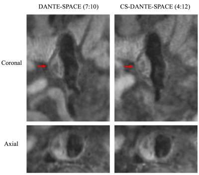
Surveillance of abdominal aortic aneurysm using accelerated 3D non-contrast black-blood MRI with compressed sensing (CS-DANTE-SPACE)
Chengcheng Zhu, Lizhen Cao, Zhaoying Wen, Sinyeob Ahn, Esther Raithel, Christoph Forman, Michael Hope, David Saloner
Although 3D non-contrast high-resolution black-blood MRI (DANTE-SPACE) is a promising tool for the surveillance of abdominal aortic aneurysm (AAA), it requires lengthy scans (~7 minutes). We implemented a compressed sensing method (CS-DANTE-SPACE) to reduce the scan time by 41% (to ~4 minutes), and tested its feasibility in 20 AAA patients undergoing routine follow-up. We found CS-DANTE-SPACE achieved accurate diameter/area measurements and intraluminal thrombus (ILT) identification, and provided better contrast and vessel sharpness. CS-DANTE-SPACE is a promising tool for AAA surveillance in the clinical setting.
|
|
4754.
 |
7 |
Imaging lower extremity vein thrombosis using a 3D FSE with novel modulated flip angle scheme and random k-space undersampling
Ling Zhang, Jie Zhang, Rui Li, Shuo Chen, Chaohong Wang, Nan-Jie Gong, Guobin Li, Ruchen Peng
Accurate detection of lower extremity vein thrombosis (LEVT) and assessment of its stage are crucial for treatment decision-making. Challenges such as long scan time and requirement for dark-blood, exist in T2-weighted MR imaging of LEVT. A 3D FSE with novel modulated refocusing flip angles and random undersampling is introduced and applied to imaging of LEVT.
|
|
4755.
 |
8 |
Late gadolinium enhancement by cardiovascular magnetic resonance, combined with right ventricle ejection fraction, for assessing the severity and predicting the prognosis of chronic thromboembolic pulmonary hypertension
Did Not Present
Ming-xi Liu, Juan-ni Gong, Xiao-juan Guo , Zhan-hong Ma, Tao Jiang, Jing An, Lu Liang, Yuan-hua Yang, Tu-guang Kuang
The aim of this study was to assess the severity and predict the prognosis of chronic thromboembolic pulmonary hypertension (CTEPH) using cardiovascular magnetic resonance with late gadolinium enhancement (LGE) combined with the right ventricle ejection fraction. Based on right heart catheterization, the percentages of the myocardial fibrosis volume (pFV>7.00%) and right ventricle ejection fraction (RVEF<34.34%) cut-off values were determined to assess the severity and predict the prognosis of 30 patients with CTEPH. The pFV and LGE are complementary to the RVEF for assessing the severity of the condition and can be a strong prognostic factor in patients with CTEPH.
|
|
4756.
 |
9 |
Volumetric black-blood fast spin echo with variable refocusing pulses for the visualization of whole-heart and great vessels
Video Permission Withheld
Markus Henningsson, Gerald Greil, Riad Zahr , Animesh Tandon, Barbara Burkhardt, Tarique Hussain
We describe the use of black-blood 3D fast spin echo with variable refocusing flip angles for whole-heart imaging. The technique allows efficient, volumetric high-resolution imaging of the heart and great vessels, and improves aortic vessel wall delineation compared to conventional 2D double-inversion recovery. It also provides complementary diagnostic information to 3D bright-blood bSSFP, particularly for the visualization of pulmonary veins. This was demonstrated in a study of 8 patients with congenital heart disease.
|
|
4757.
 |
10 |
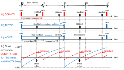
Significant Improvement of Blood Suppression in Cardiac Dark-Blood T1-TSE Imaging By Combining the T2-TSE Blood Nulling Strategy with Dummy Acquisitions for Myocardial T1-Weighting – A Comparison of Conventional and Novel TSE Techniques for Blood Signal Suppression and Overall Image Quality at 3T
Wolfgang Rehwald, Louis Klem, Stephen Darty, Nestor Mena, George Gamoneda, Raymond Kim
Cardiac dark-blood (DB) T1-turbo-spin echo (TSE) imaging frequently suffers from poor blood suppression due to its short effective TR required for good myocardial T1-weighting. We present a novel DB T1-TSE sequence that simultaneously applies a long effective TR as in T2-TSE to blood and a short effective TR as in conventional T1-TSE to myocardium, by means of dummy readouts. We ran conventional T1-TSE, T2-TSE, and novel T1-TSE sequences in 53 patients and scored the images for DB performance and overall image quality. Quality and blood suppression were drastically improved by novel T1-TSE.
|
|
4758.
 |
11 |
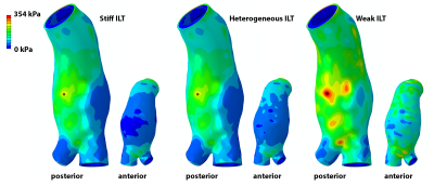
MRI-Based Stress Analyses of Abdominal Aortic Aneurysms With Resolved Intraluminal Thrombus Heterogeneity
Joseph Leach, Chengcheng Zhu, David Saloner, Michael Hope
Computational stress analyses of abdominal aortic aneurysms (AAA) are of great interest for individual aneurysm rupture risk assessment. The vast majority of patient-specific stress analyses are based on features seen at computed tomography, which is incapable of resolving material heterogeneity within intraluminal thrombus. Using T1-weighted black blood MRI, we imaged and explicitly modeled MRI-discerned intraluminal thrombus heterogeneity in multiple AAA stress analyses. Results demonstrate a limited effect of thrombus heterogeneity on the predicted vessel wall stresses, but suggest a possible role for MRI to inform thrombus material stiffness assignment in stress computations.
|
|
4759.
 |
12 |
Proton magnetic resonance spectroscopy to assess lipid content in cardiac amyloidosis
Mareike Gastl, Sophie Peereboom, Alexander Gotschy, Maximilian Fuetterer, Constantin von Deuster, Florian Bönner, Malte Kelm, Andreas Flammer, Robert Manka, Sebastian Kozerke
Amyloidosis is a multisystemic disorder frequently affecting the heart and causing heart failure. In this work, MR imaging and spectroscopy was implemented and applied to characterize myocardial structure and function as well as changes in fatty acid storage of the heart. We found that myocardial triglyceride-to-water ratio was significantly decreased in amyloidosis compared to age-and body mass index-matched controls. Myocardial triglyceride-to-water ratio showed a negative tendency with increasing markers of heart failure. It is concluded that proton spectroscopy may provide an additional biomarker to gauge progression of cardiac amyloidosis.
|
|
4760.
 |
13 |
Practical implementation of SMS bSSFP in the Heart
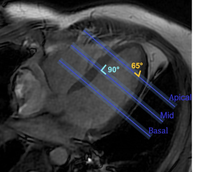
Vanessa Landes, Terrance Jao, Krishna Nayak
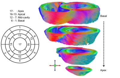
Cardiac MRI frequently relies on balanced stead-state free precession (bSSFP) for its beneficial contrast and SNR efficiency. Simultaneous multi-slice (SMS) imaging, specifically blipped controlled aliasing in parallel imaging, faces two major challenges with cardiac bSSFP: 1) artifacts from non-uniform signal in the through-slice direction and 2) spuriously excited side-lobes from imperfect multi-band excitation. We carefully evaluate both challenges using simulations and phantom experiments, and demonstrate practical SMS bSSFP imaging with 3-slice coverage (apical, mid, and basal short axis slices) at 3 Tesla, with good image quality in both transient-state and steady-state.
|
|
4761.
 |
14 |
Reconstruction of the 12-lead ECG using a novel MR-compatible ECG sensor network
Video Permission Withheld
Jesus Dos Reis, Paul Soullié, Grégory Petitmangin, Freddy Odille, Jacques Felblinger
Currently patient monitoring and sequence triggering during MR imaging are achieved using a low bandwidth ECG of 1Hz-60 Hz. Such devices provide a limited number of ECG leads and thus do not provide the same diagnostic information as the standard 12-lead ECG. In this work we developed a high-bandwidth MR-compatible ECG sensor (0.05Hz – 150 Hz) that integrates real-time signal processing for ECG denoising. Several individual sensors are combined to reconstruct a 12-lead ECG during MR imaging. The system was tested on a volunteer at 1.5T and could be used during MR-guided intervention or for inverse ECG imaging.
|
|
4762.
 |
15 |
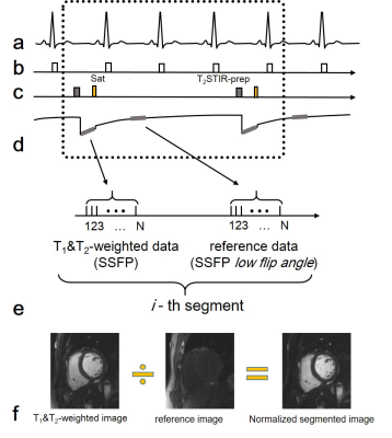
A Normalized Segmented T2STIR-bSSFP Technique for High Resolution Edema Imaging
Lixian Zou, Yanjie Zhu, Dong Liang, Xin Liu
High resolution edema image is highly desired for reducing particle volume effect (PVE) on thin myocardium. In this work, we proposed a normalized segmented T2STIR-bSSFP to improve base resolution from 192 to 320 in edema imaging as well as maintain high contrast of edema to myocardium. The results show that the proposed method can reduce PVE on thin myocardium and makes edema imaging for the right ventricle myocardium being possible.
|
|
4763.
 |
16 |
5D cardiac MRI on mouse models : preliminary results
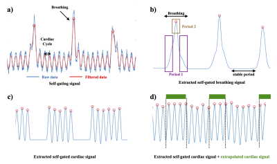
Julia Labrune, Aurélien Trotier, Emeline Ribot, Sylvain Miraux
Until now, the data acquired during breathing period are not used to obtain 4D cardiac images on mouse models. Consequently, the purpose of the work presented here is to evaluate the impact of breathing on the movement of the mouse's heart and to develop a method for reconstructing the cardiac self-gating signal during respiration. Finally, preliminary results of 5D image reconstruction (3D images during the heart rate and according to respiratory movement) will be shown in mice.
|
|
4764.
 |
17 |
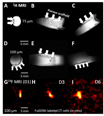
Tracking of PLGA-PFCE-labeled Cardiac Stem Cells Seeded on Novel Biodegradable Poly(3-hydroxyoctanoate) Scaffolds Implanted on the Murine Myocardium using 1H and 19F MRI/MRS
Chris Constantinides, Pooja Basnett, Barbara Lukasiewicz, Ricardo Carnicer Hijazo, Mangala Srinivas, Carolyn Carr, Ipsita Roy
Controlled administration of cardiac progenitor stem cells (CPCs), and their visualization and tracking and release, still present tremendous challenges in cellular/tissue therapy, yet fundamental and necessary tasks towards a therapeutically successful approach. In this work, we propose the synthesis and use of novel, functional biodegradable, biocompatible, Polyhydroxyalkanoate (PHA) and Poly-caprolactone (PCL) polymer blend scaffolds to: a) achieve controlled delivery of CPCs, thereby prolonging the viability, and maximizing retention of delivered stem cells to the murine myocardium, and b) the use of 19F MRI/MRS to noninvasively detect, and monitor the cells temporally.
|
|
4765.
 |
18 |
T1-mapping Cardiac-Manganese Enhanced MRI: Assessment of Calcium Homeostasis and Ischemic Injury in Mice
Nur Hayati Jasmin, Laurence Jackson, Thomas Roberts, Valerie Taylor, Mark Lythgoe, Daniel Stuckey
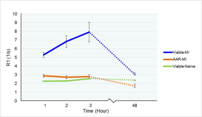
Intracellular MR contrast agents can provide essential information on cell viability. Manganese (Mn 2+) is an efficient intracellular MR contrast agent. As an analogue of Calcium, Mn 2+-induced changes in T1 and can be used as an indicator of the rate of Ca 2+ influx into cardiomyocytes in vivo and could provide additional information to that routinely acquired using late Gd-enhanced MRI. By preloaded the myocardium with Mn 2+ prior to coronary occlusion, we showed that MEMRI T1-mapping allows sensitive in vivo detection of subtle changes and accumulation of Mn 2+ in viable myocytes in the early phase of myocardial injury.
|
|
4766.
 |
19 |
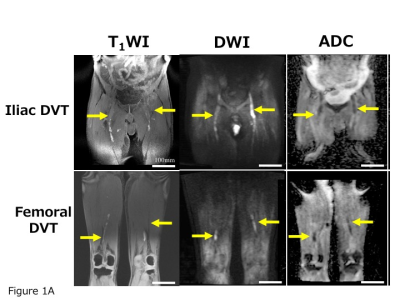
Detection of early phase of deep vein thrombosis with diffusion-weighted magnetic resonance imaging.
Yasuyoshi Kuroiwa, Atsushi Yamashita, Toshihiro Gi, Yuko Mizutani, Taketoshi Asanuma, Tosiaki Miyati, Takuroh Imamura, Yujiro Asada
To detect and characterize deep vein thrombus (DVT), we performed diffusion weighted magnetic resonance imaging (MRI) in 8 patients and a rabbit model of DVT. All patients were detected DVT as high or mixed high and iso signal intensity on diffusion weighted MRI. The rabbit venous thrombi showed high signal intensity on diffusion weighted MRI at 4 hours, or mixed iso to high and low signal intensity at 1, 2, 3 weeks. The signal intensity was positively correlated with erythrocyte contents. Diffusion weighted MRI can detect DVT and high signal intensity on the sequence may reflect early phase of DVT.
|
|
4767.
 |
20 |
Feasibility of EPI for cardiac diffusion at 7T
Elizabeth Tunnicliffe, Jesper Andersson, Aaron Hess, Matthew Robson
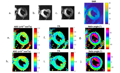
Cardiac diffusion imaging promises new insights into myocardial microstructure and integrity, but is an SNR-starved technique. Imaging at 7T may enable fewer signal averages, but most cardiac diffusion implementations use an EPI readout, which suffers from spatial distortion at higher field strengths. In this work we demonstrate distortion-corrected, EPI-based, cardiac diffusion tensor imaging at 7T using images acquired with forward and reversed phase encoding directions. We find myocardial ADC and FA consistent with previously published 3T data and that, with this acquisition protocol, distortion correction does not lead to statistically significant changes in ADC and FA.
|
|
4768.
 |
21 |
Assessing the optimal preparation strategy to minimize variability of pyruvate dehydrogenase flux measurements with hyperpolarized [1-13C]pyruvate MRS in control and type 2 diabetic rats
Kerstin Timm, Andrew Apps, Vicky Ball, Jack Miller, Michael Dodd, Damian Tyler
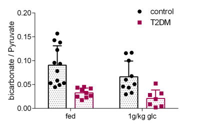
Hyperpolarized [1-13C] pyruvate MRS measures pyruvate dehydrogenase (PDH) flux in vivo in the heart through 13C-label incorporation into bicarbonate. Substrate availability modulates PDH flux; clinical protocols attempt to standardize flux with oral glucose loading prior to scanning, while rodents in preclinical studies are scanned in the fed state. We set out to establish whether feeding or glucose loading leads to more reproducible measurements of PDH flux in control rats, and found a variability of 45.1% in fed and 50.0% in glucose-loaded animals. Furthermore, glucose loading did not alter the low PDH flux seen in type II diabetic rats.
|
|
4769.
 |
22 |
Quantitative modeling and detection of flux through pyruvate dehydrogenase in human myocardium
Jeffry Alger, Jian-xiong Wang, Jeannie Baxter, Jeff Liticker, Crystal Harrison, Vlad Zaha, Albert Chen, Salvador Pena, Lucy Christie, Richard Martin, Kelley Derner, Carol Parcel, A. Sherry, Craig Malloy
Flux through pyruvate dehydrogenase, a key regulatory enzyme, may be detected in human myocardium by hyperpolarization (HP) technology. A previously-validated model for quantitation of pyruvate metabolism in isolated hearts was extended to human myocardium. HP [13C]bicarbonate and HP[1-13C]lactate were detected from heart muscle after injection of ~0.43 mL/kg of 250mM HP[1-13C]pyruvate in four healthy subjects. All subjects tolerated the procedure well. Kinetic modeling yielded a rate constant for oxidation of pyruvate to bicarbonate of 0.005 sec-1, lower than that reported for isolated rodent hearts. Assessment of PDH flux in human myocardium is feasible with hyperpolarization technology.
|
|
4770.
 |
23 |
Fast multi-slice myocardial T1 mapping (FAST1)
Li Huang, Radhouene Neji, Reza Razavi, Sébastien Roujol
Myocardial T1 mapping shows promise for the detection of cardiomyopathy. The widely-used inversion recovery based approaches, such as MOdified Look-Locker Inversion recovery (MOLLI), can only generate a single T1 map from one breathhold scan. In this study, we developed a novel FASt multi-slice myocardial T1 mapping (FAST1) approach in a single breathhold using slice-selective inversion recovery, two inversion times per slice and an advanced reconstruction approach. This technique was evaluated in a phantom as well as in healthy volunteers, and provided similar repeatability, limited precision penalty and increased spatial coverage compared to MOLLI.
|
|
4771.
 |
24 |
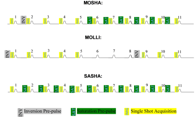
A New Myocardial T1-Mapping Technique: MOSHA
Majid Sohani, Sébastien Roujol, Andrew Powell, Andreas Maier, Mehdi Moghari
To develop an accurate and precise T1-mapping sequence, we combined the modified Look-Locker inversion recovery (MOLLI) and saturation recovery single-shot acquisition (SASHA) sequences--MOSHA. Compared to SASHA, MOSHA had similar accuracy and higher precision. Compared to MOLLI, MOSHA had better accuracy and lower precision.
|
|
Cardiovascular Image Processing
Electronic Poster
Cardiovascular
Wednesday, 20 June 2018
| Exhibition Hall |
13:45 - 14:45 |
| |
|
Computer # |
|
4772.
 |
25 |
Combined fractal and connected component analysis for characterization of cardiac fibrosis distribution in end stage renal disease patients on routine hemodialysis
Zeynep Ali, Bonnie Lam, Moriel Vandsburger
A magnetization transfer based MRI technique for measurement of cardiac fibrosis was applied to 34 end stage renal disease patients on routine hemodialysis and 19 controls. Short axis maps of changes in tissue magnetization transfer properties were combined to create 3D domains in which anatomical connectivity of voxels in the myocardium was linearized. Connected component analysis was used to isolate distinct spatially connected sub-regions of myocardium and fractal analysis of magnetization transfer changes was applied to the whole myocardium as well as these sub-regions. The measured fractal dimensions show tissue-level structural differences between the different tissue types and patient populations.
|
|
4773.
 |
26 |
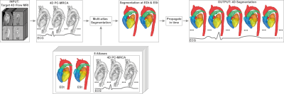
Towards automatic analysis of 4D Flow MRI: Automatic cardiac segmentation
Mariana Bustamante, Vikas Gupta, Daniel Forsberg, Carl-Johan Carlhäll, Jan Engvall, Tino Ebbers
One of the most important post-processing steps in the analysis of cardiac MR images is the segmentation of the blood pool, usually relying on manual delineation of the cardiac anatomy by an expert observer. Obtaining high quality segmentations of 4D Flow MR images acquired without the use of blood pool agents is especially challenging due to the low contrast between the blood and the myocardium present in these images. We propose an automatic multi-atlas segmentation technique that generates four-dimensional segmentations of the cardiac chambers and great thoracic vessels in 4D Flow MR images.
|
|
4774.
 |
27 |
A Simple Method for Post-Processing Correction of 3D Radial Trajectory Imperfections in 5D Cardiac MRI
Lorenzo Di Sopra, Jérôme Yerly, John Heerfordt, Davide Piccini, Matthias Stuber
Three-dimensional radial trajectories have shown important benefits for multidimensional MR imaging of the heart. However, the performance of radial scanning approaches is sensitive even to small trajectory imperfections, which can induce significant artifacts in the reconstructed images. Here, we implemented a simple and flexible post-processing technique for trajectory inaccuracy artifact compensation that does not require any pre-scan calibration or pulse sequence modification. The technique proved effective when applied to in silico and in vitro phantom datasets, and preliminarily results suggest good feasibility in vivo for fully self-gated and motion-resolved 5D cardiac imaging.
|
|
4775.
 |
28 |
Robust tissue tracking from cardiac cine MRI with deep-learning-based fully automatic myocardium segmentation
Xue Feng, Nicholas Tustison, Kun Qing, Christopher Kramer, Craig Meyer
Tissue tracking post processing from cardiac cine MRI can be used to calculate myocardial deformation parameters without additional scans. One major drawback of the processing is reduced reliability due to interference from blood and trabecular muscle signals and varying image contrast. Manual segmentation of LV myocardium can improve the robustness but is time consuming. We developed a deep convolutional neural network to automatically segment myocardium and used symmetric deformable registration to obtain the tracking information from the resulting binary masks. The segmentation and tracking worked reliably well, resulting in accurate pixel movement trajectories.
|
|
4776.
 |
29 |
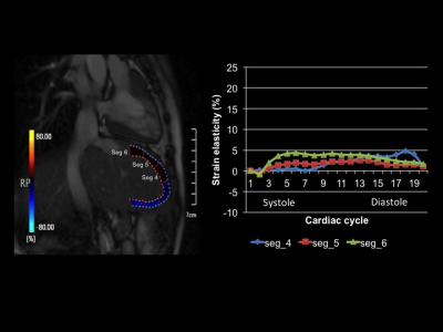
Strain elastography using a feature- tracking of cine cardiac magnetic resonance imaging: assessment of liver fibrosis in Fontan patients and tetralogy of Fallot
Ryoko Ohashi, Michinobu Nagao, Umiko Ishizaki, Yuka Matsuo, Kenji Fukushima, Yasuhiro Goto, Yumi Shiina, Kei Inai, Seiko Simizu, Yuichiro Sano, Masami Yoneyama, Shuji Sakai
We propose a new strain elastography to quantify hepatic fibrosis using feature tracking method of cardiac cine MRI, and investigate the strain elasticity in long-term post-operative patients after Fontan and intracardiac repair (ICR) for tetralogy of Fallot (TOF). The strain elasticity was lower for Fontan patients than that for TOF patients and controls, suggesting that liver fibrosis progresses after Fontan operation. The strain elastography using feature tracking method of cardiac cine MRI is a noninvasive new method for prediction of liver fibrosis.
|
|
4777.
 |
30 |
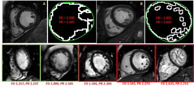
Automatic Quantification of Left Ventricular Non-compaction using Fractal Dimension and Parametric Ratio as Geometric Markers: Numerical Simulation and Clinical Evaluation
Amol Pednekar, Carter Chu, Tobias Schlingmann, Zili David Chu, Siddharth Jadhav, Cory Noel, Prakash Masand
BSSFP distinguishes the non-compacted trabeculation (NC) from compacted myocardium (C), however the current diagnostic criterion (NC/C>2.3) for left ventricular non-compaction (LVNC) suffers from subjective variability and tends to over-diagnose, especially in pediatric patients where LV trabeculation varies on a continuous spectrum. Numerical simulations estimate exponential relationship between fractal dimension (FD) and perimetric ratio (PR) with 11 times higher dynamic range for PR for the observed FD range in 30 LVNC positive and 20 negative controls. Both FD and PR indices distinguish LVNC positive from negative (p<0.0001), while the PR and FD*PR provided 6 and 11 times higher dynamic ranges respectively.
|
|
4778.
 |
31 |
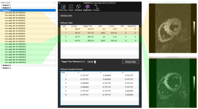
CMRDiffTools: A Processing and Analysis Tool for Cardiac Diffusion MR images
William A. Romero R., Magalie Viallon, Martijn Froeling, Christian Stoeck, Sebastian Kozerke, Elizabeth Tunnicliffe, Andrew Scott, Pedro Ferreira, Eric Aliotta, Daniel Ennis, Kévin Moulin, Pierre Croisille
CMRDiffTools is an OsiriX/Horos plug-in for simplifying and streamlining the processing and analysis tasks of Cardiac Diffusion MR images. This tool includes state-of-the-art methods in image processing of diffusion weighted images, assembled in suitable workflows to be executed in a single pipeline. The CMRDiffTools plug-in empowers the user with interactive data exploration and visualization of specific cardiac fiber metrics such as helix, transverse and sheet angle.
|
|
4779.
 |
32 |
Methodology for Non-Diseased Myocardium Part Segmentation of Delayed Enhancement 3D MRI by Using Watershed and Shape Priors
Dominika Kruk, Arnaud Boucher, Alain Lalande, Alexandre Cochet, Tadeusz Sliwa
Previous approaches on left ventricle segmentation from DE-MRI have focused on the extraction of myocardium or just diseased region in short axis orientation. However these studies are not well suited for the segmentation of non-diseased myocardium region on DE-MRI. This paper presents a novel semi-automatic segmentation method of non-diseased myocardium region segmentation on diseased patient heart from DE-MRI based on watershed algorithm with shape priors application. To assess the results segmented images were compared with gold standard images performed by an experienced user. Novel solution for DE-MRI segmentation has been proposed, which brought promising results of measured parameters.
|
|
4780.
 |
33 |
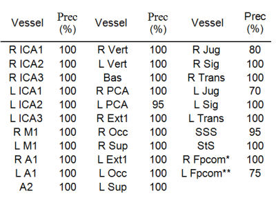
Automatic labeling of cerebral vessels in time-resolved contrast enhanced angiography
Oren Geri, Shelly Shiran, Dafna Ben Bashat
This study proposes a fully automatic labeling algorithm detecting up to 32 cerebral vessels including arteries and veins, based on 20 data sets of time-resolved contrast-enhanced angiography. The cerebral vessels include: internal carotid arteries (3-segments), middle cerebral arteries (M1), anterior cerebral artery (ACA,A1), ACA (A2), vertebral arteries, basilar artery, posterior cerebral arteries, external carotid, occipital arteries, superficial temporal arteries, internal jugular veins, and the sigmoid, transverse, superior sagittal and straight sinuses. The algorithm’s performance was validated on another 20 data sets demonstrating high precision including for the identification of the fetal posterior communicating artery, a common variant of the blood vessels.
|
|
4781.
 |
34 |
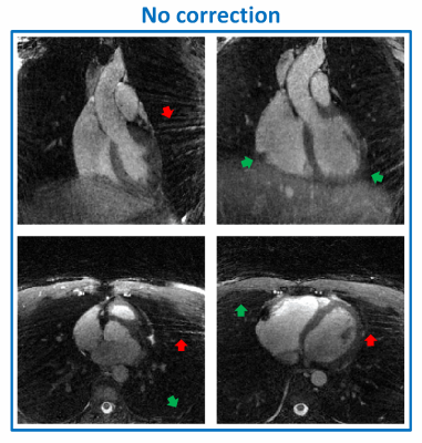
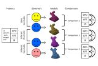
Influence of contrast, sequence, threshold, and observer on 3D segmentation of the right ventricular outflow tract for intervention planning
Video Permission Withheld
Barbara Burkhardt, Nicholas Brown, Jaclyn Carberry, Marí Nieves Velasco Forte, Nicholas Byrne, Tarique Hussain, Gerald Greil, Animesh Tandon
Three-dimensional reconstructions of cardiac magnetic resonance (CMR) datasets vary with contrast, image sequence, segmentation threshold, and manual post-processing. These influences need to be assessed before CMR-based models are used for interventional planning. Three-dimensional segmentations of the right ventricular outflow tract (RVOT) from twelve patients with three different angiography sequences were compared between and within observers using different or the same thresholds.
Thresholding was sequence-dependent and did not significantly change object volumes. Minimal diameters of 3D reconstructed RVOTs showed clinically significant variation with different thresholds and between observers.
Interventional planning should rely on source images, not three-dimensional reconstructions, for quantitative information.
|
|
4782.
 |
35 |
Aortic 4D Flow MRI Data Analysis in Bicuspid Aortic Valve Disease in Under 15 Minutes
Amer Ahmed Syed, Daniel Gordon, Jeremy Collins, Alireza Sojoudi, Qiao Wei, Xuexin Gao, Michael Scott, Bradley Allen, James Carr, Michael Markl
One of the main challenges to the efficient application of 4D flow MRI is related to cumbersome, time-consuming, and non-standardized data analysis. The purpose of this study was to test the efficiency and inter-observer reproducibility of a dedicated analysis workflow by using a 4D flow MRI tool in a cohort of 20 BAV patients. We showed that 4D flow MRI can visualize BAV mediated changes on aortic outflow and quantify associated changes in flow dynamics. We demonstrated the potential of an optimized data analysis workflow to perform standardized 4D flow MRI processing in a short time and with good-to-excellent reproducibility.
|
|
4783.
 |
36 |
Deep Learning and Compressed Sensing: Automated Image Quality Assessment in Iterative Respiratory-Resolved Whole-Heart MRI
Robin Demesmaeker, Jérôme Yerly, John Heerfordt, Tobias Kober, Pier Giorgio Masci, Dimitri Van De Ville, Matthias Stuber, Davide Piccini
We aim at creating a link between compressed sensing (CS) reconstruction and automated image quality (IQ) assessment using deep learning. An automated image quality assessment algorithm based on a deep convolutional neural regression network trained to evaluate the quality of whole-heart MRI datasets is used to assess IQ at every iteration of a respiratory motion-resolved CS reconstruction. Not only IQ evolution as assessed by the network visually correlates with the CS cost function, but the neural network is able to distinguish the image quality of different respiratory phases with high correlation to visual expert assessment.
|
|
4784.
 |
37 |
Deep Learning for Automated Medical Image Quality Assessment: Proof of Concept in Whole-Heart Magnetic Resonance Imaging
Robin Demesmaeker, John Heerfordt, Tobias Kober, Jérôme Yerly, Pier Giorgio Masci, Dimitri Van De Ville, Matthias Stuber, Davide Piccini
We aim at developing a fully automated algorithm which quantitatively gauges the quality of medical images using deep learning to mimic human perception. An automated image quality assessment algorithm based on a deep convolutional neural regression network is designed, optimized, trained, validated and tested on a clinical database of 3D whole-heart cardiac MRI scans. The algorithm was successfully trained and validated, yielding a regression performance in the range of the intra- and inter-observer agreement. These results show the relevance of deep learning concepts to image quality analysis, in particular to volumetric cardiac MR imaging.
|
|
4785.
 |
38 |
Real-World Clinical Performance of Deep Learning for Quantification and Segmentation of Biventricular Cardiac Size and Function
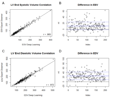
Tara Retson, Evan Masutani, Courtney Chen, Jesse Lieman-Sifry, Felix Lau, Matthieu Le, Sean Sall, Daniel Golden, Albert Hsiao
In this study of routine clinical cardiac MRIs performed for a typical range of clinical indications, we examined the effectiveness of deep learning (DL) for real-world automated quantitative analysis of cardiac size and biventricular function. We find that automated measurements correlate well with skilled readers. While the variation between DL quantification and experts lie within the range seen between experts, there remain several observed failure modes which may benefit from expert supervision. The combination of DL automation with specialist oversight may reduce the time burden of manual segmentation, improve physician efficiency, and promote technique accessibility.
|
|
4786.
 |
39 |
Cloud-based, GPU-accelerated computation of Lagrangian Coherent Structures in 4D flow enables near-instant blood flow visualization
Johannes Töger, Einar Heiberg, Christos Xanthis
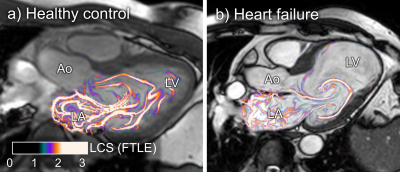
Intracardiac blood flow dynamics depicted using 4D flow magnetic resonance imaging is increasingly used for analysis of cardiac health. Lagrangian Coherent Structures (LCS) is a powerful blood flow analysis tool, with applications in diastolic dysfunction and separating flowing blood with different behavior. However, use of LCS has been limited by long computation times. Therefore, a cloud-based computation engine for LCS analysis was developed, incorporating acceleration with graphical processing units (GPU:s). A speedup factor of up to 23 times is realized, enabling sub-second LCS computation times. Furthermore, the cloud platform makes rapid LCS analysis possible without investing in expensive GPU hardware.
|
|
4787.
 |
40 |
Deep Learning Image Classification for Automatic Frequency Adjustment in High Field Cardiac MR Imaging
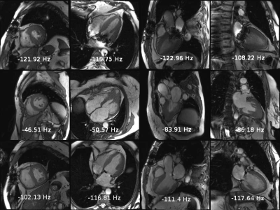
James Goldfarb, Jie Cao
Deep learning using a 3D CNN is a viable solution for the determination of the center frequency at high fields for cardiac imaging. It can quickly provide similar results to an expert observer. In the future, the strategy may be applicable to improved frequency resolution and to frequency adjustments outside of the frequency scout range. This strategy should make 3T cardiac MR imaging accessible to a wider audience.
|
|
4788.
 |
41 |
Augmenting the interpretation of cardiac MRI by biomechanical modeling: Application to tetralogy of Fallot
Radomir Chabiniok, Reagan Tompkins, Maria Gusseva, Animesh (Aashoo) Tandon, Gerald Greil, Philippe Moireau, Dominique Chapelle, Tarique Hussain
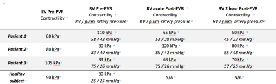
We propose connecting the acquisition and processing of cardiovascular MR (CMR) data with biomechanical cardiac modeling. Physical and physiological character predisposes biomechanical models to predictivity, and CMR data turn them into patient-specific regime. A high computational demand is addressed by applying a spatially-reduced model: the geometry and kinematics are simplified, but all other physical properties kept. The approach is illustrated by example of Tetralogy of Fallot patients, in whom we are able to access the myocardial contractility pre- and post-pulmonary valve replacement aiming at deciding on optimal therapy timing – the problem that has not been completely solved by sole CMR.
|
|
4789.
 |
42 |
Implementation of Cardiac MRF in Gadgetron for Online Reconstruction
James Ahad, Wei-Ching Lo, Jesse Hamilton, Dominique Franson, Yun Jiang, Nicole Seiberlich
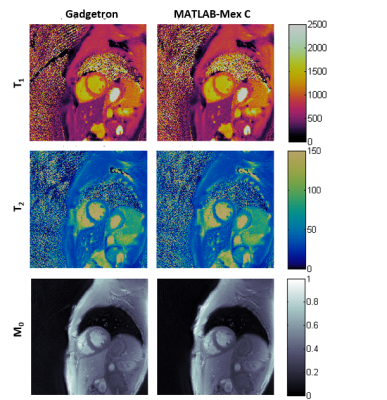
Cardiac MRF is a unique MRF technique that requires a new dictionary for each acquisition to account for heart rate variability. Dictionary simulation presents a barrier for accessibility to cardiac MRF techniques due to the lack of tools at the scanner. To this end, Bloch simulation tools were developed on the Gadgetron platform to allow for online dictionary simulation and pattern matching reconstruction. Results between prior MATLAB implementations and Gadgetron implementation were in agreement. Future work in optimization may allow for data feedback from the scanner, reducing the need for additional scan time for accurate quantification of T1 and T2 maps.
|
|
4790.
 |
43 |
Improved motion correction for myocardial T1 map and ECV map
Pan ki Kim, Chul Hwan Park, Yoo Jin Hong, Byoung Wook Choi
Chemical Exchange Saturation Transfer (CEST) has been attracting attention as a molecular imaging method to investigate myocardial muscle energetics according to creatine changes. In this study, we proposed a robust CEST imaging technique from cardiac and respiratory motion using golden angle radial readout to achieve CEST imaging at the heart of the rat. We investigated the feasibility of the proposed method for the creatine phantom and a normal rat.
|
|
4791.
 |
44 |
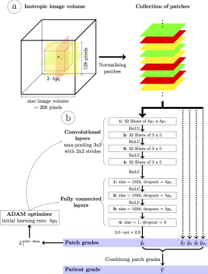
Python graphical user interface with deep learning-based segmentation for cardiac LV analysis
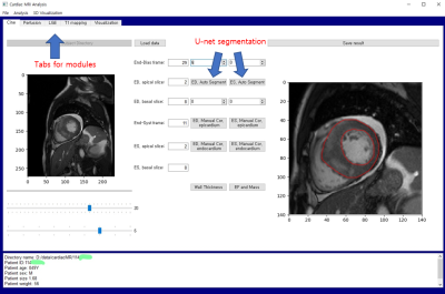
Yoon-Chul Kim, Kwanghee Choi, Yeon Hyeon Choe
The purpose of this study is to develop a deep learning algorithm for myocardial segmentation and apply it to a Python graphical user interface for cardiac MR image processing and analysis. We used a U-net architecture to simultaneously segment endocardial and epicardial borders. For training data, we used publicly available data and our internal data, both of which are from cardiac cine imaging. When the trained model was used in our Python GUI, myocardial segmentation exhibited moderate accuracy in cine data as well as in perfusion data.
|
|
4792.
 |
45 |
A powerful Matlab software platform for assessment of streamlined methods involved in quantification of MR myocardial perfusion
Clément Daviller, Thomas Grenier, Carole Frindel, Pierre Croisille, Magalie Viallon
Quantification of myocardial perfusion by MRI requires a wise combination of advanced and complex techniques from images acquisition, segmentation, deconvolution and/or modelling prior deriving perfusion indexes estimations. Accuracy/reproducibility of measures are crucial for medical diagnostic but rely on the optimization of many algorithms. Hence, the latter techniques at each step shall be made available to the community, challenged and improved for optimal state-of-the-art implementation. We propose a software platform that can easily integrate any methods addressing issues or limitations at each step of the process, integrating sharpened tools for precise assessment of individual step to global pipeline performances.
|
|
4793.
 |
46 |
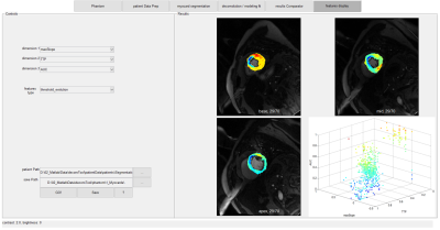
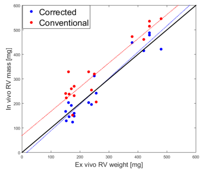
Magnetic resonance imaging of the right ventricle: a simple scheme for self-correction of partial volume effect
Bård Bendiksen, Lili Zhang, Ivar Sjaastad, Emil Espe
Magnetic resonance imaging (MRI) of the right ventricle (RV) offers important diagnostic information. However, analysis of RV MRI images is hampered by the crescent shape of the RV. In particular, pixels at the blood-myocardium border may contain signal from both blood and myocardium, hindering accurate analysis of ventricular volumes or mass. Here, we propose an easily implementable correction algorithm for analysis of standard CINE MRI image stacks, without the need for special acquisition. The proposed algorithm offers improved accuracy, in addition to more consistent RV mass estimates from CINE images of the heart in end systole and end diastole.
|
|
4794.
 |
47 |
4D Segmentation of Whole-heart Cine Cardiovascular Magnetic Resonance Imaging in Congenital Heart Disease
Jay Patel, Ruizhi Liao, Andrew Powell, Polina Golland, Mehdi Moghari
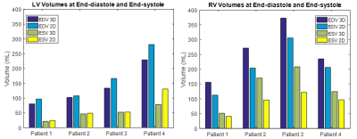
3D cine whole-heart cardiovascular magnetic resonance (MR) imaging promises to enable more accurate quantitative evaluation of cardiac function. To extract volumetric measurements of heart chambers and vascular structures from 3D cine datasets, we utilize a graph cuts segmentation algorithm and a deformable registration method. Our measurements differ from those acquired from the conventional 2D cine images, suggesting measurements of ventricular volume based on 2D cine lack accuracy. Clinical evaluation of heart function can therefore be improved by using 3D cine cardiovascular MR imaging coupled with automatic whole heart segmentation.
|
|
4795.
 |
48 |
Rigid co-registration for MOLLI, a clinical test.
Yadong Cui, Min Zhang, Xiaoqi Wang, Min Chen
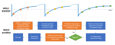
T1 quantification requires acquisition of multiple images at a sequence of delay times to derive the T1 recovery curve. Modified look-locker inversion recovery (MOLLI) is one of the most commonly used techniques. However, motion between these image acquisitions, especially at different heartbeats, often results in artifacts. In this work, we evaluated motion correction via a novel rigid co-registration based on images at a series time of delay along the T1 relaxation to eliminate the respiratory and cardiac motion artifacts.
|
|
Myocardial Tissue Characterization
Electronic Poster
Cardiovascular
Wednesday, 20 June 2018
| Exhibition Hall |
14:45 - 15:45 |
| |
|
Computer # |
|
4867.
 |
1 |
Single Acquisition Multiparametric MRI for Differentiation of Left Ventricular Scar Tissue Composition
Zahra Hosseini, Junmin Liu, Nikolaos Tzemos, Raymond Yee, Maria Drangova
Cardiac MRI plays a key role in identifying heart failure patients' response to cardiac resynchronization therapy. The identification of healthy myocardial regions for lead implantation is accomplished by means of late gadolinium enhancement MRI using exogenous contrast agents. Most MRI techniques that enable non-contrast cardiac tissue characterization are magnitude-based. MRI phase is an effective standalone source of information. However, most successful phase-based imaging is performed at lower field strength to alleviate field inhomogeneity-related artifacts. We present the application of a robust multiparametric magnitude- and phase-based myocardial mapping approach for scar tissue characterization at 3T.
|
|
4868.
 |
2 |
Temporal evaluation of acute myocardial ischemia/reperfusion injury in rats using 7.0T magnetic resonance imaging and APT102 therapeutic effect
Video Permission Withheld
Ziqian Xu, Jie Zheng, Fabao Gao
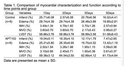
Intramyocardial hemorrhage(IMH) and microvascualr obstruction(MVO) were the serious injuries in the early myocardial infarction reperfusion period, which were independent predictors of larger infarct size, lower systolic function and the worse prognosis at follow-up. APT102 as a synthetic apyrase of the hunman nucleoside triphosphate diphosphohydrolase-3(CD39L3) can exhibit ADPase activity, prevent thrombotic reocclusion and decrease infarct size without an increased bleeding risk. The purpose of this study was to continuously detect myocardial ischemia/reperfusion injury and APT102 effect in rats by 7.0T MRI.
|
|
4869.
 |
3 |
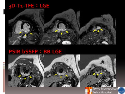
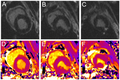
Estimation of late gadolinium enhancement of myocardial infarction with phase sensitive inversion recovery balanced steady state free precession (PSIR-bSSFP)
Takashige Yoshdia, Masami Yoneyama, Kohei Yuda, Takumi Koyano, Yuki Furukawa, Hiroe Kobayashi, Nobuo Kawauchi, Haruo Saito
The late gallium enhancement (LGE) image showing an expression amount having a significant difference for improvement wall motion or not is estimated from contrast enhancement area in patients with cardiovascular disease. However, left ventricle blood (LVB) with contrast wash-out can have similar T1 value to scar, and a fabrication sub-endocardial scar is often deluded as LVB. The phase sensitive inversion recovery balanced steady state free precession (PSIR-bSSFP) is possible to obtain black blood and contrast enhancement image. Our study of PSIR-bSSFP was propose BB-LGE that feasible visualize of myocardial hyper-enhancement area with LV blood suppression.
|
|
4870.
 |
4 |
Improvement of radiofrequency lesion visualization using 3D T1-weighted compressed sensing imaging
Pierre Bour, Valéry Ozenne, Marylène Delcey , Takeshi Kitamura, David Gonthier, Michaela Schmidt, Christoph Forman, Wadie Ben Hassen, Hubert Cochet , Pierre Jais, Bruno Quesson
Visualization of acute radiofrequency lesions in the heart is a key point to assess the endpoint of catheter-based anti-arrhythmic therapy. Albeit 3D navigated T1-weighted sequences have proven there reliability to delineate lesion cores and edema, these sequences remain too lengthy/insufficiently spatially resolved to be used clinically. In this study we investigated the benefit of combining 3D T1-weighted acquisition with compressed sensing acceleration to reduce acquisition duration while maintaining sufficient spatial resolution to visualize the core of the lesion and surrounding edema. Methods are evaluated with/without gadolinium injection with different inversion times in vivo in the heart of swine.
|
|
4871.
 |
5 |
Accelerated free breathing 3D cardiac T1? mapping in humans using compressed sensing and improved k-space reordering
Srikant Kamesh Iyer, Yuchi Han, Harold Litt, Walter Witschey
Cardiac T1ρ MRI is a parametric mapping technique for imaging myocardial fibrosis and may benefit heart disease patients unable to receive contrast agents due to poor kidney function. The purpose of this work was to develop accelerated free-breathing T1ρ mapping MRI that achieves whole heart coverage with superior spatial resolution compared to 2D methods in a reasonable scan time using compressed sensing (CS). The overall hypothesis is that 3-fold accelerated free breathing 3D cardiac T1ρ MRI is feasible.
|
|
4872.
 |
6 |
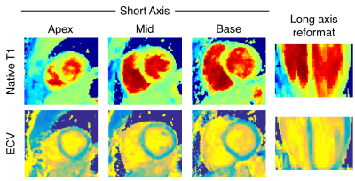
3D Whole-Ventricle, Free-Breathing, Non-ECG, Myocardial T1 and ECV Mapping with CMR Multitasking
Jaime Shaw, Anthony Christodoulou, Xiaoming Bi, Debiao Li
Myocardial fibrosis is a common pathological feature in cardiovascular diseases and can be detected with T1 and extracellular volume fraction (ECV) mapping. 3D methods are desirable to fully characterize the entire left ventricle. Typical T1 mapping methods require long scan times with respiratory navigators or repeated breath-holds which lead to patient discomfort or noncompliance. In this study, we perform 3D T1 and ECV mapping with CMR Multitasking to achieve whole-ventricle coverage in a free-breathing, non-ECG scan, producing comparable ECV to a conventional breath-hold, ECG-gated technique.
|
|
4873.
 |
7 |
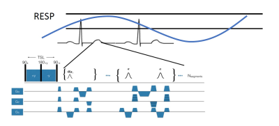
Free-breathing and ungated 3D-T1rho mapping of the heart using golden angle radial sampling and compressed sensing with low-rank constrained sparse reconstruction
Rajiv Menon, Marcelo Zibetti, Azadeh Sharafi, Li Feng, Leon Axel, Ravinder Regatte
In this study, we present a technique for T1rho mapping of myocardium using a combination of T1rho preparation, radial sampling, and low rank-based compressed sensing (CS) reconstruction. We acquire free-breathing, ungated data using a 3D T1rho prepared radial acquisition scheme with different spin lock times (TSL). After retrospectively synchronizing the data to a window in diastole, a sparse 3D k-dataset is given as input to compressed sensing reconstruction that uses a low rank constraint. Mono-exponential modeling of the reconstructed data yields the T1rho maps. The CS reconstruction results in improved images and the mean T1rho values estimated in the myocardium are consistent with literature.
|
|
4874.
 |
8 |
Myocardial fibrosis evaluated by diffusion weighted imaging and and its relationship to three-dimensional global contractile function in patients with hypertrophic cardiomyopathy
Did Not Present
Rui Wu, Dong-Ao-Lei An, Bing-Hua Chen, Ruo-Yang Shi, Wei-Bo Chen, Tong-Tong Han, Lian-Ming Wu, Jian-Rong Xu
The prognostic value of the apparent diffusion coefficient (ADC) for diffuse myocardial fibrosis and its relationship to the contractile function is still not investigated. A total of 45 HCM patients and 20 controls underwent a 3.0-T cardiac magnetic resonance imaging (CMRI), including cine, T1 mapping, and DWI. ADC values in the ECV ≥ 29.6% group were significantly increased compared to the ECV < 29.6% group. ADC values were linearly associated with 3D LV GCS, GLS, GCSR, GLSR and GRSR. Impairment of contractile function in HCM is predominantly associated with the contrast-free ADC value.
|
|
4875.
 |
9 |
Elevated NT-proBNP in a Community Cohort is Associated with Myocardial Fibrosis: the Multi-Ethnic Study of Atherosclerosis (MESA)
Did Not Present
Chia-Ying Liu, Susan Heckbert, Shenghan Lai, Bharath Amable-Venkatesh, Mohammad Ostovaneh, Robyn McClelland, Joao Lima, David Bluemke
We evaluated the relationship between cardiac MRI measures of fibrosis and NT-proBNP levels in 1334 participants in the Multi-Ethnic Study of Atherosclerosis (MESA). Univariate and multivariable regression analyses adjusting for demographics, cardiovascular risk factors, and left ventricular (LV) mass were performed to examine the association of log NT-proBNP with MRI T1 mapping indices. In the fully adjusted model, each one standard deviation increment (0.44pg/mL) of log NT-proBNP was associated with 0.62% increment in ECV (P<0.001), 4.7ms increment in native T1 (P=0.001), and 0.01 increment in partition coefficient (P<0.001). Elevated NT-proBNP is related to subclinical fibrosis in a community-based setting.
|
|
4876.
 |
10 |
Myocardial Oxygenation Using T2* BOLD Cardiac MRI with Compressed SENSE: Association with Myocardial Fibrosis and Contraction in Hypertrophic Cardiomyopathy
Michinobu Nagao, Kenji Fukushima, Eri Watanabe, Umiko Ishizaki, Yuka Matsuo, Akiko Sakai, Risako Nakao, Yasuhiro Goto, Masami Yoneyama, Shuji Sakai
We developed a new method for myocardial oxygenation using oxygen-inhalation blood-oxygen-level-dependent (BOLD) T2* cardiac magnetic resonance imaging (T2*-CMR). Myocardial oxygenation (ΔR2*, ms-1) was defined as the difference in R2* between under room-air and oxygen inhalation. ΔR2* is increased in patients with hypertrophic cardiomyopathy who had large LGE areas and low strains, suggesting that impairment of myocardial oxygenation associates with myocardial fibrosis and systolic dysfunction. In addition, T2*-CMR with compressed SENSE enables to shorten scan time and less motion artifact. This contributes to make high precision of T2* and R2* maps.
|
|
4877.
 |
11 |
Clinical validation of the T1blood-based synthetic hematocrit used to calculate the ECV on 3T MRI in patients with type 2 diabetes mellitus and hypertrophic cardiomyopathy
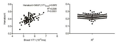
Xiaoyue Zhou, Yongning Shang, Xiaochun Zhang, Jian Wang
Calculation of the extracellular volume fraction is based on a patient’s hematocrit (Hct), which can be obtained via a clinical blood test or can be estimated synthetically according its linear relationship with the native T1 of the blood. However, the reliability of the synthetic Hct needs to be studied further. We therefore assessed the clinical validation of the synthetic Hct compared with the Hct derived from a blood test in patients with type 2 diabetes mellitus and hypertrophic cardiomyopathy.
|
|
4878.
 |
12 |
Free-Breathing Myocardial T1-Acquisition with Region-based Analysis is Feasible to Quantify Diffuse Myocardial Fibrosis
Did Not Present
Mao-Yuan Su, Yu-Sen Huang, Yeun-Chung Chang, Shun-Chung Yang, Kui-Yuan Ho, Lian-Yu Lin, Cho-Kai Wu, Wen-Yih Tseng
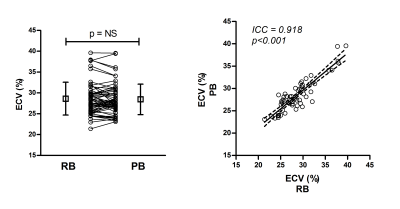
This study investigated whether free-breathing T1-acquisition with the proposed region-based (RB) method is feasible to quantify ECV as an index of diffuse myocardial fibrosis. Thirty-eight patients with non-ischemic cardiomyopathy (NICM) and 20 healthy controls were examined using both breath-hold and free-breathing myocardial T1-acquisitions. ECV was measured from free-breathing images by the RB method, which was compared with the measurement on the standard pixel-wise T1-mapping. Intraclass correlation coefficient of the two methods was 0.915. Patients with NICM showed significantly higher ECV than controls for both methods. Our results suggest that free-breathing T1-acquisition with the RB method is feasible to quantify diffuse myocardial fibrosis, and is comparable to the measurement using the standard T1-mapping.
|
|
4879.
 |
13 |
Validation of Diffusion Tensor Imaging in Diseased Myocardium
Video Permission Withheld
Irvin Teh, Darryl McClymont, Marie-Christine Zdora, Hannah J. Whittington, Katja Gehmlich, Christoph Rau, Craig A. Lygate, Jürgen E. Schneider
Diffusion tensor imaging (DTI) has been used in clinical research to identify microstructural changes in the heart following disease. However, the evidence supporting its use in the presence of abnormalities such as scar, hypertrophy and collagen infiltration is limited. Here, we investigate the application of DTI in fixed healthy, infarcted, hypertrophic and fibrotic mouse hearts, and validate these on a voxel-wise basis with high-resolution structure tensor synchrotron radiation imaging. Our findings show good agreement in helix angle estimation between the two imaging modalities across all hearts, and supports the clinical role of DTI in the presence of cardiac pathologies.
|
|
4880.
 |
14 |
Feasibility Study of Whole Heart T1 Mapping with SMS in A Single Breath Hold
Wenbo Sun, Meng Ye, Yuan Zheng, Lele Zhao, Nan Liu, Yanqun Teng, Lan Lan, Jian Xu, Haibo Xu
A novel single breath hold (BH) whole-heart T1 mapping technique with simultaneous multi-slice (SMS) imaging with improved spatial coverage and faster acquisition speed is proposed and evaluated, the initial results show that the proposed whole heart T1 mapping approach has similar results compared to conventional approaches.
|
|
4881.
 |
15 |
Spin Echo Based Cardiac Diffusion Tensor Imaging of the Unfixed, Ex-vivo Porcine Heart at 7T and 3T
Video Permission Withheld
David Lohr, Maxim Terekhov, Andreas Weng, Anja Schröder, Heike Walles, Laura Schreiber
7T, 3D, whole heart, high resolution, diffusion tensor data sets with 1.3mm isotropic voxels, unachievable in human in-vivo scans, were acquired for 8 ex-vivo pig hearts using a Stejskal-Tanner sequence with varying parallel imaging factors. ADC, FA, helix angle and secondary eigenvector angle values were analyzed and compared to a reference set of 26 ex-vivo hearts measured with the same protocol at 3T. Purpose of this study was quality assessment in DTI of the ex-vivo porcine heart at 7T. The proof of principle data acquired allows future optimization of acquisition approaches and helps translating the method to an in-vivo application.
|
|
4882.
 |
16 |
A myocardial strain phantom for cardiovascular magnetic resonance
Andrew Scott, Priyanka Sukumaran, Pedro Ferreira, Jennifer Keegan, Sonia Nielles-Vallespin, Dudley Pennell, David Firmin
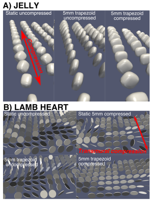
We have developed a mechanical phantom which replicates myocardial strain in blocks of jelly and sections of myocardial tissue. Initial results show the effects of strain during the diffusion time in diffusion tensor cardiovascular magnetic resonance using STEAM and the use of the phantom evaluating the DENSE strain measurement technique.
|
|
4883.
 |
17 |
Native T1 Mapping for Characterization of Acute and Chronic Myocardial Infarction in Swine: Comparison with Contrast-enhanced MRI
Did Not Present
Xi Liu, Zhi-gang Yang, Ying-kun Guo
Native T1 mapping is a novel technique that permits quantification of the T1 relaxation time of myocardial tissue without requiring exogenous contrast agents, it could be used as a reliable alternative to LGE in patients with severe renal insufficiency. The present study was conducted to determine and verify the clinical feasibility of native T1 mapping of AMI and CMI as a replacement for LGE and for the discrimination of AMI from CMI in Bama mini-pigs. In our study, native T1 mapping can accurately determine acute and chronic infarct areas as well as conventional LGE imaging, however, it cannot distinguish acute from chronic myocardial infarction.
|
|
4884.
 |
18 |
Second-order motion-compensated in-vivo cardiac diffusion tensor imaging in diastole – impact of ventricular flow, strain and trigger delay.
Robbert van Gorkum, Constantin von Deuster, Christian Stoeck, Sebastian Kozerke
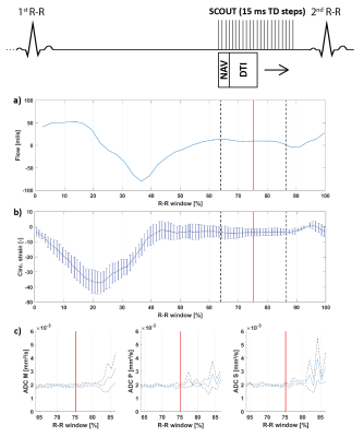
Motion-compensated spin-echo cardiac diffusion tensor imaging (cDTI) sequences suffer from signal loss if the motion pattern has higher-order motion terms than the motion compensation model of the encoding gradients. Particularly cardiac strain, ventricular flow and atrial contraction render diffusion imaging in diastole challenging. It is shown that a suitable trigger delay in the diastolic phase can be identified by taking into account ventricular flow, strain and diffusion-weighted data at several time points in diastole. This knowledge aids towards the development of spin-echo-based cDTI sequences to study dynamic myofiber changes between systole and diastole.
|
|
4885.
 |
19 |
Prognostic value of cardiac T1 mapping in Pulmonary Arterial Hypertension
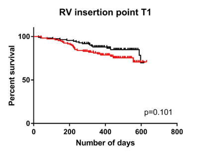
Laura Saunders, Chris Johns, Neil Stewart, Charlotte Oram, David Capener, David Kiely, Martin Graves, Jim Wild, Andy Swift
Native T1 mapping of the myocardium was performed in 223 patients with pulmonary arterial hypertension. Patients with higher right ventricular (RV) insertion point T1 were less likely to survive than patients with lower inter-ventricular insertion point T1, however when assessed in a multivariate Cox regression with other MRI markers of cardiac size and function, inter-ventricular insertion point T1 was not an independent predictor of mortality.
|
|
4886.
 |
20 |
Contrast steady state myocardial scar imaging in a chronic porcine infarct model
John Whitaker, Radhouene Neji, Rahul Mukherjee, James Harrison, Steven Williams, Henry Chubb, Louisa O'Neill, Justo Julia, John Silberbauer, Matthew Wright, Sébastien Roujol, Tevfik Ismail, Mark O'Neill, Reza Razavi
Contrast steady state (CSS) may be achieved using continuous contrast infusion following bolus. This technique was used to acquire high resolution late gadolinium enhanced (LGE) scar imaging of 8 pigs with chronic myocardial infarction. CSS imaging allowed extended acquisition to facilitate high-resolution 3D LGE imaging with improved signal-to-noise, contrast-to-noise and overall image quality. CSS may also be used in experimental studies, when total gadolinium bolus is not restricted, to directly compare imaging sequences acquired using consistent contrast distributions.
|
|
4887.
 |
21 |
An off-resonance correction for in-vivo spiral STEAM diffusion tensor cardiovascular magnetic resonance
Margarita Gorodezky, Andrew Scott, Pedro Ferreira, Sonia Nielles-Vallespin, Dudley Pennell, David Firmin
Spiral diffusion tensor cardiovascular magnetic resonance demonstrates promising results, but as with all spiral techniques is susceptible to off-resonance artefacts. The majority of off-resonance corrections rely on acquiring a separate field map based on phase differences between acquisitions with different echo times. However, bulk motion is encoded in image phase using STEAM and the motion and off-resonance induced phases are indistinguishable. Here we use a dual spiral approach in each stimulated echo to separate the motion- and off-resonance-induced phase and use this information to correct DT-CMR data.
|
|
4888.
|
22 |
Assessment of Myocardial Fibrosis in Uremic Cardiomyopathy using Cardiac MR Native T1 Mapping: A Comparison with Coronary Artery Calcium Score
Shengjia Gu, Wenjie Yang
Congestive heart failure is the leading cause of death in patients with end-stage renal disease. The process of this type of dysfunction is termed uremic cardiomyopathy. Extensive myocardial fibrosis, which leads the process, has also been shown to be a stronger predictor of death. Recently, anti-fibrosis has risen as a hot target in the treatment of uremic cardiomyopathy. Therefore, its urgent to find a more accurate and non-invasive method in order to meet the demand of clinic.In this study, we use the native T1 mapping to assess the level of myocardial fibrosis in ESRD patients,comparing with the Coronary Artery Calcium Score, in order to evaluate its accuracy in diagnosis uremic cardiomyopathy.
|
|
4889.
 |
23 |
Expanding SMS cardiac imaging with deep slice interpolation
Eric Gibbons, Akshay Chaudhari, Edward DiBella
Cardiac SMS imaging acquires multiple slices at the same time as a single slice. SMS provides increased slice coverage without a loss in temporal resolution, which is important in myocardial perfusion imaging. However, a single SMS acquisition is able to acquire three slices at a time, which is insufficient for whole heart coverage. This work expands SMS coverage by using new deep learning interpolation techniques to generate missing slices in the "gaps" between acquired slices in SMS imaging.
|
|
4890.
 |
24 |
Contrast-free 3D whole-heart magnetization transfer imaging for simultaneous myocardial scar and cardiac vein visualization
Karina Lopez, Radhouene Neji, Rahul Mukherjee, Imran Rashid, Reza Razavi, Claudia Prieto, Sebastien Roujol, Rene Botnar
A novel 3D contrast-free and motion corrected sequence for simultaneous assessment of chronic myocardial scar and coronary veins is proposed, using magnetization transfer ratio (MTR) to target the increase in collagen content associated with fibrosis. Two gradient echo datasets are sequentially acquired to obtain MTR: a reference and an off-resonance MT-weighted image. Bloch simulations and in-vivo data demonstrated that the proposed acquisition is superior to bSSFP MT-weighted sequences, yielding more consistent MTR values throughout the myocardium. Scans in patients with chronic scar confirmed the ability of MTR to localize scar, in addition to cardiac vein visualization from the MT-weighted image.
|
|
Motion & Beyond
Electronic Poster
Cardiovascular
Wednesday, 20 June 2018
| Exhibition Hall |
14:45 - 15:45 |
| |
|
Computer # |
|
4891.
 |
25 |
A comparison of motion compensated spin echo (M2SE) and stimulated echo acquisition mode (STEAM) diffusion tensor cardiovascular magnetic resonance in the in vivo assessment of hypertrophic cardiomyopathy.
Video Permission Withheld
Zohya Khalique, Andrew Scott, Pedro Ferreira, Sonia Nielles-Vallespin, David Firmin, Dudley Pennell
Diffusion tensor cardiovascular magnetic resonance (DT-CMR) interrogates myocardial microstructure, using parameters such as mean diffusivity (MD), fractional anisotropy (FA) and secondary eigenvector angulation (E2A), an index of sheetlet orientation. We compared motion compensated spin echo (M2SE) and stimulated echo acquisition mode (STEAM) and their ability to distinguish hypertrophied fibrosed myocardium (LVH+Gd+) in hypertrophic cardiomyopathy.
M2SE discriminated areas of LVH+Gd+ best through FA and MD, whilst STEAM was more sensitive to changes in E2A mobility. FA derived from both M2SE and STEAM correlated significantly with extracellular volume.
|
|
4892.
 |
26 |
Analysis of cardiac motion induced error for in-vivo cardiac DTI in different heart phases - a comparison of second-order motion compensated SE versus STEAM
Christian Stoeck, Constantin von Deuster, Robbert van Gorkum, Sebastian Kozerke
Being a single R-R interval imaging technique, second order motion compensated spin echo (M2-SE) cardiac DTI is appealing for clinical application but its implementation has so far been limited to systolic imaging. In this study we investigate signal dephasing in in-vivo cDTI at different time points within the cardiac cycle. The motion induced dephasing found for both sequences lies well within the previously reported limits for repeated measurements, when imaged in mid-to-end systole and diastole. STEAM based approaches result in consistent signal preservation across the cardia cycle, while M2-SE the exhibits a minimum in signal dephasing at around 50%-75% systole.
|
|
4893.
 |
27 |
3D Radial Free-breathing Variable Flip Angle Whole Heart Myocardial T1 Mapping
Orhan Unal, Steve Kecskemeti
A number of quantitative T1 mapping techniques have been developed for assessing myocardial pathologies. Inversion recovery (IR) techniques such as MOLLI are widely used due to higher precision and better reproducibility but tend to underestimate T1 values. The aim of this study was to evaluate the potential of new, free-breathing, variable flip angle (VFA), 3D radial and 3D hybrid-radial-Cartesian IR techniques accelerated using radial k-space undersampling for myocardial T1 quantification. Our initial results show promise and suggest that the proposed 3D T1 mapping techniques are not sensitive to B1 variations.
|
|
4894.
 |
28 |
Reconstructing real-time exercise stress cine images with multiple sets of sensitivity maps
Chong Chen, Yingmin Liu, Orlando Simonetti, Rizwan Ahmad
Motivated by the changing sensitivity maps due to exaggerated chest wall motion in real-time stress cardiac MRI (CMR), we utilize ESPIRiT-based multiple sets of sensitivity maps in a SENSE-based reconstruction method to reduce image artifact. The proposed method was tested on twelve volunteers and compared with the images reconstructed using a temporally invariant single set of sensitivity maps as well as a temporally varying single set of sensitivity maps. It was demonstrated that using multiple sets of sensitivity maps leads to significant reduction in image artifact.
|
|
4895.
 |
29 |
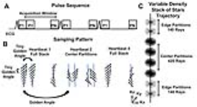
Accelerated, 3D Cine MRI with Stack of Stars k-space Sampling and Self-Navigation of Respiratory Motion for Aortic Valve Visualization.
Nivedita Naresh, Hassan Haji-Valizadeh, Bradley Allen, Matthew Barrett, Daniel Lee, Jeremy Collins, James Carr, Daniel Kim
Aortic valve disease is the most common form of valvular heart disease in the western world and accurate aortic valve visualization is very important for this reason. Accurate imaging of the aortic valve is very critical in grading and diagnosing the severity of the disease. 3D imaging may enable better visualization of the aortic valve as compared to the standard 2D techniques due to both the ability to acquire high resolution volumetric coverage and to visualize in other imaging planes. This study describes the development and evaluation of an accelerated, 3D cine MRI pulse sequence using a combination of stack-of-star k-space sampling and XDGRASP reconstruction.
|
|
4896.
 |
30 |
Influence of respiration-induced B0 variations in CSF flow quantification
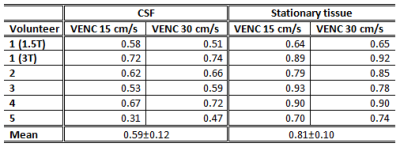
Kristina Peters, René Bastkowski, Kilian Weiss, David Maintz, Daniel Giese
Susceptibility changes and resulting B0 changes during the respiratory cycle lead to phase variations in cerebrospinal fluid phase-contrast flow measurements and might be erroneously misinterpreted as physiological flow changes. These B0 changes were analysed by acquiring flow compensated and flow encoded real-time, single-shot EPI images. The respiration-induced phase variations in phase-contrast images were found to depend on the magnetic field strength, the delay between the flow compensated and flow encoded images and the depth and frequency of respiration. Thus, respiration-induced B0 variations need to be corrected prior to analysing true respiration-dependent cerebrospinal fluid flow measured by real-time phase-contrast MRI.
|
|
4897.
 |
31 |
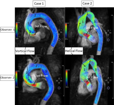
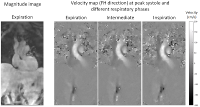
Self-gated respiratory-resolved 5D Flow MRI using the 3D spiral phyllotaxis trajectory
Monica Sigovan, Gastao Cruz, Torben Schneider, Juan Felipe Perez-Juste Abascal, Cyril Mory, Guruprasad Krishnamoorty, Rene Botnar, Philippe Douek, Claudia Prieto, Loic Boussel
Clinical diagnosis based on thoracic blood flow using three-dimensional cardiac-resolved (4D) phase contrast PC-MRI is limited by long acquisition times, due to the need of respiratory gating. In addition, gating does not provide potentially important physiological information, the variation of flow within the respiratory cycle. To address these challenges we developed a 5D (4D respiratory-resolved) PC-MRI sequence combining 3D radial k-space sampling, respiratory self-gating, and compressed sensing reconstruction. Our preliminary results demonstrate respiratory related changes in blood flow in healthy subjects. The proposed method may potentially refine diagnosis in congenital heart disease by assessing the respiratory related blood flow variations.
|
|
4898.
 |
32 |
Dynamic Auto-calibrated Multiband CAIPIRINHA: proof of principle and application to cardiac tissue phase mapping
Giulio Ferrazzi, Jean Pierre Bassenge, Clarissa Wink, Johannes Mayer, Anthony Price, Steen Moeller, Pierre-François Van de Moortele, Alexander Ruh, Michael Markl, Sebastian Schmitter
In conventional multiband CAIPIRINHA, an additional reference scan is acquired to allow the separation of the simultaneously excited slices. In this study, an acquisition-reconstruction method that makes use of the multiband data itself to calculate this reference scan is presented. The immediate implication is that the full multiband acceleration can be exploited. In addition, since the reference data has the same resolution and phase structure to multiband (i.e. it is the same data), improved slice separation is achieved. The method was implemented starting from a 2D CINE phase-contrast MR sequence, and it was used to assess velocities in the myocardium in two subjects scanned at 3T.
|
|
4899.
 |
33 |
Real life application of compressed sensing cardiac MRI for biventricular response to exercise.
Aaron Lin, Norman Morris, Helen Seale, Andrew Trotter, Benjamin Schmitt, Wendy Strugnell
Accurate assessment of left and right ventricular function using cardiac MRI plays an important role in the management of cardiac diseases. Combined with exercise stress testing, unveiling of cardiac diseases in the latent phase permits early initiation of appropriate therapy. Using compressed sensing cardiac MRI, we demonstrated dynamic quantitative biventricular functional assessment is safe and highly feasible for clinical utility.
|
|
4900.
 |
34 |
One Minute Free Breathing 3D Cardiac Cine MRI Using Data Clustering for Respiratory Self-Gating with Subject-Adaptive Gating Efficiency
Video Permission Withheld
Jing Liu, Peng Lai, Yan Wang, Zhaoying Wen, Karen Ordovas
Conventional 2D cine MRI for cardiac functional measurements requires a series of breath-holds, which is usually difficult for children or sick patients and often results in non-diagnostic images. We aim to develop a fast and reliable free-breathing 3D imaging technique, which also allows subject-specific respiratory motion compensation.
|
|
4901.
 |
35 |
3D myocardial t1 mapping using a 2D fat image navigator for respiratory motion correction
Giovanna Nordio, Torben Schneider, Gastao Cruz, Teresa Correia, Claudia Prieto, Rene Botnar, Markus Henningsson
In this study, we propose a 3D whole-heart saturation-recovery T1-mapping technique combined with a 2D fat image navigator (fat-iNAV) for respiratory motion compensation. Respiratory motion of the heart is estimated from the 2D fat-iNAVs and used to correct the T1-weighted images prior to the fitting. The delineation of the myocardium is considerably improved after motion correction, while gating permits to further improve image quality and precision (p<0.05) of the T1 maps. Future work will focus on validating the proposed technique on patients with cardiovascular disease.
|
|
4902.
 |
36 |
Motion Correction for 3D+time Cardiac MRI Perfusion Images
Apoorva Pedgaonkar, Ye Tian, Jason Mendes, Ganesh Adluru, Edward DiBella
A motion correction technique for 3D dynamic cardiac perfusion MRI using rigid and non-rigid image registrations to improve quantification of cardiac perfusion is presented. This method is unique because it employs a 3D image registration which not only resolves in plane motion but also resolves the out of plane motion for all slices in a 3D volume. This method also ensures smooth registration between all time frames of the data giving a more accurate analysis of the flow values.
|
|
4903.
 |
37 |
Propagation of Metaplastic Adipose Tissue Throughout the Scar of Hemorrhagic Myocardial Infarct is an Iron-Dependent Process: Cardiac MRI Study with Histological Insights
Ivan Cokic, Guan Wang, Xingmin Guan, Hsin-Jung Yang, Richard Tang, Diego Hernando, Scott Reeder, Rohan Dharmakumar
Lipomatous metaplasia (LM) of myocardial infarctions (MI) is typically observed in the peripheral zone of chronic MI and has been linked to major adverse clinical outcomes. To date, the mechanisms driving LM of MI remain unknown. A common feature of many disease processes associated with pathological fat accumulation is the iron-induced foam cell formation. Growing body of evidence now shows that iron deposits within hemorrhagic MI drive the prolonged recruitment of phagocytes into the infarcted territory. Herein, we investigated the spatial distribution and temporal accumulation of fatty infiltration in hemorrhagic MIs.
|
|
4904.
 |
38 |
Simultaneous T1 and T2 Mapping of the Carotid Artery with T2 and Inversion Recovery Prepared 3D Radial Imaging
Haikun Qi, Jie Sun, Huiyu Qiao, Rui Guo, Xihai Zhao, Niranjan Balu, Zechen Zhou, Chun Yuan, Huijun Chen
Multi-contrast MRI has been widely used for comprehensive characterization of atherosclerotic plaque. However, the qualitative nature and long scan time have limited its clinical application. In this study, a 3D high spatial resolution time-efficient technique, iGOAL-SNAP, is proposed, which generates different T1 and T2 contrasts in a single scan of 8min, enabling simultaneous T1 and T2 mapping. In addition, a rapid B1 correction method is presented in the fitting process. The accuracy and feasibility of iGOAL-SNAP was demonstrated using phantoms and in vivo studies.
|
|
4905.
 |
39 |
Cardiac and Respiratory Self-Gated Motion-Corrected Free-Breathing Spiral Cine Imaging.
Ruixi Zhou, Yang Yang, Roshin Mathew, Michael Salerno
We developed a free-breathing continuous-acquisition respiratory and cardiac self-gated spiral cine pulse sequence. Data was acquired using a single spiral interleaf rotated by the golden-angle in time. The cardiac self-gating signal was extracted using principal component analysis on a gridded 8x8 central region of k-space for each spiral, and the respiratory motion is derived from rigid registration for each heartbeat. Images were reconstructed with motion compensated SPIRiT using 16 seconds (2000 spirals) or 8 seconds of data. Free-breathing self-gated spiral cine imaging demonstrated high image quality providing whole heart coverage with clinical spatial and temporal resolution in under 3 minutes.
|
|
4906.
 |
40 |
Navigator-less manifold recovery of cardiac data using iterative SToRM
Did Not Present
Yasir Mohsin, Sunrita Poddar, Bijoy Thattayilath, Deidra Ansah, Mathews Jacob
Self-gated acquisitions, which rely on navigator acquisitions, can offer shortened scan time and can enable cardiac imaging of patients who cannot hold their breath; they are emerging as promising alternatives to breath-held protocols. A challenge with such schemes is the inefficiencies associated with the navigator acquisition, as well as the need for complex and heuristic processing of the navigator signals to accurately determine the cardiac and respiratory phases. The focus of this work is to introduce a navigator-less acquisition and reconstruction strategy, built upon our recent work termed as SToRM, which exploits the manifold structure of images. The proposed framework eliminates the need for navigators in SToRM, in addition to enabling spatially localized manifold modeling, where the manifold structure can vary depending on the spatial structure.
|
|
4907.
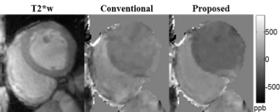 |
41 |
Initial Validation of Free-Breathing Navigator Gated Cardiac Quantitative Susceptibility Mapping via Comparison with Right Heart Catheterization Measurements
Yan Wen, Thanh Nguyen, Pascal Spincemaille, Zhe Liu, Javid Alakbarli, Jiwon Kim, Evelyn M. Horn, Meridith P. Pollie, Jonathan W. Weinsaft, Yi Wang
Our previous work has shown the feasibility of measuring differential RV-to-LV oxygen saturation with cardiac quantitative susceptibility mapping (QSM) in healthy volunteers; in this work, we present our initial validation of cardiac QSM in patients by comparing the QSM-based measurements with gold standard right heart catheter measurements.
|
|
4908.
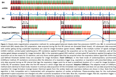 |
42 |
Reduced Specific Energy Deposition with Diagnostic Image Quality in Pediatric Population using CArdioREspiratory Synchronized (CARESync) Balanced Steady-State Free Precession Cine Imaging
Amol Pednekar, Siddharth Jadhav, Ivone Rodriguez, Connor Ho, Cory Noel, Prakash Masand
Cine-bSSFP cardiac imaging in sedated pediatric population faces two-fold challenges: 1) respiratory motion; and 2) cardiocirculatory impairment associated compromised thermoregulatory response. Prospective 63 sedated patients’ study shows that CAdioREspiratory Synchronized sequence, with prospective arrhythmia rejection and retrospective cardiac gating, reduced specific absorbed energy per slice in fixed (18.96±5.16 J/kg) by 49.36% and adaptive (24.13±9.55 J/kg) by 39.24% modes, than equivalent 4NSA sequence while maintaining spatial, temporal, and contrast resolutions possible with breath-holds, providing consistently good image quality with 21.13% increased scan time (26.38±8.15 s) per slice. Adaptive mode reduced scan time by 18% than fixed mode for favorable cardiorespiratory rates.
|
|
4909.
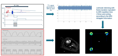 |
43 |
MRI-based retrospective gating for PET reconstruction in rats: implementation and proof of principle study
Willy Gsell, Uwe Himmelreich, Arno Nauerth, Cesar Molinos, Carlos Correcher, Antonio Gonzalez, Sven Junge, Thorsten Greeb, Ramiro Polo, Bryan Holvoet, Christophe Deroose, Michael Heidenreich
We hereby report the implementation and the preliminary results of an MRI-based retrospective gating strategy for reconstructing simultaneously acquired PET data in rats (SPECMRI). Ejection fraction extracted from MRI, SPECMRI and ECG-gated PET were all within the same range (70.0 ± 3.5%, 72.5 ± 5.4% and 70.5 ±6% for MRI, ECG-gated and SPECMRI respectively). This new technique enables to ease the animal handling (no need for ECG electrodes) and provide true synchronization of PET and MRI data.
|
|
4910.
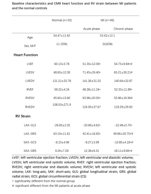 |
44 |
Assessment of right ventricular functional recovery after acute myocardial infarction by cardiac magnetic resonance
Did Not Present
Yue Gao, Zhigang Yang, Min Ma, Kaiyue Diao, Qin Zhao, Yong He
In order to clarify the assess the right ventricular (RV) function changes and recovery and the relationship with different culprit vessel for myocardial infarction (MI) patients on cardiac magnetic resonance (CMR). We prospectively recruited forty-nine MI patients and 20 healthy control, all them underwent CMR to analysis right ventricular (RV) function and global strain. Our results showed that, the RV function was related to whether the culprit vessel was RCA or not, and even RCA was obstructed the RV global strain can significantly recovery at chronic phase.
|
|
4911.
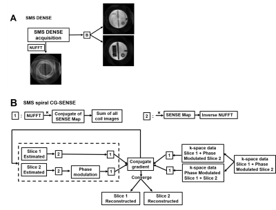 |
45 |
Simultaneous multislice spiral cine DENSE MRI
Changyu Sun, Yang Yang, Xiaoying Cai , Sophia Cui, Daniel Auger, Michael Salerno, Frederick Epstein
Spiral cine DENSE is an established method for imaging myocardial strain, however a relatively long acquisition time is a limitation. We developed a simultaneous multislice (SMS) method to accelerate spiral cine DENSE imaging. For SMS excitation we employed CAIPIRINHA phase modulation of the multiple slices. For the SMS reconstruction, we implemented a modified iterative conjugate gradient sensitivity encoding (CG-SENSE) method. Simulations and experiments in phantoms show that a 10-15% error occurs when imaging two slices simultaneously. Two-slice volunteer SMS images showed close agreement with separately-acquired single-slice images. SMS with CG-SENSE may be an effective means to accelerate spiral cine DENSE.
|
|
4912.
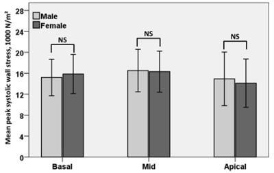 |
46 |
Effects of sex and age on CMR-derived left ventricular wall thickness and peak systolic wall stress in normal subjects
Xiaodan Zhao, Angela S Koh, Soo Kng Teo, Sen Lin, Shuang Leng, Ris Low, Yi Su, Ru San Tan, Liang Zhong
To investigate the left ventricular (LV) wall thickness, LV wall stress and their association with age and sex in a cohort of 210 healthy subjects (age: 19 to 87 years), endocardial and epicardial contour meshes from stacks of 2D short axis images were imported into our in-house software (CardioWerkz) for further analysis of 3D regional wall thickness and peak systolic wall stress (PSWS). Results indicated that mean LV wall thickness was smaller in females than males, and more positively correlated with age in female than male. Moreover, no significant difference for mean PSWS between females and males.
|
|
4913.
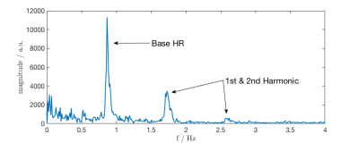 |
47 |
Model-Based Lag Free Processing of Pilot Tone Navigator Data Enables Prospective Cardiac Triggering
Mario Bacher, Peter Speier, Jan Bollenbeck, Matthias Fenchel, Matthias Stuber
Cardiac MRI acquisitions require accurate triggering, usually derived from ECG. The contactless, electromagnetic Pilot Tone navigator enables direct measurement of cardiac and respiratory motion, independently of the acquisition. Here we present an almost lag free pipeline to process the cardiac Pilot Tone signal and derive triggers from it.
|
|
4914.
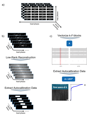 |
48 |
k-t2 ESPIRiT – Image reconstruction of respiratory motion resolved undersampled 4D Flow MRI data in a higher-dimensional subspace
Jonas Walheim, Claudio Santelli, Sebastian Kozerke
An image reconstruction algorithm termed k-t2 ESPIRiT is proposed, which constrains respiratory motion-resolved MRI to a high-dimensional subspace spanned by receiver-coil channels, cardiac phases, and respiratory motion states. Respiratory motion resolved 4D flow MRI data is reconstructed and compared to k-t ESPIRiT reconstruction of end-expiration and a standard parallel imaging protocol. Increased reconstruction accuracy compared to k-t ESPIRiT reveals that k-t2 ESPIRiT can effectively exploit redundancies between respiratory motion states to improve scan efficiency.
|
|
| Back |
| The International Society for Magnetic Resonance in Medicine is accredited by the Accreditation Council for Continuing Medical Education to provide continuing medical education for physicians. |
