|
Electronic Poster Session
Musculoskeletal |
Wednesday, 20 June 2018
Electronic PosterMusculoskeletal
5033 -5056 MSK: Cartilage
5057 -5080 MSK: Muscle 1
5129 -5152 MSK: Muscle 2 & Other Original Research
5153 -5175 MSK: Bone, Tumours & Emerging Methods
5176 -5199 MSK: Meniscus, Tendons, Ligaments & Emerging Methods |
| |
MSK: Cartilage
Electronic Poster
Musculoskeletal
Wednesday, 20 June 2018
| Exhibition Hall |
16:15 - 17:15 |
| |
|
Computer # |
|
5033.
 |
49 |
 Automated evaluation of T2 relaxation time measurements in the knee cartilage at 3T Automated evaluation of T2 relaxation time measurements in the knee cartilage at 3T
Ales Neubert, Craig Engstrom, Ilaria Croci, Shekhar Chandra, Benjamin Schmitt, Stuart Crozier, Jurgen Fripp
A software framework for fully automated analysis in 3D of regional distribution of biochemical MRI values in knee cartilage sub-regions was proposed. The framework was compared to values extracted using manual segmentations and strong agreement with the automated measures was found. The framework was applied in a preliminary analysis to assess the reproducibility of T2 relaxation time measurements at 3T with promising results. The proposed automated framework can facilitate investigations and advance the search for biomarkers of pathophysiological processes preceding the development of osteoarthritis.
|
|
5034.
 |
50 |
Depth dependence of diffusion in articular cartilage of the knee visualized at 7T
Sander Brinkhof, Qinwei Zhang, Martijn Froeling, Gustav Strijkers, Aart Nederveen, Keita Ito, Dennis Klomp
The goal of this study was to apply high-resolution diffusion weighted imaging (DWI) at 7T to assess depth dependence of diffusion in the articular cartilage in the knee. Four healthy volunteers were scanned with a diffusion prepared TSE sequence. ADC maps were consequently used to assess the depth dependence of ADC throughout the articular cartilage. The ADC value was shown to significantly increase from the bone-cartilage interface to the superficial zone. This work shows that the significant depth dependence of the ADC in cartilage layers can be observed in vivo on 7T with diffusion weighed imaging.
|
|
5035.
 |
51 |
Measurement of Acute Changes in Articular Cartilage T2 Relaxation Times Immediately After Exercise
Joanna Langner, Feliks Kogan, Bryan Haddock, Garry Gold
Increased joint loading is a known risk factor for progression of osteoarthritis (OA) of the knee. However, the acute effects of exercise and joint loading are still poorly understood. Quantitative MRI measures, such as T2 relaxation times, provide an opportunity to objectively study how exercise affects cartilage matrix organization and hydration. In this work, we evaluate the feasibility of measuring acute changes in T2 relaxation times immediately after exercise, in both knees simultaneously.
|
|
5036.
 |
52 |
T2-mapping of Femoral Cartilage 3-months Following ACL Reconstruction Surgery
Marianne Black, Katherine Young, Akshay Chaudhari, Bragi Sveinsson, Feliks Kogan, Uchechukwuka Monu, Emily McWalter, Marc Levenston, Garry Gold, Brian Hargreaves
T2-mapping can be used to detect changes in cartilage following ACL injury that are indicative of cartilage degeneration. We imaged subjects at 3-weeks and 3-months post-ACL-reconstruction surgery to obtain T2 relaxation times for the femoral cartilage of both knees. A trend in increasing T2 relaxation times was observed for the injured knee at 3-months relative to 3-weeks, although this increase was not significant, while T2 remained consistent for the contralateral knee in this time. This work demonstrates that changes to cartilage may be occurring earlier than expected in the injured knee following ACL-reconstruction surgery.
|
|
5037.
 |
53 |
T2 texture features can detect differences between the discs of patient with and without low back pain
Vahid Abdollah, Eric C Parent, Maria Beketskaia, Jeff F. Dunn
Conventional MRI is often clinically inconclusive in diagnosing the pathology of back pain, as both symptomatic and asymptomatic individuals demonstrate the same abnormal features. We hypothesized that texture driven features could be deployed to identify the underlying pathology of low back pain. Fourteen patients with chronic back pain were matched (age, weight, and gender) with 14 healthy volunteers. A grey-level co-occurrence matrix with one- to four-pixel offset and four directions was constructed to extract texture features. The texture analysis results indicated the discs of the healthy subjects were more uniform (lower contrast) than those of the participants with back pain.
|
|
5038.
 |
54 |
Compositional assessment of low-grade cartilage lesions using T2 mapping at 3 and 7 Tesla MRI: a one year follow-up study
Vladimir Juras, Markus Schreiner, Didier Laurent, Stefan Zbyn, Vladimir Mlynarik, Pavol Szomolanyi, Celeste Scotti, Joerg Goldhahn, Harry Haber, Ewa Kubiak, Stefan Marlovits, Rahel Heule, Oliver Bieri, Ivan Frollo, Siegfried Trattnig
T2 maps were assessed as a potential marker for the long-term follow-up of the patients with cartilage lesions ICRS Grade I-II in five time points (baseline, 8 days, 3, 6 and 12 months). For the T2 mapping, a 3D triple echo steady state sequence which is capable of delivering high quality high-resolved T2 maps at ultra-high field MRI was used. We observed a significant decrease in T2 values at 3T over time in superficial zone of the cartilage defect. There was no statistically significant change at 7T. T2 mapping could be used in the future as a good alternative to cartilage biopsies in clinical trials on new therapies aimed at cartilage regeneration.
|
|
5039.
 |
55 |
A composite metric R2-R1? measures an incomplete anisotropic R2 of human femoral cartilage at 3T
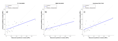
Yuxi Pang, Riann Palmieri-Smith, Thomas Chenevert
The composite relaxation metric R2-R1ρ, a potential MR imaging biomarker for detecting early cartilage degeneration, was shown as an incomplete orientation-dependent R2 at 3T. Both R2 and R1ρ mappings of femoral cartilage were performed on two subjects, and the constructed composite metrics were compared to the anisotropic R2 values that were extracted from a single 3D T2W dataset using a newly developed method. The preliminary results demonstrated that the orientation-dependent information derived from two completely different methods was comparable, implying that the diagnostically most relevant information in knee or other joint cartilage could be easily and efficiently obtained.
|
|
5040.
 |
56 |
DESS vs T1-FLASH 3D-MRI for Knee Cartilage Segmentation: An Evaluation Using Deep 3D-CNN
Archit Raj, Harsh Agarwal
Automated knee cartilage segmentation can potentially improve the clinical utility of the MRI assessment of knee osteoarthritis due to the convoluted structure of the knee cartilage in 3D. Recently deep convolutional neural network (CNN) have shown better performance for knee cartilage segmentation. Unlike other segmentation algorithms deep-CNN techniques learn the model parameters from the data itself. Therefore, this abstract proposed that deep 3D-CNN techniques can be used to determine the optimal MRI sequence for knee cartilage segmentation and demonstrated that 3D-DESS MRI have statistically better segmentation performance as compared to 3D-T1-FLASH MRI.
|
|
5041.
 |
57 |
Prediction of the mechanical properties of articular cartilage with QSM and T2* in equine post traumatic osteoarthritis model
Olli Nykänen, Juuso Ketola, Henri Leskinen, Jaakko Sarin, Nikae te Moller, Irina Mancini, Harold Brommer, Rene van Weeren, Jos Malda, Juha Töyräs, Mikko Nissi
In this study, we investigated the potential of quantitative susceptibility mapping (QSM) and T2* mapping to predict the mechanical properties of equine articular cartilage. To assess the potential of these parameters, they were used to predict the biomechanical properties of cartilage using artificial neural network (ANN) modelling of the 235 mechanical testing points in 20 equine samples representing variable tissue properties. The results indicated that both T2* and QSM correlate moderately with biomechanics (r=0.648 and r=0.652, respectively) and combining these parameters improved the correlation slightly (r=0.714). The study highlights the potential of both quantitative MRI and ANN-analysis in cartilage imaging.
|
|
5042.
 |
58 |
Simplified method for estimating chemical exchange rate in human knee cartilage from R1rho dispersion
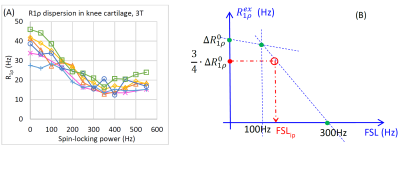
Ping Wang, John Gore
Chemical exchange between water and exchangeable protons in macromolecules in knee cartilage can be quantified by fitting R1ρ dispersion data to a model. However, acquiring the entire dispersion curve is time consuming, which therefore hampers the application in clinical practice. We propose a simple three-point method for R1ρdispersion data to estimate exchange rate. The method requires data acquired at three selected spin-locking frequencies instead of acquiring a full dispersion curve. The results show good agreement between the proposed method and measurements from the full dispersion data.
|
|
5043.
 |
59 |
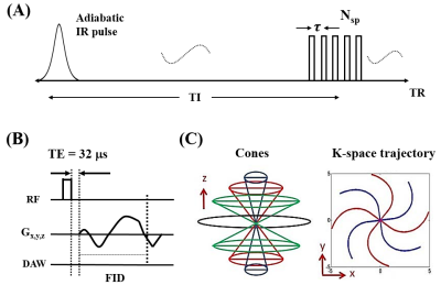
3D Ultrashort Echo Time MRI for Evaluation of Cartilaginous Endplate of Lumbar Disc In Vivo: Feasibility and Correlation with Disc Degeneration in T2 weighted Spin Echo Sequence
Yeoju Kim, Jang Gyu Cha, Gwang Pyo Hong, Juhyuen Ryu
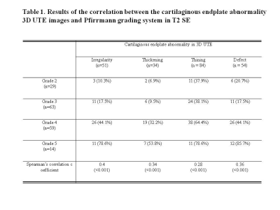
Total 165 discs of 33 patients were imaged with sagittal 3D UTE (TR = 16.1 ms, TE = 0.032 ms and 6.6 ms, echo-subtraction) with 3D cones trajectory technique and conventional sagittal T2 weighted (TR = 3232, TE = 91.4) SE in 3T MRI. Two musculoskeletal radiologists evaluated the CEP abnormalities such as irregularity, thickening, thining, and defects in the 3D UTE with consensus and correlated with the Pfirrmann grading system in the sagittal T2 SE. In result, all of the CEP abnormalities were positively correlated with the Pfirrmann grading system with statistical significance (p < 0.001).
|
|
5044.
 |
60 |
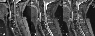
MR Evaluation of Disco-Vertebral Junction: Effects of Sequence and Parameters on SNR and CNR
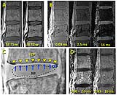
Palanan Siriwananrangsun, Tim Finkenstädt, Tailynn Chen, Michael Carl, Reni Biswas, Colin Bae, Sheronda Statum, Graeme Bydder, Christine Chung, Won Bae
The disco-vertebral junction of lumbar spine contains thin structures with short T2 values, including cartilaginous endplate (CEP) sandwiched between the bony vertebral endplate (VEP) and nucleus pulposus (NP). We have demonstrated that ultrashort echo time MRI is able to capture signal from cartilaginous endplate, and we sought to further refine the technique by characterizing contrast-to-noise ratio of these tissues when varying echo times are used. Optimal contrast between CEP and VEP was achieved with UTE source image at the shortest TE, while a balanced contrast between all tissues was achieved by Cones Subtraction imaging at a long 2nd TE.
|
|
5045.
 |
61 |
A comparative study of diffusion kurtosis imaging and T2* mapping in quantitative evalutation of lumbar intervertebral disc degeneration
Did Not Present
Feifei Zeng, Yunfei Zha, Yang Fan
The purpose of this study is to compare the diagnostic value of diffusion kurtosis imaging (DKI) and T2* mapping in assessing lumbar intervertebral disc degeneration. DKI related parameters, such as mean kurtosis (MK), mean diffusivity (MD), fractional anisotropy (FA), FA of Kurtosis (FAK) and T2* values of nucleus pulposus (NP), anterior annulus fibrosus (AAF) and posterior annulus fibrosus (PAF) were measured and correlated with Pfirrmann grades, disc level, gender and age. It was found that DKI related parameters and T2* values had significantly correlation with Pfirrmann grades, disc leveland, age, but had no significantly correlation with gender. In addition, DKI was more sensitive than T2* mapping in quantitative detection of early lumbar intervertebral disc degeneration.
|
|
5046.
 |
62 |
Comparison of DENSE-FISP and DENSE-FID for assessment of cartilage strain computation under compressive loading.
Willy Zavenbergen, Willy Gsell, Maria-Ioana Pastrama, Deva Chan, Corey Neu, Uwe Himmelreich, Ilse Jonkers
While true-FISP provides increased signal to noise ratio, it is also prone to banding artefacts due to incoherent dephasing. We thus here compare true-FISP DENSE with FID-DENSE to estimate cartilage strain under compressive loading. As FID-DENSE will be artefact free however at the expense of a lower signal to noise ratio, we investigate if DENSE-FID or DENSE-FISP will provide a more robust computation of cartilage strains under compressive loading.
|
|
5047.
 |
63 |
T1? dispersion assessment of articular cartilage in a rabbit ACL transection model
Abdul Wahed Kajabi, Victor Casula, Simo Ojanen, Mikko Finnilä, Rami Korhonen, Walter Herzog, Simo Saarakkala, Mikko Nissi, Miika Nieminen
T1ρ dispersion measurements were performed to assess early degenerative changes in articular cartilage in an experimentally induced anterior cruciate ligament transection (ACLT) rabbit model. T2 and T1ρ with spin-lock field amplitudes of 500 Hz, 1 kHz and 2 kHz were measured at 9.4 T. Cartilage mechanical properties and proteoglycan content were determined using indentation testing and quantitative histology, respectively. T1ρ dispersion identified degenerative alterations in cartilage as early as two weeks after ACL transection. The dispersion parameters were statistically correlated with biomechanical properties and proteoglycan content.
|
|
5048.
 |
64 |
T2 Relaxometry of Cartilage and Meniscus and Semi-Quantitative Assessment of the Osteoarthritic Knee using a fast 3D Quantitative DESS Scan
Susanne Eijgenraam, Akshay Chaudhari, Max Reijman, Edwin Oei, Brian Hargreaves, Garry Gold
In this study, we investigated quantitative and semi-quantitative MR imaging biomarkers of knee OA, obtained with a 5-minute DESS sequence, in increasing stages of knee OA. 54 patients were included: 20 patients with no knee OA, 18 patients with mild knee OA and 16 patients with moderate knee OA. All patients were scanned using DESS and a routine clinical knee MRI protocol. Simultaneous quantitative T2 and morphological assessment of cartilage and meniscus with a 5-minute DESS-sequence showed consistent outcomes with increasing stages of degeneration, making this sequence a useful tool for OA research.
|
|
5049.
 |
65 |
Optimizing $$$T_{1\rho}$$$ dispersion measurements for correlation time mapping in articular cartilage at 9.4 T
Hassaan Elsayed, Stefan Zbyn, Nina Hänninen, Timo Liimatainen, Mikko Nissi, Miika Nieminen, Matti Hanni
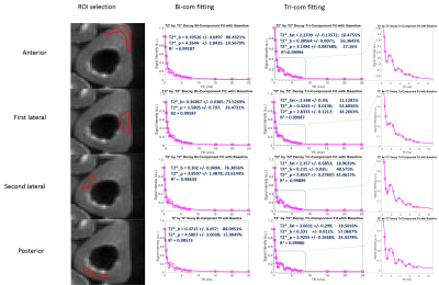
Correlation time ($$$\tau_c$$$) is an intrinsic property of molecules that can be probed from $$$T_{1\rho}$$$ dispersion measurement. The aim of this study was to evaluate the effect of low and high spin-lock frequencies (SLFs) on $$$\tau_c$$$ fitting and to find the optimum SLFs set within the typical clinical range. One bovine and two human cartilage samples were scanned to obtain $$$T_{1\rho}$$$ dispersion with 22 SLFs between 10 to 2500 Hz. $$$\tau_c$$$ maps were obtained by fitting Lorentzian function to different subsets of $$$T_{1\rho}$$$ dispersion. Our results show that $$$\tau_c$$$ fitting is significantly affected by low SLFs compared to high SLFs.
|
|
5050.
 |
66 |
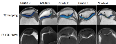
T2 mapping pseudo-color pictures and FS-FSE- PDWI in grading diagnosis of patellar cartilage damage:a retrospective study compared with arthroscopy
Shaowei Zheng, Jing Chen, Weisheng Zhang, Qingwei Song
Our institution use arthroscopy as the gold standard, apply T2mapping and FS-FSE- PDWI for the evaluation of patellar cartilage damage, to investigate value of T2 mapping pseudo-color pictures in assessment of patellar cartilage injury grading. This group of 45 cases of patellar cartilage damage, the correlations of T2mapping pseudo color grading,FS-FSE- PDWI grading and arthroscopic grading were analyzed, found the correlation between T2mapping pseudo color and arthroscopy precedes that between FS-FSE- PDWI and arthroscopy. Therefore, we believe that the T2mapping pseudo color can be used to evaluate the patellar cartilage damage, and better than FS-FSE- PDWI.
|
|
5051.
 |
67 |
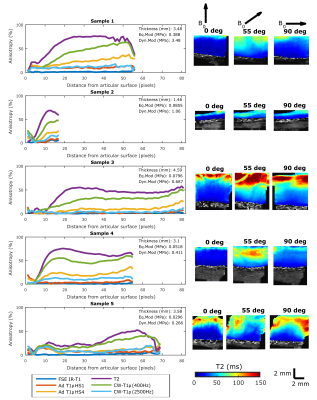
Orientation anisotropy of qMRI parameters in degenerated human articular cartilage
Nina Hänninen, Olli Nykänen, Mithilesh Prakash, Matti Hanni, Miika Nieminen, Mikko Nissi
Quantitative MR relaxation parameters have been used for evaluation of composition and structure of articular cartilage, and demonstrated to have variable sensitivity to orientation of the tissue in magnetic field. The orientation dependence of multiple relaxation parameters was assessed in human cartilage of varying degree of degeneration, and correlated with biomechanical testing. T2 and CW-T1ρ at 400Hz spin-lock demonstrated most clear anisotropy patterns which varied among the samples of different cartilage quality.
|
|
5052.
 |
68 |
Ultra-high-field (7 Tesla) MRI study of the articular cartilage in normal subjects
Giacomo Aringhieri, Massimo Marletta, Sara Toscano, Laura Biagi, Gianluigi Tiberi, Michela Tosetti, Mirco Cosottini, Virna Zampa
Ultra High Field MRI gives the opportunity to study in vivo articular cartilage with optimal signal-to-noise ratio, contrast-to-noise ratio and spatial resolution in comparison with lower field strength systems. We obtained the mean T2 and T2* maps values for under and over 50 years aged healthy subjects, both mean values increase with the subjects' age. With dedicated multichannel coils and specific cartilage sequences, it is possible to make both a qualitative and a quantitative evaluation of early and pre-clinical cartilage ageing-related changes in estimating the water content and the integrity of the cartilage matrix.
|
|
5053.
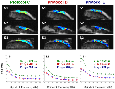 |
69 |
Accelerated T1? Dispersion Data Acquisition for Correlation Time Mapping of Cartilage at 3T
Stefan Zbyn, Mikko Nissi, Hassaan Elsayed, Victor Casula, Matti Hanni, Timo Liimatainen, Miika Nieminen
Correlation time (τc) is novel MRI parameter that can be calculated from time demanding dispersion measurements of longitudinal relaxation times in the rotating frame (T1ρ). Therefore methods able to accelerate data acquisition are needed for τc mapping of cartilage in vivo. Present results demonstrate that partial k-space acquisition and Parallel Imaging can reduce imaging time while having small influence on τc values. Although long TurboGRE readout train significantly decreases τc values, it allows the τc mapping of cartilage in clinically acceptable measurement times. The τc mapping may therefore serve for the noninvasive in vivo evaluation of cartilage at 3T.
|
|
5054.
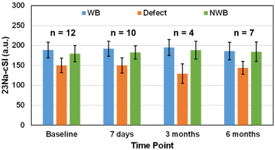 |
71 |
Sodium Imaging of Untreated Cartilage Lesions in the Knee Joint: 3-months and 6-months Follow-up Study at 7T
Stefan Zbyn, Markus Schreiner, Vladimir Mlynarik, Vladimir Juras, Pavol Szomolanyi, Didier Laurent, Celeste Scotti, Harry Haber, Joerg Goldhahn, Ewa Kubiak, Xeni Deligianni, Oliver Bieri, Stefan Marlovits, Miika Nieminen, Siegfried Trattnig
Sodium MRI was used for the follow-up of patients with cartilage lesions at 7T. MRI was obtained at baseline, 8-days, 3-months and 6-months follow-up. Regions-of-interest evaluations were performed in weight-bearing, non-weight-bearing and lesion area of femoral cartilage. Sodium values were significantly lower in lesion than in weight-bearing and non-weight-bearing regions at all follow-up measurements. On the other hand, weight-bearing and non-weight-bearing cartilage regions showed stable sodium values over the follow-up time. Sodium imaging allows noninvasive in vivo monitoring of changes in cartilage GAG content and thus can be useful for the evaluation of cartilage degeneration or cartilage regenerating therapies.
|
|
5055.
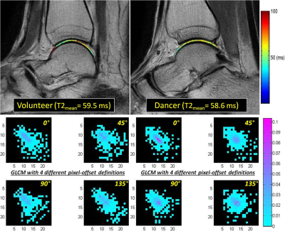 |
70 |
Texture features from T2 mapping of talar dome cartilage in normal volunteers and dancers
Hon Yu, Saya Horiuchi, Alex Luk, Adam Rudd, Jimmy Ton, Edward Kuoy, Jeff Russell, Kelli Sharp, Hiroshi Yoshioka
This study demonstrates a feasibility of texture analysis based on T2 mapping of the talar-dome cartilage. Some of the investigated texture features showed statistically significant differences between healthy volunteers and ballet dancers that are regional in nature and also very much dependent on how the spatial distribution of T2 pixels is defined during calculation of texture features. More conventional analytic approach, such as comparison based on cartilage-averaged T2 value, failed to show any difference between the groups. The results in this study demonstrate an alternative analytical approach based on texture features as surrogate variables for the evaluation of cartilage properties.
|
|
5056.
 |
72 |
Improved 3D T1rho and T2 Mapping with Synovial Fluid Suppression for the Knee Cartilage on 3T
Qi Peng, Can Wu, Xiaojuan Li, Karen Sperling
T1rho and T2 values of synovial fluid are more than twenty times larger than those of the normal cartilage tissue at 3T. The potential signal contamination originated from synovial fluid within the joints adjacent to the concerned articular cartilage could be a major source of errors associated with current T1rho and T2 mapping sequences in clinical research. In this study, we presented a long-T2-selective suppression module to null synovial fluid signal while preserve cartilage signal for acquisition. Its performance in 3D continuous-wave T1rho, adiabatic T1rho, and T2 mapping sequences was evaluated in phantom and human studies.
|
|
MSK: Muscle 1
Electronic Poster
Musculoskeletal
Wednesday, 20 June 2018
| Exhibition Hall |
16:15 - 17:15 |
| |
|
Computer # |
|
5057.
 |
73 |
Evaluation of Resting-State BOLD MRI of Calf Muscles in Healthy Volunteers and Patients with Peripheral Arterial Disease
Video Permission Withheld
Shiteng Suo, Qing Lu, Lan Zhang, Hui Tang, Qihong Ni, Suqin Li, Haimin Mao, Xiangyu Liu, Jianxun Qu, Jianrong Xu
BOLD MRI is a helpful imaging modality for assessing tissue oxygenation/perfusion characteristics in skeletal muscles. However, few studies have explored the utility of resting-state BOLD MRI in healthy subjects and patients with peripheral arterial disease (PAD). We conducted this study aimed at addressing this question, and results showed that (1) T2* was found to be independent of age factor in calf muscles; (2) T2* was useful for differentiating PAD patients with age-matched older healthy subjects, as well as mild-to-moderate PAD patients with severe ones; and (3) T2* correlated well with ankle-brachial index in PAD patients.
|
|
5058.
 |
74 |
Comparison thigh skeletal muscles between snowboarding halfpipe athletes and healthy volunteers by using quantitative multi-parameter MR imaging at rest
Did Not Present
Shinong Pan, He Sun, Mengtao Xu, Xiaoqi Wang, Menghu Wang, Baoheng Wang, Fengzhe Wang
To quantitatively investigate thigh skeletal muscles difference between snowboarding halfpipe athletes and healthy volunteers via MRI.
|
|
5059.
 |
75 |
Decreasing Water T2 based on multi-TE single-voxel MRS in fatty infiltrated skeletal muscles of patients with neuromuscular diseases
Sarah Schlaeger, Dominik Weidlich, Elisabeth Klupp, Federica Montagnese, Marcus Deschauer, Benedikt Schoser, Sarah Bublitz, Claus Zimmer, Ernst Rummeny, Jan Kirschke, Dimitrios Karampinos
Quantitative imaging techniques are emerging in the field of magnetic resonance imaging of neuromuscular diseases. Water T2 and proton density fat fraction are the most important imaging markers to assess edematous and fatty transformation in the patients’ muscle tissue. To validate the accuracy of quantitative methods 1H magnetic resonance spectroscopy can be used as a reference standard. The present study investigates water T2 of remaining muscle tissue in regions of higher proton density fat fraction in 42 patients with various neuromuscular diseases using multi-TE single-voxel MRS.
|
|
5060.
 |
76 |
Effectiveness of Diffusion-Tensor Imaging adapted to chemical-shift-encoded water-fat MRI in dystrophic skeletal muscle
Sarah Keller, Zhiyue Wang, Annette Aigner, Amir Golsari, Hendrik Kooijman, Gerhard Adam, Jin Yamamura
Diffusion tensor imaging of the skeletal muscle has been successfully applied in various conditions such as inflammation, age, and trauma. However it remains challenging in dystrophic skeletal muscle as reliable acquisition of tensor metrics is hampered due to fatty infiltration. This study aimed to assess the effectiveness of a simple clinical approach on region-of-interest localization choosing either a custom, whole muscle ROI or a selective ROI excluding areas of fatty replacement. The effect of ROI localization on tensor metrics was calculated based on mixed effect models.
|
|
5061.
 |
77 |
Do MR biomarkers for muscular fat infiltration and atrophy correlate with functionality and the DMPK CTG repeat length in myotonic dystrophy type 1?
Linda Heskamp, Marlies van Nimwegen, Guillaume Bassez, Cecilia Jimenez-Moreno, Marieke Ploegmakers, Jean-Francois Deux, Grainne Gorman, Darren Monckton, Baziel van Engelen, Arend Heerschap
Quantitative MRI provides objective non-invasive biomarkers for muscle pathology in muscular dystrophy disorders. In this work we show that MR biomarkers for muscular fat infiltration and atrophy accurately reflect clinical outcomes for disease severity and physical capacity in myotonic dystrophy type 1 (DM1) patients. Furthermore, we found that 37% of the variation in fat infiltration in DM1 patients was explained by age. Interestingly, an additional 9.7% of the variation in fat infiltration was associated with the over life time increase in the DMPK CTG repeat length, i.e. the genetic defect causing DM1.
|
|
5062.
 |
78 |
Reproducibility of Diffusion Tensor Imaging (DTI) in the hamstrings of healthy athletes
Jithsa Monte, Melissa Hooijmans, Martijn Froeling, Jos Oudeman, Johannes Tol, Mario Maas, Gustav Strijkers, Aart Nederveen
Muscle injuries are diagnosed using T2-weighted scans, but these techniques lack specificity for assessing tissue repair. DTI seems more suitable for this purpose, but reproducibility data is lacking. Therefore, the aim of this study was to determine the reproducibility of DTI, expressed as the within subject CV per DTI parameter in the hamstrings of healthy athletes. The wsCV values reported here for DTI parameters are superior or similar to previously reported wsCV. In conclusion, our protocol allows us to perform DTI on both upper legs simultaneously with an overall high SNR and high reproducibility.
|
|
5063.
 |
79 |
Combined accelerated 4D Phase Contrast and 3D Diffusion Tensor Imaging reveals a complex relation between strain and muscle architecture in contracting leg muscles
Valentina Mazzoli, Martijn Froeling, Lukas Gottwald, Nico Verdonschot, Melissa Hooijmans, Aart Nederveen, Gustav Strijkers
Skeletal muscles are geometrically complex 3D structures, and cannot be fully characterized by 2D imaging. Therefore, a complete understanding of mechanisms of force transmission and strain development in relation to muscle architecture during contraction requires a 3D approach. We measured strain rate in the lower leg using a 4D accelerated Phase Contrast protocol and 3D muscle architecture with DTI. Our 3D strain rate data revealed a planar pattern, with one negative and one positive strain rate eigenvalue. Strain rate data combined with 3D muscle architecture, suggested a complex and heterogeneous behavior of strain development during muscle contraction.
|
|
5064.
 |
80 |
Modeling skeletal muscle perfusion through application of the continuous time random walk model to diffusion-weighted images
David Reiter, Fatemeh Adelnia, Donnie Cameron, Christopher Bergeron, Richard Spencer, Luigi Ferrucci
The continuous time random walk (CTRW) model provides a flexible framework for representing complex diffusive processes, allowing for smooth interpolation between sub- and super-diffusion. This work presents supporting arguments for the application of the CTRW model to the measurement of skeletal muscle perfusion. We present model fit parameters from DW images of human skeletal muscle and compare estimates of intravascular volume fraction with previously reported values obtained using intravital microscopy.
|
|
5065.
 |
81 |
Effectiveness of fatty fraction by high-speed T2-corrected multi-echo SVS (HISTO) and multi-echo 3D DIXON (mDIXON) in rapid and robust acquisition for Duchenne Muscular Dystrophy
Xiaolei Zhu, Guijin Li, Zhiyong Li
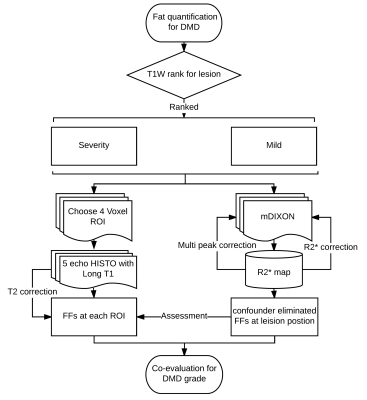
The purpose of this study is to assess a potential fat quantitative technique in Duchenne muscular dystrophy (DMD), one of High-Speed T2-corrected multi echo (HISTO) acquisition with multi echo MRS, which is a rapid way in quantifying fatty content of pelvic and thigh muscles with confound effect, and then a rapid multi echo 3D Dixon technique was performed with multi peak fitted and R2* correction method to further evaluated the un-confounded fat fraction and the region of fat content in muscles. Proposed protocol was used to quantify the accuracy of fat fraction in DMD patients; both of measurements demonstrate strong consistency in fat quantification.
|
|
5066.
 |
82 |
Quantitative water T2 mapping and 31P NMR spectroscopy to evaluate disease progression and activity in GNE myopathy patients
Harmen Reyngoudt, Julien Le Louër, Ericky Araujo, Benjamin Marty, Pierre-Yves Baudin, Jean-Yves Hogrel, Teresa Gidaro, Laurent Servais, Pierre Carlier
GNE myopathy (GNEM) is a neuromuscular disorder, characterized by distal lower limb muscle atrophy known for the relative preservation of quadriceps muscles. Quantitative NMRI including fat fraction and water T2 mapping as well as 31P NMRS were performed in 10 GNEM patients and controls. In contrast to functional and strength tests, qNMRI and 31P NMRS could detect significant changes in FF and 31P NMRS indices such as pH over the course of 1 year. More interestingly, we could also demonstrate highly significant correlations between water T2 and the rate of transformation of muscle tissue into fat, demonstrating the strength of water T2 as an indicator of disease activity.
|
|
5067.
 |
83 |
Skeletal muscle tissue characterization of Duchenne muscular dystrophy patients by 1H- and 23Na-MRI
Teresa Gerhalter, Lena Gast, Benjamin Marty, Regina Trollmann , Sophia Rügner , Stephanie Schüssler, Frank Roemer, Frederik Laun, Michael Uder, Pierre Carlier, Armin Nagel
Duchenne muscular dystrophy (DMD) is a hereditary neuromuscular disease leading to progressive muscle wasting. As there is a need to identify NMR variables as potential early sensitive indicators of dystrophic muscle response to treatment, we evaluated the sensitivity of 23Na NMR in DMD in comparison to the commonly used water T2 and fat fraction. Sodium anomalies seemed to be systematically present and precede water T2 increases and fatty degenerative changes, also in muscles that were relatively spared. Although still limited in the small number of subjects, the data supports that 23Na could be used to characterize early dystrophic muscle alteration.
|
|
5068.
 |
84 |
Assessment for Lumbar Paraspinal Muscle Activation Before and After Exercises Using BOLD and T2-Mapping Imaging
Video Permission Withheld
Bo He, Yilong Huang, Jialong Zhou, Wei Zhao, Dan Han, Weibo Chen
BOLD and T2-mapping might serve as noninvasive methods to evaluate the muscle activation of paraspinal muscles, thus providing deeper insights into muscle physiology. This has made possible the evaluation of the efficacy of early clinical exercise therapy for patients with lower-back pain.
|
|
5069.
 |
85 |
Rapid Measurement of ATP Kinetics in Human Skeletal Muscle at 7T
Jimin Ren, A. Dean Sherry, Craig R. Malloy
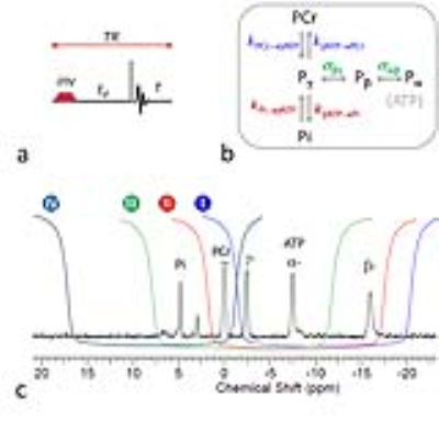
Measuring ATP energy metabolism in human subjects by conventional 31P saturation transfer (ST) is challenging because of multiple competing metabolic pathways, high SAR, and artifacts caused by the prolonged B1-saturation pulses. EKIT (exchange kinetics by inversion transfer) allows magnetization transfer (MT) to evolve in the absence of B1 pulsing and multiple pathways can be probed by inverting all spins individually, but the process is time consuming and MT effects are small. A multi-module EBIT technique (exchange kinetics by band inversion transfer) addresses these issues and provides a comprehensive picture of ATP kinetics in human skeletal muscle.
|
|
5070.
 |
86 |
Semi-automatic segmentation of individual muscles in MR images: A new tool dedicated to the follow-up of patients with neuromuscular disorders
Augustin Ogier, Linda Heskamp, Alexandre Fouré, Marc-Emmanuel Bellemare, Arnaud Le Troter, Arend Heerschap, David Bendahan
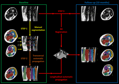
Quantitative magnetic resonance imaging can monitor intramuscular fat accumulation and has proven value for follow-up and therapy evaluation of neuromuscular disease. So far, segmentation processes of individual muscles from quantitative MRI data have been recognized as challenging in healthy subjects and even more challenging in patients for whom borders between muscles can be compromised by the disease process. We designed a semi-automatic segmentation pipeline of individual leg muscles in MR images based on automatic propagation of a minimal number of manually segmented MR slices. This segmentation pipeline allows an accurate follow-up of any MRI biomarkers in neuromuscular disorders.
|
|
5071.
 |
87 |
How reliable is DTI of the lower extremity muscles at high-field (3T) and ultra-high-field (7T) MRI?
Chiara Giraudo, Stanislav Motyka, Michael Weber, Thorsten Feiweier, Siegfried Trattnig, Wolfgang Bogner
The feasibility of DTI at 7T was already demonstrated for brain and muscles but, to date, the assessment of its reliability for calf muscles and a comparison with the reliability at 3T were still missing.Our results showed excellent ICCs(>.750) at 7T and 3T mainly for single muscles(e.g.,gastrocnemii’s tracks number).The comparison of absolute differences of the two consecutive measurements with each device demonstrated similar variability except for tracks’ number of the whole-calf(lower absolute difference at 7T;p=0.034) and FA of the gastrocnemius lateralis(lower absolute difference at 3T;p=0.032).Larger studies should further assess the overall performance of 7T for specific healthy and injured muscles.
|
|
5072.
 |
88 |
SVD Compression for Quantification of 31P Relaxation Time and Creatine Kinase Reaction Rate by 31P Magnetic Resonance Fingerprinting
Yuning Gu, Mingrui Yang, Charlie Wang, Debra McGivney, Mark Griswold, Xin Yu
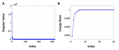
Magnetic resonance fingerprinting (MRF) provides the opportunity for efficient quantification of ATP synthesis using 31P magnetization transfer (MT) spectroscopy. However, the multi-compartment, multi-parametric nature of 31P MT experiments renders dictionary-matching computationally infeasible. In this study, singular value decomposition was employed for parameter estimation in a 31P MRF study that quantified creatine kinase activity. Such approach allowed dictionary compression by 16 fold and accelerated parameter matching by up to 80% without compromising matching accuracy. In vivo experiments on rat hindlimb (N=21) showed a 2.7-fold increase in measurement efficiency comparing to the conventional MT method using saturation transfer.
|
|
5073.
 |
89 |
Exercise-induced muscle hypoxia and re-oxygenation in the calf: A comparison between Near Infra-Red Spectroscopy (NIRS) and BOLD MRI
Christopher Conlin, Jiawei Dong, Stephen Decker, Gwenael Layec, Vivian Lee, Jeff Zhang
This study compared calf-muscle oxygenation measurements from BOLD MRI and near-infrared spectroscopy (NIRS) in a group of healthy subjects after plantar-flexion exercise. NIRS measurement of deoxyhemoglobin (dHb) was limited to the medial gastrocnemius, while BOLD imaging allowed for R2* mapping of the entire calf. Post-exercise R2* recovery dynamics in the calf indicated significant functional differences between different calf muscle groups. This advantage of BOLD makes it potentially valuable for assessing peripheral arterial disease (PAD), where impairment of muscle function can vary depending on the location of upstream stenosis.
|
|
5074.
 |
90 |
Repeatability and Reproducibility of Diffusion Tensor MRI and 2-Point Dixon Fat Fraction Measurements in the Muscle
Matthew Farrow, Ai Lyn Tan, Maya Buch, Paul Emery, Andrew Grainger, Steven Tanner, John Biglands
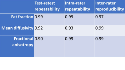
Muscle deterioration is associated with fat infiltration and alterations in muscle fibre architecture. Quantitative MRI measurements may be able to detect subtle muscle changes. However, before these can be considered for research and clinical use, the repeatability and reproducibility of these measurements must be established. 19 healthy participants had two scans separated by 30 minutes and diffusion and fat fraction measurements were obtained in the thigh. Test-retest repeatability, intra-rater repeatability and inter-rater reproducibility were assessed. Both diffusion and fat fraction measurements showed excellent repeatability and reproducibility suggesting that these measurements may be sufficiently precise to allow the study of subtle changes in muscle.
|
|
5075.
 |
91 |
Skeletal muscle acetylcarnitine in fasting and postprandial state: 1H MRS 7T pilot study.
Radka Klepochová, Martin Gajdošík, Siegfried Trattnig, Michael Krebs, Martin Krššák
Acetylcarnitine plays an important role in fat metabolism. A long TE proton magnetic resonance spectroscopy was applied for detection of skeletal muscle acetylcarnitine during the day in fasting and postprandial conditions at whole body 7T MRsystem in the vastus lateralis muscle. Our observation points towards big variations of acetylcarnitine in postprandial state and no significant changes in acetylcarnitine concentrations during the fasting. Moreover, excellent repeatability of the acetylcarnitine 1H MRS based measurement was estimated during three different days in three weeks. Our data emphasize the need for strict standardization of dietary conditions and time point for the measurement of acetylcarnitine.
|
|
5076.
 |
92 |
Muscle specific role of acetylcarnitine concentration and IMCL accumulation as a marker for long term glycemic control: 3T 1H MRS study
Radka Klepochová, Magdalena Bastian, Michael Krebs, Siegfried Trattnig, Alexandra Kautzky-Willer, Martin Krššák
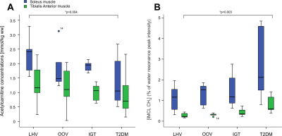
Acetylcarnitine can be observed non-invasively in 1H MRspectra in skeletal muscle and its inverse relationship to intramyocellular lipids and metabolic markers of chronic hyperglycemia was suggested. This study aimed to compare the acetylcarnitine concentrations and intramyocellular lipids content in tibialis anterior and soleus of four different groups of volunteers with broad range of glycemic control by 1HMRS on 3Tscanner. Differences in the patient phenotype were mirrored by increased intramyocellular lipids in the tibialis anterior and decreased acetylcarnitine in soleus of type2diabetes patients. This muscle specific behavior of intramyocellular metabolites could represent different fiber composition in examined muscles.
|
|
5077.
 |
93 |
Drug efficacy monitoring using magnetic resonance imaging in a cancer cachexia model
Video Permission Withheld
Ho-jin Kim, Sun Kyu Park, Jeom Yong Kim, Chul-Woong Woo, Sang-Tae Kim, Kyung Won Kim, Dong-Cheol Woo
Most studies in the field of cachexia research show photographs and H&E-stained sections of hindlimb muscles to demonstrate changes in fat/muscle volume. However, these methods do not capture the global changes in fat/muscle volume in a cachexia model. In this study, we established a cachexia animal model induced by tumor and ant-cancer drug and examined the efficacy of an anticachexia drug by monitoring fat/muscle volume using magnetic resonance imaging (MRI). Our results illustrated that MRI is a useful tool for drug development owing to its ability to monitor fat/muscle volumes in the cachexia model.
|
|
5078.
 |
94 |
Intravoxel Inchoerent Motion MRI to Evaluate Post-Occlusive Reactive Hyperemia in Calf Muscles
Alfonso Mastropietro, Simone Porcelli, Marcello Cadioli, Letizia Rasica, Elisa Scalco, Simonetta Gerevini, Mauro Marzorati, Giovanna Rizzo
The main goal of this study was to evaluate changes of IVIM parameters related to muscle perfusion changes occurring in the calf of healthy subjects before, during and after transitory ischemia of lower limb. MRI acquisitions were performed on 11 healthy volunteers on a 3T scanner. IVIM was performed on the right calf before, during and after arterial occlusion. A slight redcution of D* and fD* was observed during ischemia whereas a significant increase of D*, fD* and D was observed during hyperemia. IVIM appears as a promising tool to evaluate muscle perfusion related parameters in ischemia/hyperemia.
|
|
5079.
 |
95 |
Anisotropy of water T2* in murine skeletal muscle at rest
Aurea Martins-Bach, Ericky Araujo, Pierre Carlier
Modelling ultra-short TE (UTE) signal decay allows the extraction of multiple T2* components, an interesting approach to evaluate collagen-rich tissues with short T2 values. UTE can be promising to assess skeletal muscle fibrosis, whose non-invasive evaluation is still challenging. There are, though, indications that muscle T2* during ischemia can change when altering muscle orientation in the static magnetic field. Here we showed that muscle T2* at rest is indeed sensitive to muscle positioning in B0, with variable orientation-dependent changes in different muscles. We hypothesize that muscle structure might lead to orientation-dependent local susceptibility-induced B0 gradients, resulting in anisotropy of water-T2*.
|
|
5080.
 |
96 |
Association of thigh muscle fat infiltration with isometric strength measurements based on chemical shift encoding-based water–fat MRI
Thomas Baum, Sarah Schlaeger, Stephanie Inhuber, Florian Kreuzpointer, Michael Dieckmeyer, Friedemann Freitag, Elisabeth Klupp, Barbara Cervantes, Ansgar Schwirtz, Jan Kirschke, Dimitrios Karampinos
Chemical shift encoding-based water–fat MRI derived proton density fat fraction (PDFF) of the thigh muscles has been emerging as surrogate marker in subjects with osteoarthritis, sarcopenia, and neuromuscular disorders. However, little is known about the relationship of thigh muscle PDFF and corresponding muscle strength measurements. The present study demonstrated that PDFF measurements improve the prediction of thigh muscle strength beyond muscle cross-sectional area in healthy subjects. Thus, chemical shift encoding-based water–fat MRI can provide clinically important information and may potentially track early changes in muscles that are not severely atrophied or fattily infiltrated.
|
|
MSK: Muscle 2 & Other Original Research
Electronic Poster
Musculoskeletal
Wednesday, 20 June 2018
| Exhibition Hall |
17:15 - 18:15 |
| |
|
Computer # |
|
5129.
 |
49 |
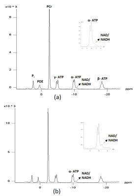
Non-invasive Detection of NADH+NAD+ in Human Muscle Using 31P MR Spectroscopy at 3T
Rajakumar Nagarajan, Miles Bartlett, Kwan-Jin Jung, Jane Kent, Nagendra Yadava
NAD+ and NADH act as coenzymes in metabolic reactions. The reduction of NAD+ to NADH is linked with generation of ATP through glycolysis and oxidative phosphorylation. The consumption of NAD+ by various signaling proteins regulates protein modification, cell fate and survival. Therefore, NAD+ and NADH measurements have the potential to inform about tissue energetics and health. Recently, some investigators have suggested that NADH and NAD+ may be detected in human muscle using 31-phosphorus MRS. However, the utility and reliability of this measure is not clear. The goals of this project were to 1) determine whether the NADH+NAD peak can be resolved in human skeletal muscle at 3T, 2) compare peak resolution with and without a decoupling technique, and 3) evaluate the reliability of this measure. Interpretation of these data and their potential for studying alterations in NAD+ and NADH homeostasis in human muscle remain to be determined.
|
|
5130.
 |
50 |
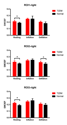
Altered Microcirculation and Oxygenation of Skeletal muscle in Type 2 Diabetes Mellitus (T2DM) Rhesus Monkeys with Non-contrast MRI Perfusion and Oximetry Techniques
Yushu Chen, Li Gong, Yu Zhang, Wen Zeng, Jie Zheng, Fabao Fao
In this study, the altered microcirculation and oxygenation of skeletal muscle in T2DM rhesus monkeys were evaluated by non-contrast skeletal muscle MR perfusion and oximetry techniques. We found that the perfusion of skeletal muscle decreased, especially in fast-twitch fiber muscles, and with an air-cuff caused muscle hyperemia, the ability to reperfusion in slow-twitch muscle is higher than in fast-twitch muscle. The oxygen extraction fraction of skeletal muscle significantly increased in all skeletal muscle angiosomes. These results suggest the diverse adaptation of slow- and fast-twitch skeletal muscles to T2DM.
|
|
5131.
 |
51 |
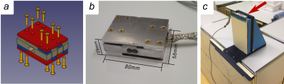
OpenForce MR: A Low-Cost Open-Source MR-Compatible Force Sensor
Video Permission Withheld
Francesco Santini, Oliver Bieri, Xeni Deligianni
This work presents an open design for a low-cost (100 USD) MR-compatible force sensor to be used in the context of dynamic muscle MRI/MRS. The sensor is both based on commercial nonmagnetic components and custom manufactured parts. The electronics is realized using Arduino and a prototype software interface is presented. The sensor is calibrated using a commercial dynamometer and shows good linearity in the sensitivity. The setup is proven to function in an MR environment without disturbing the signal acquisition.
|
|
5132.
 |
52 |
Multi-Nuclei Functional Imaging of the Skeletal Muscle during Synchronized Electrical Stimulation: Interleaving 1H Velocity Mapping with 31P Spectroscopy
Video Permission Withheld
Francesco Santini, Dirk Fischer, Oliver Bieri, Xeni Deligianni
In this work, we present a novel multi-nuclei interleaved acquisition sequence that acquires three-directional phase contrast images and phosphorus spectra during muscle exercise. The sequence is combined with electrical muscle stimulation to achieve a comprehensive investigation method of the functionality of the skeletal muscle.
|
|
5133.
 |
53 |
Diffusion, perfusion, and T2 analyses of lower leg muscles after intermittent pneumatic compression
Video Permission Withheld
Kento Furihata, Tosiaki Miyati, Naoki Ohno, Yuki Hiramatsu, Yurina Ohta, Toshifumi Gabata
Although the IPC supposedly changes regional muscle functions, this mechanism has not been clarified. Therefore, to quantitatively assess the effect of IPC, we simultaneously acquired functional information on diffusion, perfusion, and transverse relaxation time (T2) for lower-leg muscles before and after IPC using single-shot diffusion echo-planar imaging (SSD-EPI) with different b-values and echo times (TE). IPC reduces water molecule diffusion in the soleus muscle. Our method makes it possible to simultaneously obtain regional functional information on diffusion, perfusion, and T2 in lower-leg muscles before and after IPC.
|
|
5134.
 |
54 |
3D Fiber Aligned Strain Rate: Application to Unilateral Limb Suspension Induced Atrophy
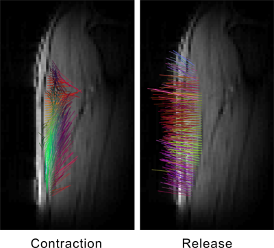
Usha Sinha, Vadim Malis, Shantanu Sinha
2D and 3D strain rate imaging has been recently introduced to study local tissue deformations. The strain rate tensors are represented in the principal basis while further relevant physiological information can be obtained by extracting 3D strain rate tensors in the muscle fiber basis; the latter is determined by diffusion tensor imaging. Here, we present the methodological developments to extract 3D fiber aligned strain rate images and application to study atrophy induced by Unilateral limb suspension. FASR indices were much smaller than those extracted in the principle basis and further studies are required to understand FASR changes with suspension.
|
|
5135.
 |
55 |
Signal-to-Noise Assessment for Diffusion Tensor Imaging of Muscles Using Single Data Set
Zhiyue Wang, Jin Yamamura, Sarah Keller
The DTI metric are prone to bias if the SNR is too low, such as in muscular dystrophies where fat infiltration decreases the partial volume of the muscle. DTI is usually acquired using parallel imaging so the noise level from the background air space is unreliable for SNR assessment. SNR can be measured using a difference image method from 2 image sets acquired repeatedly, but in many studies only one data set is acquired. Here high-pass k-space filtering method for measuring SNR of muscle DTI from single data set was optimized using a difference image method as reference.
|
|
5136.
 |
56 |
Reproducibility of calf-muscle perfusion measurements from dynamic contrast-enhanced MRI
Christopher Conlin, Jiawei Dong, Stephen Decker, Nan Hu, Mariya Chadovich, Michelle Mueller, Lillian Khor, Christopher Hanrahan, Gwenael Layec, Vivian Lee, Jeff Zhang
This study examined the reproducibility of calf-muscle perfusion measurements from DCE-MRI following plantar-flexion exercise, which is a promising technique for assessing calf-muscle function and viability. In a group of healthy subjects, the same post-exercise DCE-MRI protocol was repeated on two different days and calf-muscle perfusion measurements were compared between the two visits. High correlation and agreement of perfusion between visits was observed for the posterior calf muscles, demonstrating that a plantar-flexion exercise protocol followed by DCE-MRI is suitable for achieving precise measurements of calf-muscle perfusion.
|
|
5137.
 |
57 |
Deformable registration of calf muscle MRI using an improved Demons approach
Sarath Chintalapati, Christopher Conlin, Gwenael Layec, Stephen Decker, Nan Hu, Jiawei Dong, Christopher Hanrahan, Michelle Mueller, Lillian Khor, Vivian Lee, Jeff Zhang
Lower-extremity peripheral arterial disease (PAD) as a major clinical problem and MRI, which measures multiple aspects of the function of the calf muscles, such as muscle perfusion and oxygenation has not been significantly used for this. The reason being no efficient scanning and processing to compare data acquired during the course of treatment. Here we propose to register the calf MRI images using a modified Demons registration method which is more efficient. This method involves first a fast rigid registration and then the Demons deformable registration, by first applying rigid translation and rotation which substantially improved the registration performance for the calf muscle images.
|
|
5138.
 |
58 |
The interplay of MRS measured skeletal muscle acetylcarnitine, mitochondrial function and glucose availability
Anne Tonson, Robert Wiseman, Ronald Meyer, Taylor Ann Callahan, Ashley Lang, Jill Slade
Recently resting muscle acetylcarnitine content (AC) has been proposed as a marker for peripheral insulin resistance. However, muscle oxidative capacity and glucose availability may largely contribute to interindividual AC fluctuations independent of peripheral insulin sensitivity. In this study we monitored resting muscle AC in healthy subjects using 1H MRS in response to carbohydrate ingestion and examined the relationship of fasting muscle AC to muscle oxidative capacity measured by 31P MRS. Our results show a strong relationship between mitochondrial capacity and fasting muscle AC and also show that carbohydrate ingestion causes a rapid sharp decline in muscle AC.
|
|
5139.
 |
59 |
Rectus capitis posterior minor and nuchal ligament work as a mydural bridge complex-from 3D MR Imaging in vivo
Mei-yu Sun, Ai-lian Liu, Qing-wei Song, Li-Zhi Xie, Sheng-bo Yu, Hong-jin Sui
Myodural bridges (MDB) are soft tissue connections crossing the cervical epidural space to link suboccipital muscles, ligament and cervical dura. It may transmit tensile force from its connection components to dura mater which correlates with spinal cord circulation and chronic headache. The MDB was first described as a dense connective tissue bridge located between the rectus capitis posterior minor (RCPmi) muscle and the spinal dura. RCPmi mass has been suggested as a biomechanical contributor to injury severity in mild traumatic injury and cervicogenic headache. The fibers from nuchal ligament (NL) have also been demonstrated to attach to the cervical dura mater and may affect the RCPmi muscle mass. However, the correlation between RCPmi and NL remains unclear and is investigated in this work.
|
|
5140.
 |
60 |
A static MR follow-up study of injured levator ani muscle recovery
yujiao zhao, wen shen, zhizheng zhuo
The abnormal structure or function of Levator ani muscle(LAM) is the basis for pelvic floor dysfunction disease. Our study is to assess the recovery of injured LAM resulting from vaginal delivery by using static MRI.The primiparas who presented LAM injury at six weeks after delivery were brought into MRI follow-up study(reviewed at three months and six months).54 pubovisceralis injury with edema in a bilateral summary, there was significant difference among different postpartum time points. To summarise, the injured LAM has the ability to recover after delivery. LAM edema may exaggerate the true severity and extent of LAM injury.
|
|
5141.
 |
61 |
Quantitative and qualitative analysis of paraspinal back muscle with focus on fat content using CT and MRI in asymptomatic volunteers
Eun Kyung Khil, Jung-Ah Choi, Eunjin Hwang, Sabrilhakim Sidek, Jang Gyu Cha, Il Choi
MRI and CT can be reliably used for qualitative and quantitative analysis of para-spinal back muscles in healthy volunteers, especially regarding fat content. Good correlation was found between the two methods. Female gender and older age were associated with higher fat content of para-spinal back muscles.
|
|
5142.
 |
62 |
Reproducibility of MR elastography of lumbar paraspinal muscle in healthy volunteers
Video Permission Withheld
Chia-Hui Chen, Chien-Kuo Wang
The abnormal tissue stiffness of lumbar paraspinal muscle in symptomatic patients can be diagnosed by palpation in clinical practice. MRE can measure tissue stiffness quantitatively and noninvasively. The objective of this pilot study was to evaluate the reproducibility of MRE of lumbar paraspinal muscles in healthy volunteers. The strong positive relationship between 2 MRE exams was demonstrated by the Pearson's correlation coefficient.
|
|
5143.
 |
63 |
Variation of Strain Rate with Force Output in the Medial Gastrocnemius During Isometric Contractions in Young and Senior Subjects
Shantanu Sinha, Vadim Malis, Usha Sinha
Strain rate tensor mapping can be conveniently computed from velocity encoded phase contrast imaging. It provides a tool to explore local tissue deformations including the magnitude and directions of the principal axes of deformations. The study of the variation of strain rate indices with force output (% Maximum Voluntary Contraction (MVC) can provide additional information similar to stress-strain relationships measured at the whole muscle level. Here, we present the methodological developments to extract 2D Strain rate indices as a function of %MVC in 6 normal young (3) and senior (3) subjects. An approximately linear variation of SR indices with %MVC force was seen in the range of 20-50% MVC in young and senior subjects.
|
|
5144.
 |
64 |
Muscle Activation using 3D Cones Sodium, T2-Weighted Imaging, and T2 Mapping: Comparison of Techniques
Logan Thorneloe, Neal Bangerter, Victoria Violette, Clint Frandsen, Michael Mendoza, Wayne Johnson, Grayson Tarbox
We present a comparison of muscle functional magnetic resonance imaging (mfMRI) techniques in order to determine the most accurate method of measuring muscle activation via MRI that is practical and easy-to-use in numerous research environments.
|
|
5145.
 |
65 |
Diffusion tensor imaging of the human calf in healthy and diseased subjects during plantar-flexion exercise
Masoud Edalati, Christopher Sorensen, Mary Hastings, Mohamed Zayed, Michael Mueller, Jie Zheng
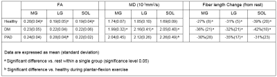
This study aims to elucidate diffusion variations between human healthy and diseased calf muscles. Subjects were assigned into three groups: healthy, diabetes mellitus (DM), and peripheral artery disease (PAD). DTI echo planar imaging was performed at rest and ankle plantar-flexion to provide fractional anisotropy (FA), mean diffusivity (MD), and fiber length for calf medial gastrocnemius (MG), lateral gastrocnemius (LG), and soleus (SOL). Our initial results revealed noticeable diffusion indices variations from resting to contraction of the above muscles between healthy and diseased groups and demonstrated a good accordance with previous healthy studies in the literature.
|
|
5146.
 |
66 |
Real-time MRI of the larynx: detecting phonation contrasts
Sarah Johnson, Marissa Barlaz, Shuju Shi, Ryan Shosted, Brad Sutton
The present study assesses the ability of rt-MRI to detect subtle laryngeal configuration changes during varying phonation contrasts. One subject lay supine within a 3T Siemens Trio scanner while producing a variety of phonation types including breathy, modal, and creaky voice. An analysis of axial and coronal slices of the larynx detected predictable changes at the ventricular folds, vocal folds and arytenoid cartilages. We conclude that rt-MRI of the larynx may have further application in the study of phonation in both research and clinical settings as a non-invasive measure of laryngeal function.
|
|
5147.
 |
67 |
Can 5-Minute 3D Isotropic Turbo Spin-Echo MR Imaging Replace 2D Standard Knee MR Imaging at 3T?
Seung hee Han , Na Hye Han, Won-Hee Jee, Joon-Yong Jung, Jun-Pyo Myoung, Yoonho Nam, Young In, Won-Hee Jee
2D TSE intermediate(IM) or T2-weighted images have been traditionally used to evaluate internal derangements of knee. However partial volume effect due to relatively thick image section and gap and long acquisition time due to impossible reformation is limitation of standard 2D TSE technique. Newly developed 3D isotropic T2 or IM weighted image is made by isotropic voxel data, which allows 3D reformation and reduce acquisition time. The purpose of this study is to compare the image quality and diagnostic performance of 5-minute sagittal fat-suppressed 3D isotropic turbo spin-echo sequence (MSK VIEW) and 2D standard knee magnetic resonance (MR) imaging at 3T.
|
|
5148.
 |
68 |
Rapid enhanced SPI imaging using inductively coupled local coils for metal artifact reduction
Alexander Storm, Kilian Stumpf, Tobias Speidel, Jan Paul, Jan Hövener, Volker Rasche
As a pure phase encoding technique, Single Point Imaging (SPI) has a great potential to reduce metal-induced artifacts, at the cost of long acquisition times normally not suited for clinical use. In this contribution, we present an approach combining SPI with reduced field-of-view imaging and SPARSE-SENSE reconstruction techniques, by using inductively coupled coils (ICC) for local signal boosting and multi-element receive coils. Initial phantom and in-vivo results show reduced metal artifacts at clinically acceptable scan times with the proposed techniques.
|
|
5149.
 |
69 |
MAVRIC SL Compressed Sensing STIR Imaging using a Total Generalized Variation (TGV) Reconstruction
Suryanarayanan Kaushik, Graeme McKinnon, Matthew Koff, Hollis Potter, Kevin Koch
3D Multi-spectral imaging sequences like MAVRIC SL have been used overcome susceptibility artifacts caused by metallic hardware. Several studies have attempted to reduce long scan times by using parallel imaging and compressed sensing, but PD images are commonly displayed with evident blurring. MAVRIC STIR images, due to their sparseness, may be more amenable to compressed sensing acceleration. The current work focuses on accelerating MAVRIC SL STIR images using compressed sensing and a total generalized variation reconstruction (TGV).
|
|
5150.
 |
70 |
Comparison of DESS and conventional clinical sequences for imaging the lumbosacral plexus
Daehyun Yoon, Adam Bartret, Peter Cipriano, Brian Hargreaves, Sandip Biswal, Amelie Lutz
Peripheral nerve imaging with MRI has gained increasing attention for non-invasive detection of nerve diseases. Unfortunately, the current performance of clinical sequences (T2-weighted fast spine echo with fat saturation and other supporting sequences) in spatial resolution, fat-saturation, and nerve-vessel distinction is often insufficient to make a convincing diagnosis. We conducted a radiologic review of lumbosacral plexus images from 3D double-echo in steady state (DESS) in comparison with images from conventional sequences in our current clinical protocol. Our results demonstrate the improved nerve visualization with the DESS sequence with comparable sensitivity to pathology.
|
|
5151.
 |
71 |
A New Contrast Mechanism for Parametric Segmentation of MRI Using Alternating RF Pulses in bSSFP Sequence
Joonsoo Kim, Hyunseok Seo, Sungpil Jung, JaeMoon Jo, Seohee So, HyunWook Park
Magnetic resonance imaging (MRI) technology offers the most important diagnostic information for musculoskeletal (MSK) disorders, in comparison to other imaging modalities (1). However, human MSK system contains a lot of fat, by which the important tissue signals from the cartilage and the synovial fluid are obstructed. Even with fat suppression techniques, it is still difficult to distinguish tissues having similar proton density (PD), T1, and T2 values. In this abstract, a new contrast enhancing method for the color MR image based on parametric segmentation is introduced, using the modified bSSFP pulse sequence.
|
|
5152.
 |
72 |
Pressure-Triggered Gated MRI Acquisition of a Vibrating Scaled Vocal Fold Model
Grayson Tarbox, Cassandra Smith, Bradley Bolster, Scott Thomson, Neal Bangerter
In this work, we present a technique for MR gated imaging of a scaled model of the vocal folds vibrating in the 13-17 Hz frequency range. The vocal fold model was scaled to roughly 4X human size to decrease the frequency of vibration and make gating feasible. The model contained an embedded grid of markers such that biomechanical motion of the laryngeal model could be accurate tracked across multiple phases of the periodic vibrational cycle.
|
|
MSK: Bone, Tumours & Emerging Methods
Electronic Poster
Musculoskeletal
Wednesday, 20 June 2018
| Exhibition Hall |
17:15 - 18:15 |
| |
|
Computer # |
|
5153.
 |
73 |
Inversion-Recovery sat-UTE sequence for short-T2 structures positive contrast generation and quantification
Lucas Soustelle, Ericky Caldas de A. Araujo, François Rousseau, Jean-Paul Armspach, Pierre G. Carlier, Paulo Loureiro de Sousa
Short-T2 structures such as myelin and cortical bone often requires the use of inversion-recovery modules in UTE sequences to provide a selective contrast in the components of interest. The sat-UTE sequence allows for an effective slice selection, and avoid issues found in commonly used 2D IR-UTE sequences concerning the use of reshaped half-radiofrequency pulses to achieve a minimal echo time. In this work, we propose to make use of an Inversion-Recovery-prepared sat-UTE sequence to provide a short-T2 positive contrast and quantification in the white matter and in the cortical bone of a fixed mouse head.
|
|
5154.
 |
74 |
MR Imaging of Patients Receiving Anterior Cervical Discectomy and Fusion Surgery Using MAVRIC-SL-STIR Technique
Renjie Yang, Yunfei Zha, Yu Zhang, Changsheng Liu, Yang Fan
Metal implants are now very common in modern joint and spine surgeries. However, conventional MR images are significantly compromised by implant-induced magnetic susceptibility artifacts. A novel metal artefacts reduction technique, termed MAVRIC-SL was proposed. The purpose of this study is to evaluate its clinical feasibility and diagnostic value in patients after anterior cervical discectomy and fusion surgery compared with routine 2D FSE images at 3 T. As a result, although the image quality of MAVRIC-SL is limited at 3 T, it can still provide important additional diagnostic information through substantially reduced metal artefacts.
|
|
5155.
 |
75 |
Ultra-short echo time (UTE) imaging with Three-component fitting analysis of human cortical bone
Xing Lu, Saeed Jerban, Michael Carl, Wenhui Yang, Annette Drygalski, Eric Chang, Jiang Du
Increased cortical porosity is a major cause of the decreased strength of osteoporotic bone, which can be evaluated by MRI based bone water components analysis. The chemical shift caused by fat in the bone may lead to incorrect estimation of water components with a bi-component exponential model. Thus, we propose a tri-component fitting method for accurate bound and pore water quantification incorporating a multi-peak spectral modeling of fat. Nine human cortical bone samples were studied. Our results suggest improved curve fitting and additional information when a tri-component analyses is used.
|
|
5156.
 |
76 |
Image derived arterial input function using popliteal artery for [18F]-sodium fluoride (NaF) PET/MRI
Bryan Haddock, Feliks Kogan, Audrey Fan, Charlotte Suetta, Garry Gold
This study evaluates the feasibility of performing dynamic [18F]-sodium fluoride (NaF) kinetic studies of the knee using PET/MRI with a focus on determining a robust method to produce an image derived input function (IDIF) using MRI images. Input functions are created using an angiography sequence to isolate the popliteal arteries and create intravascular ROIs centred in the artery to measure blood activity. 12 subjects were given two injections 75 min apart and the derived IDIFs were compared for reproducibility and accuracy. The resulting IDIFs had high reproducibility and gave values matching blood samples and literature values. Given its accuracy and robustness, this technique is well suited for clinical PET/MRI examinations.
|
|
5157.
 |
77 |
Time interleaved multi-gradient-echo imaging with UTE and non-UTE sampling for simultaneous PDFF, T2* and magnetic susceptibility mapping of cortical bone
Sophia Kronthaler, Maximilian Diefenbach, Stefan Ruschke, Jakob Meineke, Holger Eggers, Peter Boernert, Dimitrios Karampinos
Recently, quantitative magnetic susceptibility mapping (QSM) is gaining attention in the context of probing bone microstructure with potential clinical application in the assessment of bone health. Particularly, in bone applications the presence of multiple chemical species including short and long T2* components embedded in the bone environment poses several challenges including rapid signal decay, fieldmap estimation and chemical species separation. Using non-UTE QSM leads to signal voids in cortical bone regions complicating QSM. Therefore, the present study investigates the application of a stack-of-stars time-interleaved multi-gradient echo sequence including UTE and regular echo sampling for bone QSM.
|
|
5158.
 |
78 |
Prognostic value of dynamic MRI in assessing femoral head vascularity of male smokers.
Did Not Present
lei hu, yang fan
Smokers were at a higher risk of osteonecrosis of the femoral head (ONFH). In this study we aim to evaluate prognostic value of dynamic magnetic resonance imaging in assessing the femoral head vascularity of male smokers. 80 young adult men of whom 40 were smokers and 40 were nonsmokers underwent routine MR and DCE MR examination. The images and data were analyzed. In this study, we found that there was no difference of T1WI and T2WI between smokers and non-smokers. Ktrans and Kep of femoral head in the smoking group were higher compared to the control group. DCE MR can be a potential tool to detect the early change of femoral head vascularity of male smokers.
|
|
5159.
 |
79 |
Correlation study between micro-architecture of trabecular bone and magnetic resonance transverse relaxation times at multiple spatial resolutions
DongKyu Lee, Youngkyu Song, Bumwoo Park, Hwapyung Cho, Gyunggoo Cho, HyungJoon Cho
The effects of magnetic resonance (MR) imaging resolutions on the correlations between standard trabecular structural indices and MR transverse relaxation-times (T2 and T2*) were evaluated by performing Monte Carlo proton diffusion simulations, ex vivo experiments with defatted human trabecular specimens, and bovine knee trabecular samples with intact bone marrow via 7T system. T2 relaxation-time robustly represented the trabecular micro-architecture, such as trabecular spacing and number, while T2* was vulnerable with degrading spatial resolution. T2 relaxation times may facilitate the radiation-free diagnosis to assess osteoporotic fractures and therapy response for deep trabecular areas within a feasible scan time on a 7T system.
|
|
5160.
 |
80 |
Femoral Head Perfusion Color Mapping using DCE-MRI in Slipped Capital Femoral Epiphysis: Preliminary Experience
Kojiro Ono, Hirofumi Watanabe, Yasuhiro Oikawa, Takumi Okubo, Akira Shirayama
There is no established method for diagnosing femoral head necrosis before surgery, because it is difficult to predict AN due to SCFE. In this study, we propose a scheme and optimization to evaluate the femoral head perfusion in children with SCFE. Our objective is to optimize DCE-MRI and to provide useful color maps for diagnosing the femoral head perfusion.Femoral head perfusion color mapping using DCE-MRI in SCFE is useful for decision of surgical treatment method. Color maps of positive enhancement integral and maximum slope of increase contributes to evaluation of perfusion status.
|
|
5161.
 |
81 |
Imaging Scoliosis Using Zero Echo Time MRI
Chien-Yuan Lin, Hsiao-Ling Lin, Ya-Lin Fang, Hung-Ta Wu, Chi-Kuang Feng, Wan-You Guo
A high resolution, rapid scanning and three-dimensional zero-echo time (ZTE) protocol for cortical bone of whole spine imaging was established in this study. It provides computed tomography-like bone contrast and offers the similar result in the measurement of Cobb angle with conventional radiography, suggesting that the detection scoliosis and measurement of curvature using ZTE is possible, providing a potential alternative radiation-free diagnostic option that is especially relevant to scoliosis patients who need to undergo repeated examinations for evaluating the progress of spinal curvature.
|
|
5162.
 |
82 |
Estimating vertebral bone marrow triglyceride unsaturation based on the extraction of the olefinic peak in short-TE STEAM MRS using a constrained fitting model
Jan Syväri, Stefan Ruschke, Michael Dieckmeyer, Daniela Franz, Hans Hauner, Jan Kirschke, Thomas Baum, Dimitrios Karampinos
The measurement of vertebral bone marrow triglyceride unsaturation using single-voxel STEAM MRS serves as a potential biomarker for bone health. The required quantifiction of the olefinic fat peak (OFP) is often prevented by an overlapping water signal. The aim of this study was to investigate the feasibility of measuring the OFP by defining a quantification reliability measure (QRM) based on the comparison of two constrained triglyceride models. Result: The feasibility of estimating the OFP depends on the water linewidth and proton density fat fraction, increases with the QRM and may not be achievable if the water peak and OFP are not intercepting.
|
|
5163.
 |
83 |
Sex dependence of age-related vertebral bone marrow PDFF and T2 relaxation time changes in a cohort of nearly 200 subjects using multi-TE single-voxel MR spectroscopy
Jan Syväri, Stefan Ruschke, Michael Dieckmeyer, Daniela Franz, Hans Hauner, Jan Kirschke, Thomas Baum, Dimitrios Karampinos
Fat quantification of vertebral bone marrow (VBM) has been often performed with single-TE MRS. Using MRS the differences in the T2-decay of water and fat is neglected and therefore the extracted fat fraction (FF) measure comprises T2-weighting. The aim of this study was to examine differences between T2-weighted FF and proton density fat fraction (PDFF) using multi-TE single-voxel MRS and to relate the observed differences with changes in T2-relaxation of water with age in 197 subjects. The T2-relaxation of water remained constant in males and showed an age dependence in females resulting in amplified gender differences of T2-weighted FF with age compared to PDFF.
|
|
5164.
 |
84 |
What is the role of MRS-based bone marrow fatty acid unsaturation level compared to fat fraction in predicting vertebral bone strength?
Michael Dieckmeyer, Stefan Ruschke, D Anitha, Jan Kirschke, Dimitrios Karampinos, Subburaj Karupppasamy, Thomas Baum
In addition to bone marrow fat content, bone marrow composition is attracting growing interest as an advanced biomarker in the investigation of osteoporosis and bone metabolism. Finite-element analysis of CT imaging data allows non-invasive assessment of bone fracture risk. The present vertebral specimen study showed that vertebral bone marrow fatty acid unsaturation level (UL) is positively correlated with finite-element analysis based failure load and confirmed the negative correlation between bone marrow fat fraction and failure load. However, UL estimation did not improve failure load prediction beyond bone marrow fat fraction in this in-vitro study.
|
|
5165.
 |
85 |
Comparison of cortical bone visualization in the hip using ZTE and in-phase 3D gradient echo MRI sequences at 3T
Aiming Lu, Krzysztof Gorny, Joel Felmlee, Stephen Broski, Benjamin Howe
Visualizing cortical bone with MRI is challenging, as it appears as signal void in conventional MRI. Both the “black bone” technique and the zero-TE (ZTE) sequence have been proposed for cortical bone visualization. While the “black bone” technique potentially provides better cortical bone to soft tissue contrast, the ZTE sequence enables differentiation from air. This work aimed to compare the two techniques for visualizing cortical bone in the hip. Our results show that cortical bone visualization was compromised using the “black bone” technique due to chemical shift artifacts, while ZTE MRI delivered excellent depiction of the cortical bone.
|
|
5166.
 |
86 |
Cortical Bone Imaging Using Ultrashort Echo Time (UTE) Phase Sensitive Dual Inversion Recovery Subtraction Technique
Yajun Ma, Wei Zhao, Adam Searleman , Nikolaus Szeverenyi, Jiang Du, Graeme Bydder
Imaging of cortical bone is of fundamental importance in clinical MR but signals are low even with ultrashort echo time (UTE) sequences and contamination with high signals from long T2 muscle and fat is a problem 1. Here we propose a new phase sensitive dual inversion recovery subtraction method to obtain pure cortical bone images with zero signal from surrounding long T2 muscle and fat.
|
|
5167.
 |
87 |
Quantitative Dynamic Contrast-enhanced MR Imaging in Different Arterial Input Functions and Measurement Dimensions: Which Method Performs Best in differentiation of Malignant from Benign Soft Tissue Tumors?
Jimin Yoon, Won-Hee Jee, Jun-Pyo Myoung, Joon-Yong Jung, Chan Kwon Jung, Yang-Guk Chung
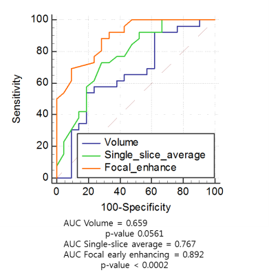
This study was designed to evaluate the reliability of quantitative dynamic contrast-enhanced magnetic resonance imaging (DCE-MRI) according to the different arterial input functions (AIF) at different measurement dimensions in differentiating malignant from benign soft tissue tumors at 3T. Quantitative DCE-MRI parameters of either benign or malignant tumors were obtained in three different measurement dimensions: focal early enhancing area, single-slice average, and whole tumor volume. They were calculated using three different population-averaged AIF (fast, intermediate and slow) and one of the three AIF of the lowest Chi-square, using SyngoVia software. The result showed quantitative DCE-MRI may be reliable and accurate in differentiating malignant from benign soft tissue tumors at 3T, particularly from focal early enhancing area using intermediate or fast AIF.
|
|
5168.
 |
88 |
Differentiation of Pulmonary From Non-Pulmonary Spine Metastases Using Conventional DCE Kinetic Analysis and Machine Learning
Ning Lang, Yang Zhang, Enlong Zhang, Jiahui Zhang, Daniel Chow, Peter Chang, Melissa Khy, Hon Yu, Huishu Yuan, Min-Ying Su
DCE-MRI of the spine was analyzed to differentiate metastasis from lung cancer (N=30) and other tumors (N=31, 9 breast, 6 prostate, 7 thyroid, 6 liver, 3 kidney). Using DCE parameters measured from the tumor ROI, CHAID decision tree classification selected the wash-out slope of -6.6% and wash-in SE of 98% as thresholds, which could achieve diagnostic accuracy of 0.79. In machine learning, the enhanced tumor on DCE image was segmented automatically by using the normalized cut algorithm. The Convolutional Long Short Term Memory (CLSTM) network with all 12 sets of DCE images as the input could yield accuracy of 0.75-0.84.
|
|
5169.
 |
89 |
Texture Analysis Based Separation of Osteosarcoma and Ewing’s Sarcoma using Diffusion Weighted MRI
Video Permission Withheld
Esha Baidya Kayal, Devasenathipathy Kandasamy, Jayendra Alampally, Sameer Bakhshi, Raju Sharma, Amit Mehndiratta
Osteosarcoma(OS) and Ewing’s sarcoma family of tumors(ESFT) are the most common primary malignant bone tumor in children and adolescents. As the patient’s age, presenting signs & symptoms and qualitative imaging features are common in both, there could be a diagnostic dilemma that can only be resolved by biopsy & histopathological assessment. Texture analysis in Diffusion Weighted Imaging(DWI) might provide additional useful information as DWI captures early cellular changes within tumors. We computed textural features in patients with OS and ESFT. Experimental results revealed that textural features provided significant discrimination between OS and ESFT; thus might be useful as diagnostic marker.
|
|
5170.
 |
90 |
Texture Analysis of Apparent Diffusion Coefficient (ADC) Maps of Vertebral Bone Marrow in Patients with Multiple Myeloma: Correlation with Clinical Parameters and the Revised International Staging System
Video Permission Withheld
Qin Wang, Huadan Xue, Tianyi Qian, Alto Stemmer, Shuo Li, Zhaoyong Sun, Zhengyu Jin
This study aimed to correlate textural parameters derived from apparent diffusion coefficient (ADC) maps of bone marrow to clinical parameters and the revised international staging system (R-ISS) in patients with multiple myeloma (MM). Sixty-one patients with MM underwent whole-body DWI. Texture analysis of ADC maps on vertebral bodies extracted five parameters, including mean, standard deviation, entropy, skewness and kurtosis. These parameters were significantly correlated with clinical parameters of disease burden. Entropy showed the highest diagnostic capacity for discriminating R-ISS stage III from stage I/II. ADC-based textural parameters may serve as adjunct tools for detecting MM patients with advanced R-ISS stages.
|
|
5171.
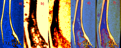 |
91 |
MRI, CT, and histomorphometry co-registration studies: analysis of murine tibia morphology before and after decalcification process
Marilena Tauro, William Dominguez-Viqueira, Jan Poleszczuk, Conor Lynch, Gary Martinez, Mikalai Budzevich
In this work we evaluate the relative merits of MRI and CT to “gold standard” histopathology assessment, with regard to bone tumor metastases. We also investigate the profound effect that decalcification process has on tumor/bone morphology. MRI shows differences in tumor shape before and after decalcifications, however the differences in total tumor volume were smaller than 5 percent. Co-registered microCT images and MR images exhibit displacement of bone-matrix after decalcification. The question of co-registration between all three modalities remains open, however we believe our algorithm minimizes registration errors.
|
|
5172.
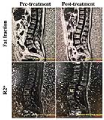 |
92 |
Evaluation of the therapeutic effect on multiple myeloma patients using IDEAL IQ
Did Not Present
Yichen Ma, Li Wang, Yanhua Tang, Yang Fan, Tao Jiang
Multiple myeloma is the second most common hematologic malignancy which occurs commonly in elderly patients. The alteration of bone marrow microenvironment caused by multiple myeloma may lead to the change of fat content and T2* of the bone. IDEAL IQ is a technique which can simultaneously and quantitatively calculate the fat fraction and R2* value of given tissues. The purpose of this study is to investigate the prognostic value of IDEAL IQ technique for the Blood treatment under M protein test on multiple myeloma.
|
|
5173.
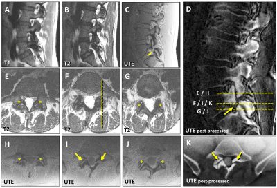 |
93 |
From Bench to Bedside: Improved Detection of Spondylolysis Using 3D Cones Ultrashort Time to Echo (UTE) MR Technique
Tim Finkenstaedt, Suraj Achar, Ibraheem Algarni, Michael Carl, Palanan Siriwanarangsun, Nirusha Abeydeera, Reni Biswas, Sheronda Statum, Christine Chung, Won Bae
Spondylolysis, defined as a bony defect in the pars interarticularis of the vertebral arch, is a frequent condition in the young population in which exposure to ionizing radiation is a major concern. In practice, CT imaging is performed in addition to MRI if spondylolysis is suspected. 3D Cones-based UTE MR techniques providing contrast and resolution similar to CT may have overcome the issue of conventional MR-sequences that do not yield sufficient contrast of short T2 tissues near the pars defect. Our study shows in an exemplary manner that detection of spondylolysis with UTE sequences is feasible in the clinical setting.
|
|
5174.
 |
94 |
Fat Saturation in Musculoskeletal MRI using Spectral Heterogeneity Adaptive RF Pulses (SHARP)
Video Permission Withheld
Venkata Veerendranadh Chebrolu, Matthew Frick, Peter Kollasch, Benjamin Schmitt, Vibhas Deshpande, John Grinstead, Daniel Spence, Joel Felmlee, Kimberly Amrami
In this work we propose the use of adaptive RF pulses for achieving uniform fat-saturation (fat-sat) that is robust to B0 and B1 heterogeneity often observed in musculoskeletal MRI. A prototype with Spectral Heterogeneity Adaptive RF Pulses (SHARP) was developed to improve the quality and functionality of fat-sat. SHARP was shown to provide fat-sat with higher robustness to B0 and B1 heterogeneity compared to SPIR and SPAIR fat-sat methodologies.
|
|
5175.
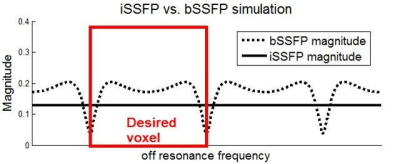 |
95 |
Water/fat separation imaging at 7T using 3D integrated SSFP
Kaibao Sun, Zhentao Zuo, Huilou Liang, Jing An, Danny J.J. Wang, Rong Xue
Balanced SSFP (bSSFP) has been used as a new alternative for water-fat separation imaging due to its fast acquisition and high SNR efficiency. However, it displays characteristic banding artifacts in the presence of field inhomogeneity, especially at the 7T ultrahigh-field MRI system. Complex combination of two phase-cycled bSSFP was used to alleviate the artifact at 3T, but would take more scan time. Integrated SSFP (iSSFP), which was modified from bSSFP, was introduced to separate water and fat while removing banding artifacts in shorter scan time. iSSFP also inherited the advantages of relatively high SNR and bright fluid. 3D iSSFP with high spatial resolution may have great potential to separate water and fat in the clinical application of 7T ultrahigh-field MRI.
|
|
MSK: Meniscus, Tendons, Ligaments & Emerging Methods
Electronic Poster
Musculoskeletal
Wednesday, 20 June 2018
| Exhibition Hall |
17:15 - 18:15 |
| |
|
Computer # |
|
5176.
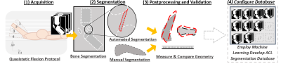 |
97 |
Protocol for in vivo acquisition and fully-automated segmentation of the anterior cruciate ligament
Laura Lane, Gabriel della Maggiora, Nicolás Schlotterbeck, Cristián Montalba, Pablo Besa, Sebastián Irarrazaval, Alvaro Burdiles, Pablo Irarrazaval
In this preclinical study we propose a protocol for rapid 3D imaging and fully automated segmentation to create a standardized healthy ACL image database. The segmentation problem of the ACL is particularly challenging due to its poor contrast. Our protocol demonstrated promising fully-automated segmentation of the ACL. Thus, allowing us to have a 3D computational model of the ACL. Ongoing experimentation explores dynamic imaging of the ACL in motions of flexion-extension. Such work will improve understanding of in vivo knee mechanics with potential to inform treatment of different injuries related to the ACL.
|
|
5177.
 |
98 |
CPPD-diseased, Normal and Degenerated Menisci: Biomechanical and quantitative MRI-derived Properties
Tim Finkenstaedt, Reni Biswas, Nirusha Abeydeera, Palanan Siriwanarangsun, Robert Healey, Sheronda Statum, Won Bae, Christine Chung
Calcium pyrophosphate crystal deposition (CPPD) disease of the knee is a frequent condition in the elderly population that might cause chondrocalcinosis of the fibrocartilaginous menisci. Controversy exists whether chondrocalcinosis of the menisci is a cause or consequence of osteoarthritis. Thus, it would be desirable to determine whether CPPD-afflicted menisci exhibit altered biomechanical properties and if they correlate with quantitative MR values. Purpose of this study is to investigate if CPP deposits alter the biomechanical properties as well as the UTE values of menisci compared to grossly normal and degenerated human menisci.
|
|
5178.
 |
99 |
Pre-operative MRI for Predicting Clinical Outcome Following Rotator Cuff Tendon Repair Surgery
Richard Kijowski, Kevin Lian, Peter Thurlow, Gang Li, Michael Tuite, Fang Liu
This study was performed to determine whether pre-operative MRI could provide prognostic information regarding clinical outcome following rotator cuff tendon repair surgery. The study group consisted of 141 patients undergoing surgical repair of a full-thickness rotator cuff tendon tear who completed the Disabilities of Arm, Shoulder and Hand (DASH) and Short Form (SF-12) surveys before and one year after surgery. The pre-operative MRI was reviewed to determine tear size, tear retraction, torn tendon tendinosis, and composite muscle atrophy. There was a significant inverse association (p<0.05) between post-operative DASH and SF-12 scores and tear width, tendon retraction, and torn tendon tendinosis.
|
|
5179.
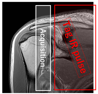 |
100 |
Usefulness of non-contrast perfusion using arterial spin labeling (ASL) for the differential diagnosis of shoulder pain
Katsumi Nakamura, Fumihide Arakami, Tomoko Nishi, Kenichi Inoue, Akiyoshi Yamamoto, Asaka Aso, Mitsue Miyazaki
We evaluated the usefulness of non-contrast perfusion obtained using arterial spin labeling (ASL) for the assessment and the differential diagnosis of shoulder disorders. Twenty consecutive patients were divided into 4 groups. Group 1 (partial tear or less) had no enhancement at the subacromional region; whereas, group 2 (full thickness tear of rotator cuff) showed significant enhancement due to bursitis. Group 3 (calcific tendinitis) and group 4 (osteoarthritis) also showed well enhancement. ASL perfusion could provide the additional physiological information to the morphological routine shoulder MRI without use of contrast materials, which is valuable for the management of shoulder disorders.
|
|
5180.
 |
101 |
Quantitative MRI in patients with Greater Trochanteric Pain Syndrome and Asymptomatic Volunteers: an Exploratory Study on T2* and T1 mapping.
Joseph Dadour, Guillaume Gilbert, Bernard Leduc, Roger Vadeboncoeur, Jean Rémillard, Nathalie Bureau
Greater Trochanteric Pain Syndrome is a debilitating and highly prevalent disorder in women. Quantitative short-TE MRI sequences might overcome conventional MRI limitations in characterizing and staging gluteus tendinopathy. Ten patients and 10 asymptomatic volunteers were evaluated with hip MRI. Manual segmentations of the hip tendons were done to map T1 and T2* relaxation times. Statistically significant results were found for the T1 values of the lateral gluteus medius tendon, and for the T2* values of both parts of the gluteus medius, and the gluteus minimus tendons, indicating very promising results.
|
|
5181.
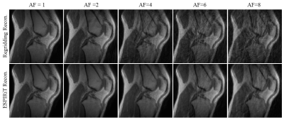 |
102 |
Feasibility of Quantitative Ultrashort Echo Time Magnetization Transfer (UTE-MT) Modeling Based on Undersampled Acquisitions and ESPIRiT Reconstruction
Yanchun Zhu, Yajun Ma, Jie Yang, Yinghua Zhao, Yaoqin Xie, Eric Chang, Jiang Du
Recently the ultrashort echo time magnetization transfer (UTE-MT) technique with two-pool modeling has shown promise as a clinically compatible quantitative technique which is resistant to the magic angle effect. However, the UTE-MT quantitative technique requires multiple acquisitions, which result in suboptimal acquisition times. The purpose of this study is to assess the feasibility of undersampled acquisitions for quantitative UTE-MT modeling applications.
|
|
5182.
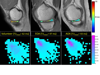 |
103 |
Texture features from T2 and T1? mapping of meniscus in normal and osteoarthritic knee joints
Hon Yu, Shoichiro Takao, Shigeo Hagiwara, Hiroshi Yoshioka
This study demonstrates a feasibility of texture analysis based on T2 and T1ρ mapping of the meniscus. Some of the investigated texture features showed statistically significant differences for both the medial and lateral meniscus between normal volunteers and patients with osteoarthritic knees. One particular texture feature (contrast) based on T2 was also able to differentiate patients with advanced from early knee osteoarthritis for the lateral meniscus as well. The results in this study demonstrate an alternative analytical approach based on texture features as surrogate variables for the evaluation of meniscus properties using T2 and T1ρ mapping.
|
|
5183.
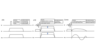 |
104 |
Comparison of ZTE vs UTE vs Cones for Ultrashort TE MSK Imaging
Michael Carl, Yajun Ma, Jiang Du
We compared different center out 3D radial trajectories and assess their advantages and disadvantages for short T2 MSK imaging. We found that while ZTE and Cones may provide some unique capabilities, UTE represents a good compromise between the two.
|
|
5184.
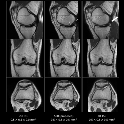 |
105 |
Super-resolution Reconstruction of Knee MRI
Pieter Van Dyck, Floris Vanhevel, Eline De Smet, Paul Parizel, Jan Sijbers, Ben Jeurissen
Clinical knee MRI is most commonly performed with 2D-turbo-spin-echo (TSE) sequences in multiple imaging planes. Although these multi-slice sequences provide excellent tissue contrast and high in-plane spatial resolution, they use relatively thick slices, which leads to partial volume effects. This study used super-resolution reconstruction (SRR) to reconstruct isotropic high-resolution knee MR images. Our results indicate that SRR of knee MRI is feasible and compares favorably to direct high-resolution TSE acquisition in terms of image quality and acquisition time. SRR provides an attractive alternative for isotropic 3D-MRI of the knee.
|
|
5185.
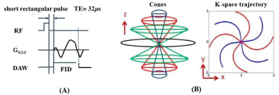 |
106 |
Single- and Bi-component Analyses of T2* Relaxation in Knee Tendon and Ligament by Using 3D Ultrashort Echo Time Cones (UTE Cones) Magnetic Resonance Imaging
Yinghua Zhao, Yajun Ma, Yanchun Zhu, Shaolin Li, Jiang Du
For patellar tendon (PT), anterior cruciate ligament (ACL) and posterior cruciate ligament (PCL) of the knee joint, the in vivo application of the bound and free water mapping techniques is still limited. In this study, we aimed to develop 3D multi-echo fat saturated ultrashort echo time Cones (3D FS-UTE-Cones) imaging protocol for fast volumetric mapping of free and bound water components of whole knee joints in a clinical 3T scanner. The results showed that, for PT, PCL and ACL, the short and long T2* components and their fractions can be characterized by 3D FS-UTE-Cones acquisitions with bi-component T2* analysis, accompanying with superior resolution and short scanning time.
|
|
5186.
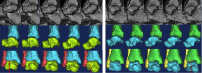 |
107 |
Novel MR coil hardware and fast 3D imaging sequence in evaluating ankle kinematic instability
Did Not Present
Yuqing Zhao, Zheng Zhang, Xuanhui Wei, Jiayu Zhu, Ling Ji, Nan-Jie Gong, Huishu Yuan
A specially designed Ankle & Foot coil that adapts Achilles Tendon Boot is utilized to multiple angle imaging of ankle kinematic instability. With the 3D fast spin echo sequence with compressed sensing based acceleration technique, multi angle high resolution ankle imaging can be achieved in less than 20 minutes. Images from the volunteer with a history of ankle injuries showing anterior talar translation and rotated internally. The integration of novel coil and compressed sensing based acceleration technique enabled ankle kinematic imaging into clinical routine.
|
|
5187.
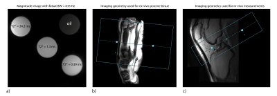 |
108 |
Influence of fat saturation on 3D -UTE Cones T2* mapping of tendon tissue
Paul Baron, Dirk Poot, Stephan Breda, Edwin Oei, Juan Hernandez-Tamames
T2* mapping of tendon tissue is important to assess tissue pathology. Fat contamination however may lead to bias in quantification of T2*. The application of fat saturation (FS) to reduce signal contamination is not straight forward, as this may influence the signal of species with short T2* and broader line-widths. Therefore, the influence of FS on T2* was investigated in tendon tissue. No influence of FS on T2* was observed in ex-vivo tendon tissue. However, in-vivo results suggest fat-contamination was present and FS may be beneficial to reduce T2* quantification bias.
|
|
5188.
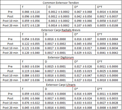 |
109 |
Application of Intravoxel incoherent motion diffusion imaging in evaluation of tendon perfusion
Michael Liu, Patrick Quarterman, Sachin Jambawalikar, Tony Wong
Altered perfusion may be a marker of tendon damage. Measurable perfusion characteristics using non contrast techniques such as Intravoxel Incoherent Motion Diffusion Weighted imaging may be a useful technique for assessing perfusion characteristics of tendon.
|
|
5189.
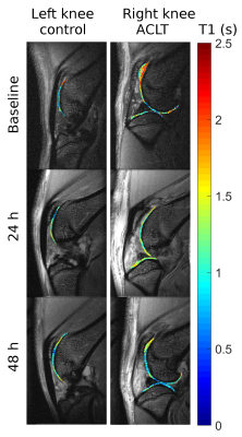 |
110 |
Imaging low grade inflammation in post-traumatic osteoarthritis
Amparo Ruiz, Alejandra Duarte, Mark Milne, Zahid Dewan, Len Luyt, José Raya
Chronic inflammation has been identified as a major driver of joint degradation after injury. MRI is very limited to assess chronic inflammation with high specificity. Here we propose a novel contrast agent that targets the interaction of hyaluronan with RAMM cell receptors that represent an important signaling pathway for chronic inflammation. We validated the contrast agent with optical imaging and showed its value as an MRI contrast agent.
|
|
5190.
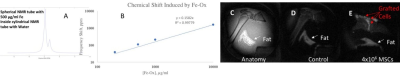 |
111 |
New Tissue Saturation Method for the Detection of Therapeutic Cells in a Knee Joint
Djaudat Idiyatullin, Michael Garwood, Sergey Magnitsky
Synapsis. Mesenchymal Stem Cells (MSCs) have a high potential for a treatment of bone diseases. We develop new acquisition protocol for the detection of therapeutic cells in knee joints. Labeling MSCs with iron oxide particles not only reduce T2* but also induce the resonance frequency shift of labeled cells. This shift enabled us to implement tissue saturation scheme and detect distinct hyperintense signal from grafted cells. New protocol allowed us to detect and quantify therapeutic cells for six days after implantation which was not possible before. Proposed protocol opens new opportunities for in vivo monitoring of cell therapy of bone disorders.
|
|
5191.
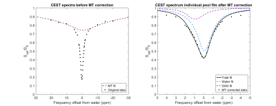 |
112 |
High resolution 1 mm isotropic semi continuous wave gagCEST of articular cartilage using parallel transmit at 7T
Sander Brinkhof, Vitaliy Khlebnikov, Fredy Visser, Hans Hoogduin
The purpose of this work was to implement gagCEST imaging in articular cartilage at 7T using a parallel transmit setup. Two alternating sets of four transmit channels were used in quadrature mode to create a semi continuous excitation pulse. Using this setup, an isotropic 1 mm spatial resolution could be achieved with a low coefficient of variation of 1.05% (@0.9ppm) within an acquisition time of 5 minutes. To the best of our knowledge, this is the highest gagCEST resolution shown so far in literature.
|
|
5192.
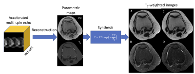 |
113 |
SyntheticTSE: Accelerated multicontrast spin-echo knee MRI using a combination of Bloch-simulation models, compressed sensing and simultaneous multislice acquisition
Akio Yoshimoto, Noam Ben-Eliezer, Mary Bruno, Steven Baete, Michael Recht, Ricardo Otazo
Clinical knee MRI examinations usually consist of a series of 2D acquisitions with different contrasts and orientations. In this work, we propose to simplify and shorten the examination by performing multi-contrast imaging using a single acquisition by directly reconstructing parametric maps (PD and T2) and then synthesizing the contrasts of clinical interest (PD-weighted and T2-weighted with and without fat suppression). The proposed SyntheticTSE method uses a Bloch-simulation signal model for parametric mapping and a combination of compressed sensing and simultaneous multislice acceleration. Initial feasibility is demonstrated using retrospective undersampling of multi-spin-echo knee dataset
|
|
5193.
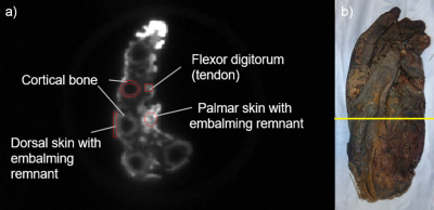 |
114 |
Effects of Reduced Dead Times on the SNR of Tissue with Short T2* values
Agazi Samuel Tesfai, Johannes Fischer, Ali Caglar Özen, Ute Ludwig, Michael Bock
MRI of tissues with short T2* can be realized with ultra-short echo time (UTE) sequences. The effect of lowering TE on relative SNR is calculated and evaluated. The limiting factor for dead time and, consequently, low TE is determined for hardware components of clinical MR system and compared to benchmark results.
|
|
5194.
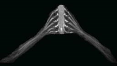 |
115 |
3D nerve-sheath signal increased with inked rest-tissue rapid acquisition of relaxation enhancement imaging(3D SHINKEI) in traumatic brachial plexopathy
Did Not Present
Shanshan Wang, Guangbin Wang, Weibo Chen
3D SHINKEI MRN with high spatial resolution ,is an extremely useful modality to image the traumatized brachial plexus and helps to accurately image the complete brachial plexus and localize as well as describe the lesions from the root still the terminal nerves.
|
|
5195.
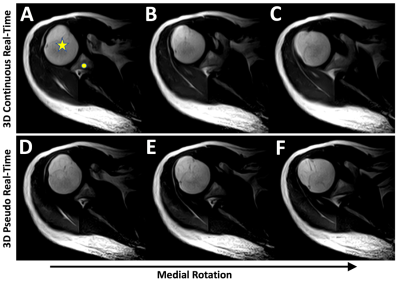 |
116 |
4D MSK MRI: Feasibility of Continuous and Pseudo Real-Time Imaging in the Shoulder and the Wrist
Mitsue Miyazaki, Won Bae, Cheng Ouyang, Dawn Berkeley, Sheronda Statum, Christine Chung
For dynamic evaluation of musculoskeletal joints including the shoulder and the wrist, continuous and pseudo real-time techniques were developed in 4D (3D acquisition and time) using single shot FSE (SSFSE). Effective echo time (TEeff) of SSFSE was optimized for the contrast at each joint. Advantages of 4D over existing 2D techniques are volume acquisition with thinner slices, crucial in cases of target structures moving out of a single 2D imaging slice. Both continuous and pseudo real-time images permitted depiction of joint movements with good contrast; however, pseudo real-time gives slightly sharper images as compared to continuous real-time images.
|
|
5196.
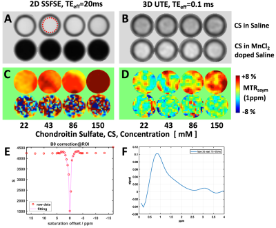 |
117 |
Comparison of gagCEST Effects in Phantoms and Bovine Musculoskeletal Specimens Using 2D Single-Shot Fast Spin Echo and 3D Ultrashort Time-to-Echo at 3-Tesla
Mitsue Miyazaki, Cheng Ouyang, Nirusha Abeydeera, Dawn Berkeley, Reni Biswas, Sheronda Statum, Won Bae, Christine Chung
3D UTE and 2D single-shot fast spin-echo (SSFSE) gagCEST techniques were used to image varying concentrations of chondroitin sulfate (CS) in long or short T1 solutions. Echo time (TE) in SSFSE and UTE ranged from 10-60 ms, and 0.1-8 ms, respectively. Effect of TE on asymmetric magnetization transfer ratio (MTRasym) was examined around 1.0 and 3.5 ppm. For long T1 saline phantom, we found a general increase in MTRasym with concentration, with similar slopes over different TEs, for either technique. For short T1 MnCl2 doped phantom, while UTE could acquire gagCEST signal unlike SSFSE, no consistent trends were found.
|
|
5197.
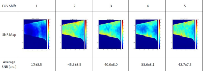 |
118 |
SNR Optimization of Simultaneous Multi-Slice Accelerated TSE Sequences for Diabetic Foot Imaging
Yauheni Rudzevich, Kenneth Buckwalter, Chen Lin, Dingxin Wang
To optimize Simultaneous Multi-Slice (SMS) accelerated TSE sequences for ultra-fast imaging of patients with diabetic neuropathy, a phantom study was performed to determine the optimal Field of View Shift (FOVS) which is a key parameter for SMS, but depend on other imaging parameters and coil configuration. SNR maps for different FOVS were measured using a subtraction method combined with shifting ROI. The optimal selections of FOVS based on quantitative SNR measurement are in good agreement with the selections derived from visual evaluation of clinical images by radiologists. Such agreement provides additional confidence.
|
|
5198.
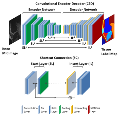 |
119 |
Fully-Automated Segmentation of Knee Joint Anatomy using Deep Convolutional Neural Network Approach
Fang Liu, Zhaoye Zhou, Richard Kijowski
A new fully-automated approach was proposed using a deep convolutional encoder-decoder (CED) network combined with 3D fully-connected conditional random field (CRF) and 3D simplex modeling for performing efficient and accurate multi-class musculoskeletal tissue segmentation from MR images. The deep learning-based segmentation method could be used to create 3D rendered models of all knee joint structures including cartilage, bone, tendon, meniscus, muscle, infrapatellar fat pad, and joint effusion and Baker’s cyst which may be sources of pain in patients with knee osteoarthritis. The results of our study serve as a first step to provide quantitative MR measures of musculoskeletal tissue degeneration in a highly time efficient manner which would be practical for use in large population-based osteoarthritis research studies.
|
|
5199.
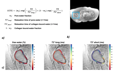 |
120 |
Ultrashort Echo Time-MRI of proximal femoral cortical bone in patients with osteoporosis
Dimitri MARTEL, Mary BRUNO, H.Michael BELMONT, Amit SAXENA, Gabrielle TURYAN, Stephen HONIG, Ravinder R. REGATTE, Gregory CHANG
Ultra-short echo time MRI (uTE-MRI) allows exploration of the role of cortical bone properties in osteoporosis. OP can be primary or can also be induced by drugs such as glucocorticoids (glucocorticoid-induced osteoporosis, GIO).No previous studies have assessed cortical porosity in GIO patients compared to OP patients.Our aim was to perform uTE-MRI of cortical bone of the proximal femur (a common site of fracture) in GIO patients and compare the results to those of OP patients using a bi-exponential fitting procedure of uTE images.
|
|
| Back |
| The International Society for Magnetic Resonance in Medicine is accredited by the Accreditation Council for Continuing Medical Education to provide continuing medical education for physicians. |
