|
Electronic Poster Session
Contrast Mechanisms |
Wednesday, 20 June 2018
Electronic PosterContrast Mechanisms
4986 -5009 Quantitative Susceptibility Mapping
5010 -5032 Physiological Techniques
5081 -5104 Electrical Property Imaging
5105 -5128 Advancements in CEST Methodology & Applications |
| |
Quantitative Susceptibility Mapping
Electronic Poster
Contrast Mechanisms
Wednesday, 20 June 2018
| Exhibition Hall |
16:15 - 17:15 |
| |
|
Computer # |
|
4986.
 |
1 |
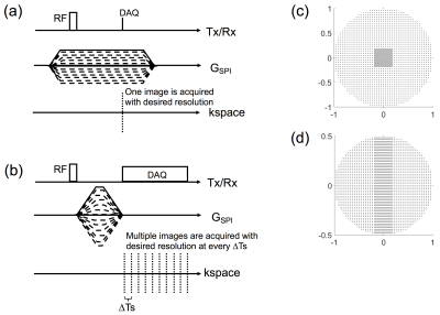
 True Phase Quantitative Susceptibility Mapping Using Continuous Single Point Imaging: A Feasibility Study True Phase Quantitative Susceptibility Mapping Using Continuous Single Point Imaging: A Feasibility Study
Hyungseok Jang, Xing Lu, Eric Chang, Jiang Du
Quantitative susceptibility mapping (QSM) has recently been in the limelight as a novel contrast mechanism to provide quantification of apparent magnetic susceptibility based on phase information in MR images. In this study, we explore feasibility of single point imaging (SPI) for QSM. SPI, also known as constant time encoding or pure phase encoding, provides true phase information not affected by phase evolution during readout, which is beneficial for accurate QSM. We propose a new and efficient SPI acquisition scheme for QSM, termed Continuous SPI, where SPI images are continuously obtained with extremely high temporal resolution in a single scan.
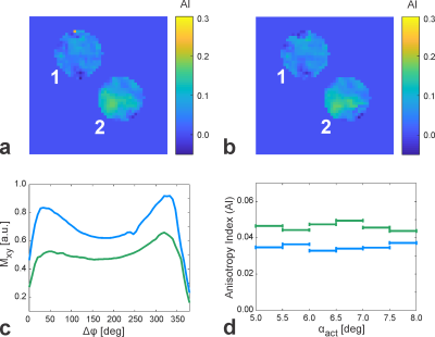
|
|
4987.
 |
2 |
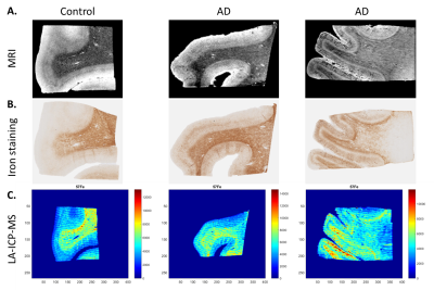
Quantitative MRI and laser ablation-inductively coupled plasma-mass spectrometry imaging of iron in post-mortem frontal cortex of Alzheimer patients
Marjolein Bulk, Walid Abdelmoula, Wim Wiarda, Itamar Ronen, Jouke Dijkstra, Louise van der Weerd
Previous imaging studies reported iron-induced T2* or phase contrast changes in the cortex of post-mortem brain tissue of patients with Alzheimer’s disease (AD), but comparison with a gold standard is lacking. This study used laser ablation-inductively coupled plasma-mass spectrometry (LA-ICP-MS) as a gold standard for iron in post-mortem brain tissue of controls and AD patients and investigated the correlation between LA-ICP-MS, quantitative MRI (R2*, phase, and QSM) and histology. R2* and QSM showed the highest correlation with iron content; the correlation of phase with iron was weaker, probably due to its high orientation dependence.
|
|
4988.
 |
3 |
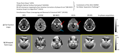
Fat Correction of MRI Phase Images for Accurate Susceptibility Mapping in the Head and Neck
Anita Karsa, Shonit Punwani, Karin Shmueli
Susceptibility Mapping (QSM) is increasingly applied in parts of the body where fatty tissue is present. QSM uses the phase of the complex MRI signal which contains both susceptibility-, and chemical-shift-induced components. For accurate QSM, the latter need to be suppressed. Here we compared a range of different fat-correction strategies for QSM in head-and-neck images. Techniques providing reliable fat-fraction maps also gave similar susceptibility values in fatty fascia. However, some of these methods were not robust to the choice of echo times. In-phase imaging was found to be the best candidate for robust fat-correction in QSM of the head-and-neck.
|
|
4989.
 |
4 |
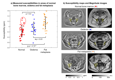
Bone Marrow Susceptibility as a Marker of Bone Mineral Density in Spondyloarthritis
Anita Karsa, Timothy Bray, Alan Bainbridge, Shonit Punwani, Margaret Hall-Craggs, Karin Shmueli
New bone formation (causing spinal fusion) and bone loss are both key features of spondyloarthritis, and contribute to significant morbidity and disability. However, these processes are difficult to monitor using conventional MRI, which provides minimal information about bone mineral density (BMD). Here, we show that bone marrow susceptibility can be used as a marker of BMD, using data from a fat-water-bone phantom and from subjects with spondyloarthritis. Susceptibility values are significantly increased in areas of fat metaplasia compared to normal marrow, suggesting that this lesion represents a form of local bone loss, which could be monitored using susceptibility mapping.
|
|
4990.
 |
5 |
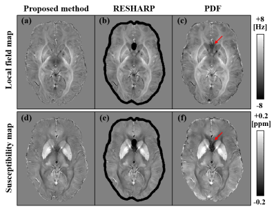
Whole Brain Background Field Removal using Spherical Mean Value Filtering and Local Polynomial Approximation for Quantitative Susceptibility Mapping
Toru Shirai, Ryota Sato, Takenori Murase, Yoshitaka Bito, Hisaaki Ochi
We propose a novel background-field removal method to recover a local field of a brain edge. The proposed method consists of two steps. First, the background field of the brain edge is calculated from the total field by using local polynomial approximation. Second, the local field of the whole brain is calculated by regularization that enables sophisticated harmonic artifacts reduction for phase data (RESHARP) processing with the constraint term of the background field of the brain edge. The results from a human brain experiment showed that the method is useful for calculating the local field and susceptibility maps of the whole brain.
|
|
4991.
 |
6 |
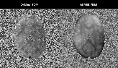
High resolution frequency difference mapping using ASPIRE phase combination
Korbinian Eckstein, Siegfried Trattnig, Simon Robinson
Frequency difference mapping (FDM) is a promising new method for investigating tissue microstructure. To date, images have been of quite low resolution and noise because of the need to use monopolar readout and two difference operations. We propose an improved approach – ASPIRE-FDM – which is based on a recently developed coil combination method which removes both phase offsets and phase gradients in readout direction in more efficient bipolar acquisitions. The resulting maps have drastically reduced noise and make feasible a four-fold reduction in voxel volume compared to prior work.
|
|
4992.
 |
7 |
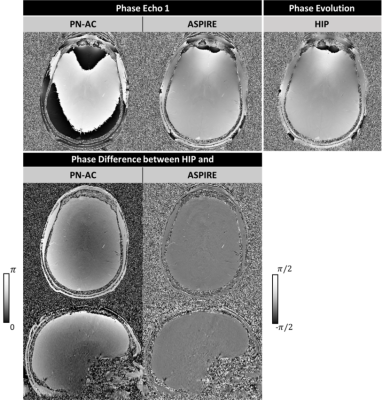
An assessment of the ‘Prescan-Normalize Adaptive Combine’ approach to combining phase images from multi-channel coils at 3T
Korbinian Eckstein, Siegfried Trattnig, Simon Robinson
The combination of data acquired with array coils often leads to phase artifacts. The recently-introduced method ‘Prescan-Normalize Adaptive Combine’ (PN-AC) is assessed in terms of non-ΔB0-related contributions and reproducibility with different head positions and compared with a robust multi-echo phase combination approach (called ASPIRE) which yields only ΔB0-related phase. PN-AC was found to generate low noise phase images but introduce non-ΔB0-related contributions to the combined phase. It was robust to motion between the prescan and the main acquisition other than the introduction of arbitrary background phase.
|
|
4993.
 |
8 |
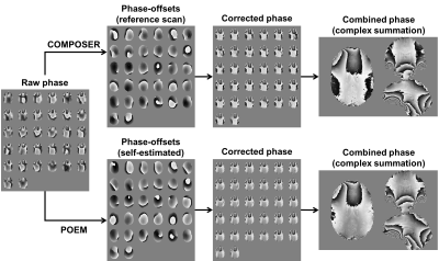
Multi-channel phase combination for quantitative susceptibility mapping at 7T using Phase-Offsets Estimation from Multi-echoes (POEM) method
Hongfu Sun, Jon Cleary, Rebecca Glarin, Scott Kolbe, Bradford Moffat, Roger Ordidge, G. Bruce Pike
A Phase-Offsets Estimation from Multi-echoes (POEM) method is proposed to combined the phase-arrayed coil at 7T for quantitative susceptibility mapping (QSM). The method demonstrates equivalent or better results than adding a conventional reference scan for both single-orientation and multi-orientation QSM.
|
|
4994.
 |
9 |
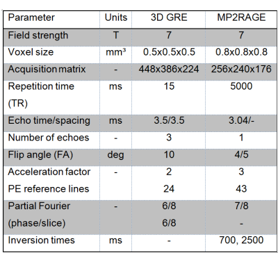
Extracting more: Multi-echo MP2RAGE at 7T enables simultaneous T1w, quantitative T1, T2* and susceptibility mapping from a single acquisition
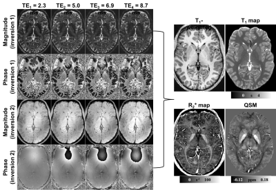
Hongfu Sun, Jon Cleary, Rebecca Glarin, Yuhan Ma, Kieran O'Brien, Scott Kolbe, Bradford Moffat, Roger Ordidge, G. Bruce Pike
We demonstrate the feasibility of extracting simultaneous T1w, quantitative T1, T2* and susceptibility mapping from a single Multi-echo MP2RAGE scan of 10min at 7T. After optimizing the multi-channel phase combination, QSM from the Multi-echo MP2RAGE is comparable to that from a standard GRE sequence, with the benefits of additional parametric maps.
|
|
4995.
 |
10 |
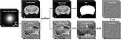
Shading Artifact Suppression using Relaxation Map and Machine Learning-based Region Detection for Quantitative Susceptibility Mapping
Video Permission Withheld
Taichiro Shiodera, Takashi Watanabe, Tomoyuki Takeguchi, Naotaka Sakashita, Masao Yui, Samir Sharma
We propose a dipole inversion method for improving quantitative susceptibility mapping. In conventional methods, shading artifacts often occur near the longitudinal fissure (LF) region of the estimated susceptibility map. Here, we propose an algorithm for LF region detection and regularized inversion, to reduce the shading artifacts. The LF region is automatically detected using information from the T2* map as well as training datasets via machine learning. The proposed method eliminates shading artifacts near the LF region in the susceptibility maps, while also showing negligible change in regions that do not suffer from shading artifacts, such as the basal ganglia.
|
|
4996.
 |
11 |
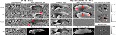
Considerations in Quantitative Susceptibility Mapping using Echo Planar Imaging
Beata Bachrata, Korbinian Eckstein, Siegfried Trattning, Simon Robinson
Phase images from EPI and GE acquisitions differ due to the divergent acquisition schemes and reconstruction steps. The effect of these is investigated both in measured phase and estimated susceptibility values. We show that non-ΔB0-related phase is present in phased array data combined with the Virtual Receiver Coil and Roemer approaches and that this influences estimated susceptibilities. For EPI, data can be combined using a multi-echo “ASPIRE” GE prescan, leading to minimal non-ΔB0-related phase. There was large variability of in-vivo EPI fieldmaps due to physiological noise and non-linearities in phase evolution in both GE and EPI data.
|
|
4997.
 |
12 |
Accelerating 3D whole brain quantitative susceptibility mapping acquisition using compressed sensing
Nian Wang, Gary Cofer, Yi Qi, G. Allan Johnson
To evaluate the feasibility of compressed sensing (CS) for accelerating quantitative susceptibility mapping (QSM) acquisition at a high spatial resolution in mouse brains (22.5 μm3, isotropic). This preliminary study shows that CS can be applied to significantly reduce the acquisition time of GRE MRI of mice brains at 9.4 T without losing apparent accuracy in quantitative susceptibility values.
|
|
4998.
 |
13 |
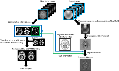
Simultaneous Analyses and Reconstruction of Quantitative Susceptibility Mapping and Voxel-based Morphometry Using Magnetization-prepared Spoiled Turbo Multiple Gradient Echo
Video Permission Withheld
Hirohito Kan, Nobuyuki Arai, Satoshi Tsubokura, Masato Yamada, Yuta Nishiwaki, Kyosuke Mizuno, Harumasa Kasai, Yasujiro Hirose, Yuta Shibamoto
Quantitative susceptibility mapping (QSM) and voxel-based morphometry (VBM) analyses are helpful in detecting an abnormal iron overload and the regional change of volume in the gray and white matters, respectively. This abstract presents a novel method for simultaneous analyses of QSM and VBM using magnetization-prepared spoiled turbo multiple gradient echo sequence with inversion pulse for QSM sequence which provides 3D T1-weighted structural images for VBM and multi-echo phase images for QSM on single scan.
|
|
4999.
 |
14 |
A multi-scale approach to quantitative susceptibility mapping (MSDI)
Julio Acosta-Cabronero, Carlos Milovic, Cristian Tejos, Martina Callaghan
We propose a new QSM algorithm, namely multi-scale dipole inversion (MSDI), which builds on the nonlinear MEDI (nMEDI) framework incorporating two additional features: (i) improved error control through dynamic phase-reliability compensation across harmonic scales and (ii) scale-specific use of the morphological prior. MSDI is the first algorithm to rank in the top-10 for all performance metrics evaluated in the 2016 QSM Reconstruction Challenge. It also demonstrates lower variance than nMEDI in a reproducibility test.
|
|
5000.
 |
15 |
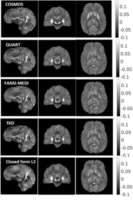
Quantitative susceptibility mapping Accelerated Reconstruction Technique (QUART): A novel Split Bregman based approach for rapid reconstruction of quantitative susceptibility maps
Srikant Kamesh Iyer, Brianna Moon, Walter Witschey
This abstract presents a novel Split Bregman (SB) based approach to enable rapid minimization of the quantitative susceptibility reconstruction formulation that includes a weighted least squares fidelity constraint and a total variation (TV) penalty. The purpose of this approach is to develop a rapid minimization technique that does not need complex matrix factorization or computation of matrix preconditioners to accelerate convergence. Rapid minimization is achieved by the application of two variable substitutions, one to the weighted fidelity constraint and the other to the total variation term. Minimization of the cost functional is achieved by the novel combination of FISTA based iterative re-weighting and soft thresholding.
|
|
5001.
 |
16 |
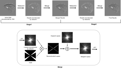
Reducing Streaking Artifacts in Quantitative Susceptibility Mapping via Deep Learning
Jie Liu, Yida Wang, Yang Song, Haibin Xie, Jianqi Li, Guang Yang
In this study, we proposed a new approach to reduce streaking artifacts in quantitative susceptibility mapping via deep learning. It combined two convolutional neural networks to reduce streaking artifacts from classic threshold-based k-space division (TKD) . The proposed method achieved impressive performance both visually and statistically.
|
|
5002.
 |
17 |
Susceptibility-Weighted Imaging on a Compact 3T Scanner with High-Performance Gradients
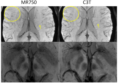
Yunhong Shu, Shengzhen Tao, MyungHo In, Joshua Trzasko, Erin Gray, John Huston III, Matt Bernstein
Susceptibility-weighted imaging (SWI) uses a 3D multi-echo gradient-echo sequence with unipolar gradient lobes to acquire all echoes, with fly-back gradients inserted in-between. These fly-back gradients reduce acquisition efficiency and increase echo spacing. We demonstrated that the high gradient performance available on a compact 3T system can reduce the echo spacing in the multi-echo readout by reducing the pulse-width of the fly-back gradient, which consequently allows a greater number of echoes to be sampled. The increased number of echoes translates into reduced noise and improved small vessel conspicuity in SWI acquired on the compact 3T compared to the conventional whole-body system.
|
|
5003.
 |
18 |
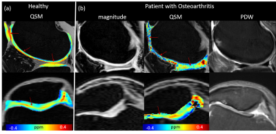
Quantitative Susceptibility Mapping of Articular Cartilage in Patients with Osteoarthritis
Hongjiang Wei, Huimin Lin, Yuyao Zhang, Weibo Chen, Fuhua Yan, Chunlei Liu
Osteoarthritis (OA) is a multifactorial degenerative joint disease and is the most common form of arthritis which characterized by degenerative changes in the cartilage, menisci, ligaments and bone. The purpose of this study is to evaluate the magnetic susceptibility changes in knee diseases, such as patients with bone marrow lesions and cartilage loss. Clear susceptibility contrast was observed between bone marrow lesions and surroundings. The multilayer pattern of the damaged cartilage was lost compared with that of healthy subjects. QSM may provide a new way to improve the characterization of tissue microstructure in the knee joint related to osteoarthritis.
|
|
5004.
 |
19 |
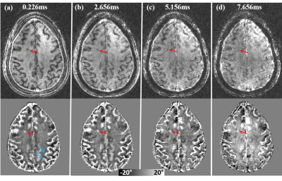
GRE Phase Contrast of the Brain at Ultra-Short TE in Patients with Multiple Sclerosis
Hongjiang Wei, Peng Cao, Roland Henry, Peder Larson, Chunlei Liu
The purpose of this study was to determine whether gradient-echo proton images of the brain exhibit any phase contrast at UTE and whether this contrast can be used to improve the characterization of tissue microstructure in the brain. Our data demonstrated that UTE images of the brain can attain strong phase contrast even at a TE of 226ms by using off-resonance RF pulses for selective saturation. UTE phase in an MS patient shows a strong contrast with surrounding white matters.
|
|
5005.
 |
20 |
Imaging Human Brain Cortical Substructure with Quantitative Susceptibility Mapping at 7 T
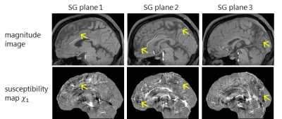
Hongjiang Wei, Berkin Bilgic, Kawin Setsompop, Boris Keil, David Feinberg , Chunlei Liu
High-field MRI combined with high-density motor-cortex coil and a novel QSM algorithm allows in vivo brain cortex imaging with high contrast-to-noise ratio and spatial resolution (0.15x0.15x0.65 mm3). Our work revealed up to six apparent myelin/iron layers in human cortex in vivo determined by the underlying magnetic susceptibility and complex vasculature in the cortical areas. Cortical susceptibility imaging provides high-resolution quantitative measurements of cortical cytoarchitecture and vasculature.
|
|
5006.
 |
21 |
In vivo quantitative susceptibility mapping detection of ß-amyloid targeted by curcumin-conjugated magnetic nanoparticles
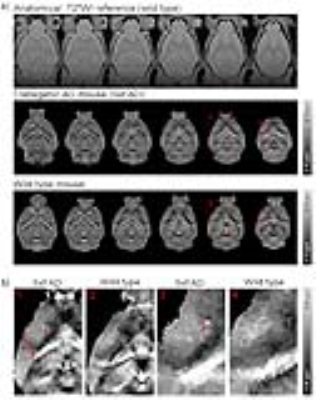
Celia Dong, Mengye Lyu, Anthea To, Ed Wu
At present, there is a lack of reliable in vivo diagnostic methods to visualize and quantify the Alzheimer's disease (AD) pathologies. β-amyloid (Aβ) plaques are the hallmarks of AD brains. Recently, we have designed a novel curcumin-conjugated magnetic nanoparticles (Cur-MNPs) that target Aβ pathologies. Quantitative susceptibility mapping (QSM) offers the possibility of quantifying Cur-MNPs in vivo as a quantitative surrogate marker for Aβ pathologies. In this study, we optimized the QSM acquisition and processing procedure for mouse brains in vivo. Furthermore, we investigated the ability of QSM in detecting Cur-MNPs targeted Aβ pathologies in vivo in transgenic AD mouse models.
|
|
5007.
 |
22 |
Quantitative assessment of melanoma metastases at 7T: Susceptibility and T1 mapping
Sina Straub, Frederik Laun, Martin Freitag, Heinz-Peter Schlemmer, Mark Ladd, Till Schneider
Susceptibility values and T1 values of melanoma metastases measured at 7T are shown. There is an ongoing discussion about whether melanin or hemorrhage dominate the contrast mechanisms observed in melanoma metastases. Susceptibility maps as well as T1 maps benefit from the high contrast and high resolution available at 7T, though we observed paramagnetic susceptibility in areas corresponding to vessels or hemorrhagic events, but no general relation between susceptibility values and T1 values of individual metastases
|
|
5008.
 |
23 |
Clinical Implementation of Quantitative Susceptibility Mapping: Experience Across Multiple Sites and Scanner Platforms.
Pascal Spincemaille, Zhe Liu, Shun Zhang, Matteo Ippoliti, Marcus Makowski, Richard Watts, Ludovic de Rochefort, Vijay Venkatraman, Patricia Desmond, Brian Kopell, Patrice Péran, Yi Wang
In recent years, quantitative susceptibility mapping (QSM) has undergone a series of technical improvements and found applications in an expanding array of diseases. To support this ongoing process of development and validation, it is important to automate the computationally intensive susceptibility reconstruction on the scanner after the acquisition of gradient echo data. In this work, an online QSM reconstruction system for a variety of scanner platforms with low cross-site ROI standard deviation is demonstrated.
|
|
5009.
 |
24 |
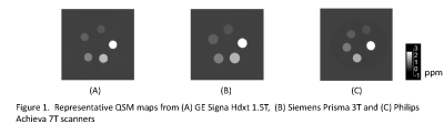
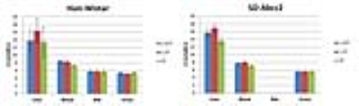
Multi-site reproducibility of quantitative susceptibility mapping at 1.5, 3 and 7T
Kofi Deh, Keigo Kawaji, Deb Horng, Marjolein Bulk, Louise Van Der Weerd, Pascal Spincemaille, Thanh Nguyen, Yi Wang
Quantitative susceptibility mapping (QSM) is increasingly being applied to quantitative research on disease conditions including intracerebral hemorrhage, liver iron overload and bone mineral quantification. These applications require knowledge of the reproducibility of QSM of sources with high susceptibility, which has not been previously reported. Here, we investigate the agreement between QSM maps generated from gradient-echo scans acquired at multiple sites using multiple platforms.
|
|
Physiological Techniques
Electronic Poster
Contrast Mechanisms
Wednesday, 20 June 2018
| Exhibition Hall |
16:15 - 17:15 |
| |
|
Computer # |
|
5010.
 |
25 |
Gadoxetate relaxivities increase significantly after hepatic uptake at clinical field strength impacting kinetic modelling for liver function analysis
Video Permission Withheld
Gregor Jost, Gunnar Schuetz, Hubertus Pietsch
Schuhmann-Giampieri1 reported r1 of gadoxetate to be significantly higher in liver tissue compared to blood at 0.47T. Gadoxetate relaxivities at 1.5T, 3T and 4.7T have since then been reported for water and plasma2, but not hepatocytes. We here present r1 values for gadoxetate in hepatocytes at 1.5T and 3T to complement existing data. r1 values of gadoxetate after uptake into hepatocytes are about 2x higher compared to blood. There is no relevant field strength dependency over a wide range (0.47T - 3T). This has implications on kinetic modelling of dynamic gadoxetate enhanced MRI data for determination of liver transporter activity.
|
|
5011.
 |
26 |
Exploring the Origin of Asymmetries in the Balanced SSFP Profile at 9.4 Tesla: Microstructure Anisotropy or Chemical Exchange?
Rahel Heule, Moritz Zaiss, Philipp Ehses, Klaus Scheffler
The balanced steady-state free precession (bSSFP) profile is known to carry information about the tissue-dependent frequency content in a voxel. There has been strong evidence that the observed asymmetries in the bSSFP profile of white matter depend on the tract orientation with the largest asymmetries occurring in tracts perpendicular to B0. Recently, it was demonstrated that the bSSFP sequence can be used for chemical exchange detection based on profile asymmetries arising in isotropic probes of two exchanging pools. In this work, we explore the question whether exchanging species might contribute to the bSSFP profile asymmetry observed in white matter.
|
|
5012.
 |
27 |
Towards Quantification of Manganese Deposition in the Human Brain in the Presence of Iron: A Calibration Study
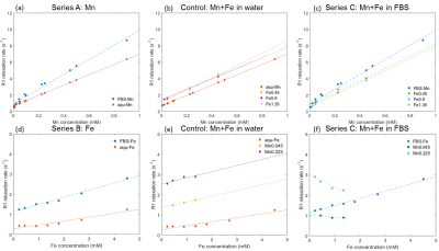
Chien-Lin Yeh, Ulrike Dydak
Welders are exposed to high levels of manganese (Mn) and iron (Fe). Since Mn is neurotoxic, being able to quantify brain Mn deposition by MRI is of high interest. This phantom calibration study is a first step towards understanding the combined effect of Mn and Fe on R1. Our results show that the presence of serum in the solution requires a Mn-Fe interaction term to improve the fit, likely due to the competing binding to the protein. The presence of Fe reduces the R1 effect of pure Mn, which would lead to an underestimation of brain Mn if Fe was neglected.
|
|
5013.
 |
28 |
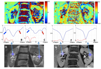
Comparison of B0 versus B0 and B1 field inhomogeneity correction for glycosaminoglycan chemical exchange saturation transfer imaging
Anja Müller-Lutz, Alexandra Ljimani, Julia Stabinska, Hans-Jörg Wittsack, Christoph Schleich
Glycosaminoglycan CEST (gagCEST) has been shown to be a useful tool for assessing changes in glycosaminoglycan concentrations in cartilage. However, accurate B0 referencing is necessary for observing reliable gagCEST. Here we investigate whether the quality of in vivo gagCEST images can be further improved by correction of both B0 and B1 inhomogeneity by using a novel water shift and B1 (WASABI) mapping method. We present results from 20 healthy volunteers acquired at 3T. A comparison with the outcome of WASSR B0 mapping shows that the application of the WASABI maps to inhomogeneity artefact correction is advantageous to gagCEST imaging.
|
|
5014.
 |
29 |
Dephasing and diffusion in blood vessel networks: an exact solution of the Bloch-Torrey-equation
Lukas Buschle, Felix Kurz, Heinz-Peter Schlemmer, Christian Ziener
The relaxation in blood vessel networks depends on susceptibility and diffusion effects around vessels. In 1994, Yablonskiy and Haacke developed a geometrical model of vessels and analyzed the gradient echo signal for negligible diffusion. Many approximative methods were developed generalizing this model in the limits of small and large diffusion effects. Important methods like vessel size or architectural imaging are based on these works. Here, we provide an exact solution of the Bloch-Torrey-equation in the model of Yablonskiy and Haacke for arbitrary diffusion effects that allows a validation of previously developed methods.
|
|
5015.
 |
30 |
An improved blood pool MRI agent with dinuclear structure: characterization and in vivo bio-distribution
Alberto Fringuello Mingo, Francesca La Cava, Luigi Miragoli, Enzo Terreno, Enrico Cappelletti, Luciano Lattuada, Sonia Colombo Serra, Luisa Poggi, Fabio Tedoldi
A novel dinuclear gadolinium(III) chelate containing two moieties of DTPA, covalently conjugated to deoxycholic acid is presented. The product was synthesized and characterized in vitro (analysis of relaxometric properties in different media) and in vivo (blood pharmacokinetic and MRI bio-distribution). The complex showed a much higher relaxivity than Gd-DTPA and other dinuclear complexes proposed in literature. Moreover, it displayed a strong interaction with human serum albumin in three binding sites. This property translates in a long blood elimination half time (130min in rats) making the product an optimal blood-pool agent.
|
|
5016.
 |
31 |
Comparison of cardiac output calculated from low dose Ultra-fast DCE MRI to the values measured from cardiac MRI
Shiyang Wang, Yue Zhang, Xiaobing Fan, Dianning He, Milica Medved, Tatjana Antic, Ambereen Yousuf, Gregory Karczmar, Aytekin Oto
Accurately measuring arterial input function (AIF) is essential for quantitative dynamic contrast enhanced (DCE) MRI. The indicator dilution principle was used to verify the accuracy of AIF measured at iliac arties following injection of low dose (0.015 mmol/kg) gadolinium contrast media. The cardiac output from cardiac MRI (COCMRI) were compared with the cardiac output from Ultra-fast DCE-MRI (CODCE). Results demonstrated that the CODCE were consistent with the ‘gold standard’ COCMRI. This demonstrates that the low dose ultra-fast DCE-MRI can be used to accurately measure the AIF.
|
|
5017.
 |
32 |
Principal Process Analysis of dynamic GlucoCEST MRI data
Stefano Casagranda, Marco Pizzolato, Francisco Torrealdea, Xavier Golay, Timothé Boutelier
GlucoCEST is an MRI contrast enhancement technique sensitive to the concentration of sugar in the tissue. Because of a difference in metabolism, it is thought that tumors consume more sugar than normal tissue. However, glucose metabolism is complex and depends on many processes, which are all important to understand the origin of the measured signal. To achieve this goal we apply here a process analysis method to a deterministic system describing the metabolism of glucose in the tissue.
|
|
5018.
 |
33 |
NOE contributions to the z-spectrum of human blood
Andrew Carradus, Simon Shah, Olivier Mougin, Hans Hoogduin, Penny Gowland
This study investigates the properties of relayed NOE effects in the blood by fitting z-spectra acquired using continuous wave saturation to the analytical solutions of the Bloch-McConnel equations. We systematically model for an increasing number of NOE peaks based on previous reports, and find that adding in two NOE peaks at -3.5ppm and -1.7ppm significantly improves the fit, but adding further peaks does not. Furthermore, we fit for the exchange rates and T2s of these pools, and find that the peak at -3.5ppm has exchange rate=21.5Hz and T2=0.9ms and the peak at -1.7 ppm has exchange rate=8.6Hz and T2=3ms.
|
|
5019.
 |
34 |
Non-rigid atlas registration for improved quantitative assessment of rat brain regions with limited inherent anatomical contrast
Matthew Tarasek, Jeannette Roberts, Deirdre Cassidy , Desmond Yeo, Randall Carter, Brian Bales
We present a method for 3D non-rigid, feature-based atlas registration to images that contain limited inherent anatomical MR contrast. This method can be used to standardize ROI identification and may be applied to any multi-functional imaging technique to provide increased quantitative registration accuracy. Here quantitative T1 measurements were used to test the accuracy and reproducibility of the method. Overall, data analysis performed with atlas registration provides a 2-fold reduction in standard deviation and 4-fold increase in reproducibility versus data analysis performed without registration.
|
|
5020.
 |
35 |
Phenols as Diamagnetic T2-exchange Magnetic Resonance Imaging Contrast Agents
Jia Zhang, Yuguo Li, Stephania Slania, Nirbhay Yadav, Jing Liu, Rongfu Wang, Jianhua Zhang, Martin Pomper, Peter van Zijl, Xing Yang, Guanshu Liu
To further explore the ability of using diamagnetic compounds for generating MRI contrast, we chose phenol as a model compound and systematically characterized its T2-exchange (T2ex) ability. We designed a library of phenol-based compounds and determined the effects of chemical modification on their proton exchange rate (kex) and chemical shift (?ω) and consequently, their T2ex contrast. A large ?ω is favorable for generating strong T2ex contrast, while there is an optimal kex at each specific ?ω. Finally, as an example of biomedical application, we demonstrated the label-free detection of tyrosinase activity using T2ex MRI.
|
|
5021.
 |
36 |
Nanomicelles – A Blood Pool Contrast Agent for MRI
Vassily Vorobiev, Lindsey Crowe, Yohan van de Looij, Lothar Helm, Samuel Espy, Andrej Babic, Jean-Paul Vallée, Eric Allémann
A blood pool MRI contrast agent for free breathing MR angiography is highly desirable in particular for pediatric cardiovascular exams in order to improve both image quality and patient comfort. Apnea in infants and be propofol induced, or via intubation, which is invasive and limits acquisition to short protocols. We present here initial in-vivo studies in mice of a newly developed nanomicelles gadolinium contrast agent. Persistent vascular enhancement is observed with no toxicity. Micelles also open the way to functionalization for theranostic applications.
|
|
5022.
 |
37 |
Investigating the nature of distinct regions in brain tissue affected by stroke with principal component analysis of multi-b valued diffusion data
Ana-Maria Oros-Peusquens, Omid Nikoubashman, Martin Wiesmann, N. Jon Shah
A data-driven, observer-independent segmentation of brain regions affected by stroke is proposed. It is based on PCA analysis of a multi b-value diffusion trace acquisition and offers fast, high-SNR visualisation of affected areas. Substructure of these areas is easily identified and can be automatically delineated using clustering algorithms.
|
|
5023.
 |
38 |
Reducing T2-related bias in mq-BOLD derived maps of Oxygen Extraction Fraction by 3D acquisition
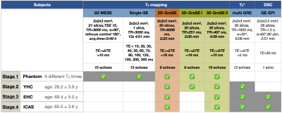
Stephan Kaczmarz, Jens Goettler, Andreas Hock, Dimitrios Karampinos, Claus Zimmer, Fahmeed Hyder, Christine Preibisch
Multi-parametric quantitative BOLD (mq-BOLD) measurements have been successfully applied in several studies to assess the vascular oxygenation, however, calculated relative Oxygen Extraction Fraction (rOEF) shows unphysiological systematic elevations. We suspect biased T2-measurements to be the main source of error. Therefore, we present an optimized 3D-GraSE T2-mapping sequence and its evaluation in four stages within phantoms, young healthy controls, elderly controls and internal carotid artery stenosis (ICAS) patients. We found significant T2-decreases, fully consistent with reference-values, and thereby significantly decreased rOEF-values by ~25 % towards physiologically more realistic values. Additional clinical value was demonstrated by detecting focal rOEF-increases in an ICAS-patient.
|
|
5024.
 |
39 |
Comparison of Labeling Capacity for Protamine-sulphate-conjugated and FuGENE-labeled Progenitor Cardiac Stem Cells using Perfluorocarbon Nanoparticle Labels for In Vivo Murine Cardiac 19F MRI/MRS
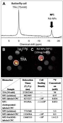
Chris Constantinides, Akhilesh Rai, Mangala Srinivas, Lino Ferreira, Carolyn Carr
Despite prior work, a direct comparison of the labeling of progenitor cardiac stem cells (CPCs) using protamine-sulphate (PS)-conjugated or FuGENE-labeled perfluorocarbon nanoparticle labels (based on their respective cellular uptake mechanisms), and their capacity to achieve direct cardiac 19FMRI on the same animal species, is still lacking. We report herein improved in vivo cardiac 19FMRI performance for FuGENE-labeled compared to PS-labeled CPCs.
|
|
5025.
 |
40 |
High rotating frame relaxation MRI mapping for detecting ischemia in rats
Eleni Demetriou, Hanne Laakso, Timo Liimatainen, Silvia Mangia, Shalom Michaeli, Xavier Golay
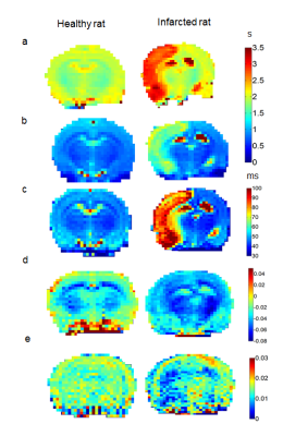
The goal of this work is to evaluate novel MRI technique entitled Relaxation Along a Fictitious Field (RAFF) in the rotating frame of rank n (RAFFn, with n=4) in its ability to detect ischemia, specifically when tuning the sensitivity of RAFF4 to exchange processes of amide protons. Relaxation maps were thus measured in healthy and infarcted rats at 24h post stroke, and were compared with other conventionally used quantitative MRI modalities. RAFF4 robustly detected infarcted regions with enhanced contrast as compared to T1 relaxation and MTR asymmetry, thus offering a sensitive novel MRI marker for quantifying tissue abnormalities during ischemia.
|
|
5026.
 |
41 |
Direct D2O MRI for quantifying tissue permeability: application to Vitreous
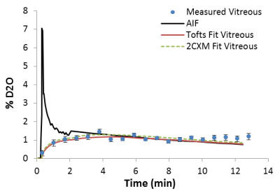
Shengwen Deng, Eric Muir, Shiliang Huang
D2O is a freely diffusible trace for calculating blood flow and give more accurate than limited diffused Gadolinium based contrast agents. A few publications from over twenty years ago used MRI or single-voxel NMR spectroscopy to assess D2O (or 17O water) for tissue perfusion. However, these studies never investigated the application of D2O MRI to altered permeability. Our current study explore the dynamic direct D2O MRI to map tissue permeability, and applied it to map the water exchange in rodent vitreous.
|
|
5027.
 |
42 |
Performance of ultrafast DCE-MRI for diagnosis of prostate cancer
Aritrick Chatterjee, Dianning He, Xiaobing Fan, Shiyang Wang, Teodora Szasz, Ambereen Yousuf, Federico Pineda, Tatjana Antic, Melvy Mathew, Gregory Karczmar, Aytekin Oto
This study aimed to test high temporal resolution DCE- MRI for different zones of the prostate and evaluate its performance in the diagnosis of prostate cancer (PCa). High temporal resolution (~2.2s) was achieved by modestly decreasing spatial resolution, increasing sensitivity encoding and partial Fourier factors. Our results show that PCa had significantly faster signal enhancement and washout rates than normal tissue. DCE-MRI with higher temporal resolution may capture clinically useful information for PCa diagnosis that would be missed by low temporal resolution DCE-MRI. This new information could improve the performance of mpMRI in prostate cancer detection.
|
|
5028.
 |
43 |
Is the Parker arterial input function necessary to model the second pass for dynamic contrast enhanced MRI? - A simulation study
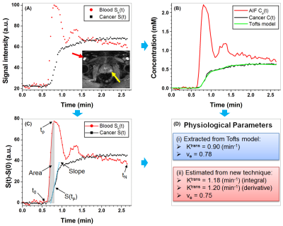
Dianning He, Lisheng Xu, Wei Qian, Xiaobing Fan
Accurately modeling arterial input function (AIF) is important for dynamic contrast enhanced (DCE) MRI. Simulations were performed comparing nine population AIF models to the Parker AIF. Effects of AIF second pass with and without adding noise onto extracted physiological parameters were evaluated with n=1,000 randomly generated physiological parameters (Ktrans and ve) used to calculate contrast agent concentration curves using the Tofts model and Parker AIF. Results demonstrated that the six-parameter linear function plus bi-exponential function AIF model was almost equivalent to Parker AIF. Effects of the second pass were small, unless noise with signal-to-noise ratio was <10 dB.
|
|
5029.
 |
44 |
Relaxivity of Ferumoxytol at 1.5T and 3.0T
Gesine Knobloch, Timothy Colgan, Curtis Wiens, Xiaoke Wang, Tilman Schubert, Diego Hernando, Scott Reeder
Ferumoxytol (Feraheme, AMAG, Waltham, MA) is an iron supplement that has shown promise as an off-label alternative contrast agent for MRI. Optimization of imaging and dosing protocols requires accurate knowledge of the relaxation characteristics of ferumoxytol, which are currently not well understood. Therefore, the purpose of this work was to measure the r1, r2 and r2* relaxivity of ferumoxytol. Studies were performed at 1.5T and 3.0T over a range of concentrations, in saline, human plasma, and human blood, all at body temperature.
|
|
5030.
 |
45 |
R2-Star & DCE-Perfusion MRI based Novel Approach for Classification of Intra Tumoral Susceptibility Signal (ITSS) into Haemorrhage, Non-Leaky Vessels and Leaky Vessels in Glioblastoma
Anup Singh, Rupsa Bhattacharjee, Prashant Budania, Pradeep Kumar Gupta, Rakesh Kumar Gupta, Sunita Ahlawat
Susceptibility-weighted-imaging (SWI) demonstrates intra-tumoral-susceptibility-signal (ITSS) which could be a combination of haemorrhage and vasculature. True biological classification is necessary to understand the tumor-viability, aggressiveness and angiogenesis. This study develops a novel quantitative approach which combines SWI, R2-Star-relaxivity and DCE-MRI parameters for segmenting ITSS and its further classification into biological-behavior-based sub-categories. After analysis of 128 ITSS from 25 high-grade-glioblastoma patients, we found haemorrhages have higher R2-Star and lower rCBV values compared-to vessel ITSS. Leakage parameter Ve from tracer-kinetic analysis is found as differentiator between leaky and non-leaky-vessels. Proposed approach enables automatic-classification of ITSS into haemorrhage, non-leaky (passive) and leaky (aggressive) vessels.
|
|
5031.
 |
46 |
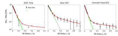
Formalin fixation significantly changes cell membrane permeability in cortical brain tissue
Ruiliang Bai, Xihui Ju, Peter Basser
Ex vivo, formalin-fixed biological tissue sample are often used to validate MRI methods for in vivo applications. However, since fixation may alter the chemical and physical properties of tissue, including the cell membrane permeability, it is important to know how and whether fixation changes in water microdynamics and potentially affects MRI signals. In this work, we studied the transmembrane water exchange kinetics in live rat brain cortical tissue in vitro, and following formalin fixation. We found that the fixation process can significantly increase the transmembrane water exchange kinetics by increasing the cell membrane permeability to water.
|
|
5032.
 |
47 |
A compact solution for physiological parameters from ultrafast prostate DCE-MRI may reduce random and systematic errors
Xiaobing Fan, Dianning He, Aritrick Chatterjee, Shiyang Wang, Milica Medved, Federico Pineda, Ambereen Yousuf, Tatjana Antic, Aytekin Oto, Gregory Karczmar
The Tofts pharmacokinetic model requires multiple calculations for analysis of dynamic contrast enhanced (DCE) MRI. This can result in error propagation that reduces the accuracy of pharmacokinetic measurements. Here, we present a new compact solution for estimating physiological parameters based on changes in signal intensity, without the Tofts model. Human prostate DCE-MRI data were analyzed to compare physiological parameters estimated from proposed compact solution with the Tofts model. The Ktrans and ve from the compact solution correlated strongly with values from the Tofts Model. Bland–Altman plots showed moderate to excellent agreement between the compact solution and the Tofts Model.
|
|
Electrical Property Imaging
Electronic Poster
Contrast Mechanisms
Wednesday, 20 June 2018
| Exhibition Hall |
17:15 - 18:15 |
| |
|
Computer # |
 |
5081.
 |
1 |
Electro-Magnetic Property Mapping Using Kalman Filtering with a Single Acquistion at 3.0 T and 7.0 T MRI
Han-Jae Chung, Jong-Min Kim, You-Jin Jeong, Jeong-Hee Kim, Chulhyun Lee, Chang-Hyun Oh
The phase-based Electro-Magnetic (EM) MR property imaging such as Quantitative Susceptibility Mapping (QSM) and MR Electric Properties Tomography (MREPT) shows great potential clinically. The main post-processing steps in QSM and MREPT are high-pass filtering and Laplacian of MR images. They, however, cause severe artifacts and noise during conventional calculations. In this work, we propose a novel reconstruction method of EM property MRI using Kalman filter algorithm and show the utility of the proposed method by comparing the imaging results.
|
 |
5082.
 |
2 |
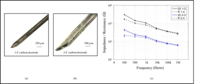
Development of Carbon Electrodes for Current Density Mapping during DBS
Neeta Ashok Kumar, Munish Chauhan, Rosalind Sadleir
We used MR phase mapping techniques in a preclinical DBS model to image current distributions nearby deep brain stimulation electrodes. To avoid safety issues and artifacts associated with imaging typical platinum-iridium (Pt-Ir) DBS leads, we developed custom carbon electrodes. We compared carbon electrode performance to size-matched Pt-Ir and clinical DBS electrodes at 7 T, using uniform phantoms and fixed brain tissue. Artifacts surrounding carbon electrodes were smaller than for Pt-Ir electrodes. Current density distributions derived from phase images were similar for both electrode types in uniform phantoms and fixed tissue.
|
|
5083.
 |
3 |
In vivo Current Density and Conductivity Tensor Imaging of Human Brain During TACS using DT-MREIT
Munish Chauhan, Aprinda Indahlastari, Aditya Kasinadhuni, Christopher Saar, Bakir Mousa, Kevin Castellano, Thomas Mareci, Rosalind Sadleir
Knowledge of the electrical properties of brain tissue is key to developing better understanding of whole brain function. In this study, we present the first in vivo images of anisotropic conductivity distribution in the human head, measured at a frequency of ~10 Hz. We used MREIT techniques to encode phase changes caused by transcranial AC current flow (TACS) within the head via two independent electrode pairs. These results were then combined with DTI data to reconstruct full anisotropic conductivity distributions in 5 mm-thick slices of the brains of two participants. Conductivity values recovered in the study were broadly consistent with literature values.
|
|
5084.
 |
4 |
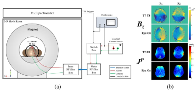
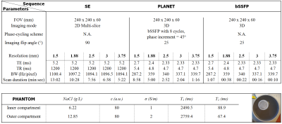
Sequences for transceive phase mapping: a comparison study and application to conductivity imaging
Soraya Gavazzi, Stefano Mandija, Cornelis van den Berg, Yulia Shcherbakova, Mick Bennis, Jan Lagendijk, Lukas Stalpers, Hans Crezee, Astrid van Lier
Electrical properties imaging relies on accurate transceive phase determination. We explored the use of PLANET, an ellipse fitting approach on phase-cycled bSSFP data, for transceive phase mapping for the first time. We compared its accuracy, precision and time-efficiency with conventional SE and bSSFP techniques. Additionally, we reconstructed conductivity maps based on these techniques. We found that bSSFP and PLANET were as accurate as SE, but more precise. Also, bSSFP was the most time-efficient. Nevertheless, banding artefacts corrupting bSSFP transceive phase were, instead, intrinsically removed by PLANET. PLANET had clinically acceptable scan-time and was generally more suitable for conductivity mapping.
|
|
5085.
 |
5 |
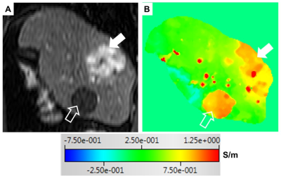
Noninvasive Assessment of Electrical Conductivity Characteristics of Normal and Diseased Liver Using Electric Properties Tomography
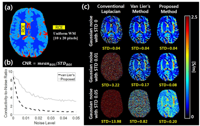
Khin Tha, Ulrich Katscher, Kinya Ishizaka, Kohsuke Kudo, Hiroki Shirato
The feasibility of Electric Properties Tomography (EPT) in distinguishing between the normal and diseased liver tissues was evaluated. A 2D steady state free precession sequence was used to acquire the RF transceive phase needed for a simplified version of EPT; and a total of 10 dynamic sagittal scans of the liver were obtained under single breath-hold, in 10 healthy volunteers and 11 patients with hepatic lesions. Despite the need of technical improvements, noninvasive electrical conductivity assessment of the liver by EPT was possible. Its potential utility in identifying hepatocellular carcinomas with intratumoral necrosis and/ or high vascularity was also shown.
|
|
5086.
 |
6 |
Diffusion Tensor Magnetic Resonance Electrical Impedance Tomography versus Magnetic Resonance Conductivity Tensor Imaging
Mehdi Sadighi, Figen S. Oktem, B. Murat Eyuboglu
In this study, recently proposed diffusion tensor magnetic resonance electrical impedance tomography (DT-MREIT) is compared with magnetic resonance conductivity tensor imaging (MRCTI) using simulated measurements generated by means of a finite element model. Both methods are used to reconstruct conductivity tensor images of an anisotropic conductivity distribution. In DT-MREIT, extra cellular conductivity and diffusivity ratio (ECDR) is recovered from its transverse gradient. In MRCTI, the conductivity tensor is reconstructed from two current profiles by using anisotropic Bz sensitivity (ABzS) method with a stronger regularization. Reconstructed conductivity images suggest that MRCTI provides better accuracy than DT-MREIT, at lower SNR levels.
|
|
5087.
 |
7 |
Transceive Phase Corrected Contrast Source Inversion-Electrical Properties Tomography
Peter Stijnman, Stefano Mandija, Patrick Fuchs, Rob Remis, Cornelis van den Berg
Contrast Source Inversion Electrical Properties Tomography (CSI-EPT) is an integral-based method that aims to reconstruct tissue electrical properties through an iterative minimization procedure. This method requires complex $$$B_1^+$$$ data as input. In practice, however, the transmit phase cannot be measured in MRI-experiments. Only the transceive phase can be calculated from MR-measurements. In this work, the CSI-EPT reconstruction algorithm is reformulated to take the transceive phase into account. This transceive phase correction opens the possibility to exploit higher sensitivity of EPT at higher field strengths with regular quadrature setups. Additionally, for the first time CSI-EPT reconstructions from MR-measurements are shown.
|
|
5088.
 |
8 |
Evaluating Validity of MREPT Assumptions for 21.1 T
Ghoncheh Amouzandeh, Jens Rosenberg, Frederic Mentink-Vigier, Nastaren Abad, Samuel Grant
This study examines conductivity mapping using MR Electrical Properties Tomography (MREPT) at ultra-high field (21.1 T). The accuracy of reconstructing conductivity using the complex B1+ field (Full-form) versus only the B1+ phase (Phase-based) is evaluated. Phantoms containing different NaCl concentrations were tested to compare these reconstructions with actual conductivities measured by dielectric probe at 900 MHz. Also, these methods were evaluated for experiments acquired with volume and surface coil configurations operated in either linear or quadrature transceiver. Conductivity maps of Full-form versus Phase-based MREPT from in vivo MCAO rats were acquired, with both providing similar variations across the ischemic brain.
|
|
5089.
 |
9 |
bSSFP Phase Correction and its use in MREPT
Safa Ozdemir, Yusuf Ider
Balanced steady state free precision (bSSFP) has various advantages, namely high speed, high SNR, motion insensitivity and eddy current compensation. However, due to the B0 inhomogeneity, the so called "banding artifact" occurs at certain frequency regions. In this paper, phase correction method for the bSSFP sequence is proposed utilizing B0 and T2 maps. As an application, acquired B0 insensitive phase maps are used to obtain artifact-free conductivity maps.
|
|
5090.
 |
10 |
Implementation of Conductivity Tensor Imaging (CTI) using MRI
Nitish Katoch, Bup Kyung Choi, Saurav Sajib, Hyung Joong Kim, Oh In Kwon, Eung Je Woo
Electrical conductivity is a passive material property primarily determined by concentrations of charge carriers and their mobility. The macroscopic conductivity of biological tissue at low frequency may exhibit anisotropy related with its structural directionality. When expressed as a tensor and properly quantified, the conductivity tensor can provide diagnostic information of numerous diseases. Imaging of conductivity distributions inside the human body requires probing it by externally injecting conduction currents or inducing eddy currents. Here we propose a novel method to reconstruct conductivity tensor images using an MRI scanner without any current injection.
|
|
5091.
 |
11 |
A Fast and Dedicated First-Order Differencing EPT Reconstruction Method
Patrick Fuchs, Stefano Mandija, Peter Stijnman, Wyger Brink, Cornelis van den Berg, Rob Remis
A new method for reconstructing electrical properties from $$$B_1^+$$$ data based on Maxwell's equations in an E polarized field (found in the midplane of a birdcage coil) is presented. This first-order EPT (foEPT) method uses first order spatial derivatives as opposed to the second order Helmholtz based MR-EPT methods and is thus less susceptible to noise. Furthermore, the method does not rely on any homogeneity assumptions. The method is validated using an in-vivo phantom measurement and compared to an MR-EPT reconstruction. FoEPT conductivity reconstructions show less noise-amplification and less boundary artefacts compared to Helmholtz-based MR-EPT reconstructions.
|
|
5092.
 |
12 |
An Explicit Method for MR-Based Electrical Properties Reconstruction Free from Their Boundary Values
Motofumi Fushimi, Takaaki Nara
This paper presents a new explicit reconstruction method for Magnetic Resonance Electrical Properties Tomography (MREPT) in a circular region of interest (ROI) that does not require EP values on the boundary of the ROI. Starting from the complex form of Maxwell's equations, we solved the D-bar equation of the electric field with the Neumann boundary condition. The proposed method reconstructs EPs successfully without giving any knowledge of EP values on the boundary of the ROI. To extend the method to, for example, a rectangular ROI, is our future work.
|
|
5093.
 |
13 |
LCF Artifact Elimination in cr-MREPT using Phased-Array Receive Coil
Gulsah Yildiz, Yusuf Ider
Convection-reaction equation based MREPT (cr-MREPT)1 conductivity images suffer from LCF artifact at low convective field (LCF) regions. Padding2 method has been proposed to overcome this issue but it requires additional acquisition, prolonging the total time. In this paper, we propose using data from different channels of phased-array receive coil to eliminate LCF artifact without requiring extra acquisitions.
|
|
5094.
 |
14 |
Automated Seed Selection for Gradient-based Electrical Properties Tomography and Its in vivo Validation in the Brain
Yicun Wang, Pierre-Francois Van de Moortele, Bin He
Electrical Properties Tomography (EPT) retrieves tissue electrical conductivity and permittivity at Larmor frequency which potentially provides diagnostic information and facilitates subject-specific local SAR estimation. Gradient-based EPT (gEPT) significantly alleviates boundary artifact encountered by conventional EPT methods, yet its implementation requires subjective assignment of integration seed points. In this study, we developed an automated seed selection strategy based on locally calculated conductivity values, and evaluated the effect of seed number for human brain imaging. This new strategy was validated in eight healthy subjects to produce robust and accurate results, paving the path for an unbiased and fully-automated process for EP quantification.
|
|
5095.
 |
15 |
Global Maxwell Tomography with Match Regularization for accurate electrical properties extraction from noisy B1+ measurements
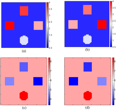
Jose Serralles, Athanasios Polimeridis, Luca Daniel, Daniel Sodickson, Riccardo Lattanzi
We introduce a new regularization approach, “Match Regularization”, and show that in tandem with Global Maxwell Tomography (GMT) it enables accurate, artifact-free volumetric estimation of electrical properties from noisy B1+ measurements. We demonstrated the new method for two numerical phantoms with completely different electrical properties distributions, using clinically feasible SNR levels. Estimated electrical properties were accurate throughout the volume for both phantoms. Our results suggest that GMT with match regularization is robust to noise and can be employed to map electrical properties in phantoms and in vivo experiments.
|
|
5096.
 |
16 |
In-vivo validation of water content Electrical Properties Tomography reconstructions in white matter using independent MR-EPT measurements
Stefano Mandija, Petar Petrov, Jord Vink, Sebastian Neggers, Peter Luijten, Cornelis van den Berg
MR-Electrical Properties Tomography (MR-EPT) can provide accurate mean conductivity values in large homogeneous tissues such as the white matter, provided sufficient erosion to avoid boundary regions. Water-content-EPT (wEPT) has been recently proposed to reconstruct EPs on a voxel-to-voxel basis. However, wEPT uses an empirical model calibrated with literature EPs values, assumed correct, obtained from ex-vivo probe measurements. In this work, the validity of the model employed in wEPT is verified for white matter conductivity reconstructions by using in the wEPT model calibration the mean white matter conductivity value obtained from in-vivo MR-EPT reconstructions as an independent modality.
|
|
5097.
 |
17 |
Error Analysis of Helmholtz-based MR-Electrical Properties Tomography
Stefano Mandija, Alessandro Sbrizzi, Ulrich Katscher, Peter Luijten, Cornelis van den Berg
The numerical error arising from the computation of spatial derivatives using finite difference kernels is investigated for Helmholtz-based MR-Electrical-Properties-Tomography conductivity reconstructions. We show that this numerical error is one major cause of limited accuracy in Helmholtz-based MR-EPT reconstructions, even if mitigation strategies such as Gibbs ringing correction and Gaussian apodization in k-space are adopted. Ultimately, large derivative kernels lead to more noise-robust conductivity reconstructions, at the cost of more spatially-extended boundary errors. If boundaries are not explicitly taken into account during reconstructions, the accuracy of MR-EPT is severely hampered, particularly for spatially convoluted tissues such as the human brain.
|
|
5098.
 |
18 |
Spatial resolution of Full cr-MREPT: 2D and 3D evaluation
Yusuf Ider, Celik Boga, Gulsah Yildiz
Determining the spatial resolution (SR) of Magnetic Resonance Electrical Property Tomography (MREPT) is important for assessing its utility in clinical applications. This study aims at finding the SR of Full cr-MREPT which yields images without internal boundary artifact. It is shown by simulations and experimental results that SR in general is determined by the resolution of the MR data. With noise-free simulation data SR is 2-2.5 pixels. With noisy and real data it may go up to 4-4.5 pixels due to Low Pass filtering and regularization. SR appears to be equal in all three directions.
|
|
5099.
 |
19 |
A Dictionary-Based Method for Conductivity Tensor Mapping
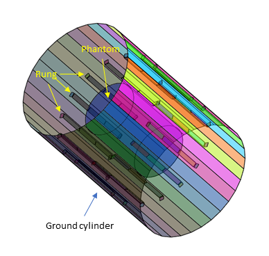
Kathleen Ropella-Panagis, Scott Peltier, Douglas Noll
Measuring conductivity tensors provides an additional layer of information as to how tissues in the body conduct electric current. Tissues with anisotropic conductivity values may include white matter tracts and muscle. Measurement of the tensor requires the object to rotate with respect to the main magnetic field of the MRI scanner, but the degree of rotation is severely limited in human subjects. We propose a dictionary-based approach that provides an estimate of the tensor given small rotation angles of the object.
|
|
5100.
 |
20 |
In Vitro Imaging of Therapeutic Effect of Curcumin on Liver Cirrhosis using MR-based Electrical Conductivity Imaging Method
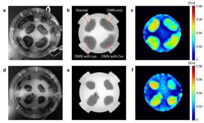
Bup Kyung Choi, Nitish Katoch, In Ok Ko, Ji Ae Park, Jin Woong Kim, Hyung Joong Kim, Oh In Kwon, Eung Je Woo
Curcumin has been used for the treatment of inflammatory diseases in oriental medicine, and its anti-inflammatory effect was recently reported. In this feasibility study, hepato-protective effect of curcumin was imaged in rat liver cirrhosis model, which was induced with dimethylnitrosamine (DMN). Magnetic resonance (MR)-based electrical conductivity imaging method was applied to evaluate tissue condition associated with protective effect. From electrical conductivity images, damaged liver tissues by DMN showed decreased conductivity than normal liver tissues. In contrast, cirrhotic tissues with curcumin treatment showed increased conductivity which was similar to normal tissue.
|
|
5101.
 |
21 |
Contrast source inversion global Maxwell tomography: a technique for electric properties MR imaging without phase information.
Alessandro Arduino, Oriano Bottauscio, Luca Zilberti
The possibility to perform MR imaging of the electric properties relying only on the measurable magnitude of transmit sensitivity, without any hypothesis on its phase, is an extremely interesting task pursued by the scientific community in the last years. Here, the adoption of the contrast source inversion technique in the context of the global Maxwell tomography is proposed. The lack of phase information affect the numerical minimisation procedure by introducing local minima in the cost functional. The convergence of the method is restored by the adoption of multi-channel transmit coils, which can increase the data by measuring multiple transmit sensitivities.
|
|
5102.
 |
22 |
Dictionary-based Electric Properties Tomography for brain conductivity imaging
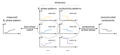
Ulrich Katscher, Max Herrmann, Thomas Amthor, Christian Findeklee, Mariya Doneva
Electric Properties Tomography (EPT) derives tissue conductivity and permittivity according to the Helmholtz equation via the second derivative of the measured complex B1 map, or by iteratively solving the corresponding forward problem. This abstract presents a different type of EPT reconstruction: the measured B1 map is compared locally with entries of a dictionary, which are small B1 maps of a priori known electric properties. This "dictionary-based EPT" (db-EPT) could be able to solve the transceive phase problem as well as the boundary problem of EPT. This study demonstrates the feasibility of db-EPT by measuring brain conductivity of healthy volunteers.
|
|
5103.
 |
23 |
Evaluation of the Noise Behavior of Gradient-based vs. Helmholz-based Reconstruction of Electrical Properties Tomography in Simulation
Yihe Hua, Ileana Hancu, Seung-Kyun Lee, Teck Beng Desmond Yeo, Jiaen Liu
Electrical properties tomography (EPT) is a promising technique that has the potential to generate high resolution images of tissue electrical properties in vivo. One limitation of EPT is its high sensitivity to noise in the measured data. In this study, a comparison was performed between the so-called gradient-based EPT (gEPT) algorithm and the Helmholtz-based EPT method in a simulation. The result suggests significantly improved performance using gEPT and provides useful insight into the noise behavior of various EPT algorithms for optimization of the algorithm design.
|
|
5104.
 |
24 |
Mapping the Brain’s water content and low-frequency electrical properties through T1-weighted MRI imaging
Cornelia Wenger, Catherine Tempel-Brami, Hadas Hershkovich, Moshe Giladi, Zeev Bomzon
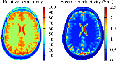
This study investigates the possibility to extend the water-content based electrical properties tomography (wEPT) technique to lower frequencies. The wEPT approach assumes that electrical properties (EP) of brain tissues can be estimated from water-content (WC) which is derived from two T1-weighted MRIs. Adapted wEPT model parameters were evaluated from ex-vivo measurements of calf brain tissue samples. We performed wEPT estimations in an in-vivo rat brain tumor model, followed by ex-vivo measurements of brain extracted samples. Results predict good correlation between WC ex-vivo measurements and in-vivo wEPT estimations. Yet, mapping EPs with wEPT at lower frequencies needs further investigation.
|
|
Advancements in CEST Methodology & Applications
Electronic Poster
Contrast Mechanisms
Wednesday, 20 June 2018
| Exhibition Hall |
17:15 - 18:15 |
| |
|
Computer # |
|
5105.
 |
25 |
Spin-lock Imaging of 3-o-Methyl-D Glucose (3oMG) in Brain Tumors
Zhongliang Zu, Xiaoyu Jiang, Junzhong Xu, John Gore
We evaluated the ability of spin-lock imaging to detect the uptake of 3-o-methyl-D-glucose (3oMG) in normal brain and brain tumors in animals. We used $$$\triangle R_{1\rho}^{diff}$$$ to isolate the contribution from only the injected agent. We found that $$$\triangle R_{1\rho}^{diff}$$$ in tumors increased rapidly after injection, whereas intact brain showed a gradual increase up to 1h. $$$\triangle R_{1\rho}^{diff}$$$ was significantly different between tumors and contralateral normal tissues
|
|
5106.
 |
26 |
Chemical Exchange Rotation Transfer imaging of Phosphocreatine in Muscle
Zhongliang Zu, Eugene Lin, Elizabeth Louie, Xiaoyu Jiang, Christopher Lankford, Bruce Damon, Mark Does, John Gore, Daniel Gochberg
CEST imaging of Creatine has been reported, whereas selective mapping of PCr in vivo has not been implemented. We found that CEST imaging of Creatine may be influenced by other molecules in muscle, but CERT imaging of PCr is more specific and thus should be a better indicator of changes of flux through the CK reaction.
|
|
5107.
 |
27 |
Chemical Exchange Saturation Transfer imaging of prostate cancer at 3T: Repeatability, and initial results of an acquisition and multi-pool analysis protocol
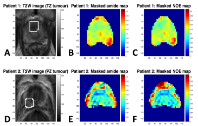
Vincent Evans, Francisco Torrealdea, Marilena Rega, Mina Kim, Mrishta Brizmohun Appayya, Arash Latifoltojar, Shonit Punwani, Xavier Golay, David Atkinson
An optimised acquisition and post-processing protocol for multi-pool Lorentzian analysis of CEST data in the prostate at 3T is described. The repeatability of the technique is evaluated in five healthy volunteers and the contrast observed between healthy tissue, TZ tumour and PZ tumour in two prostate cancer patients is evaluated.
|
|
5108.
 |
28 |
Toward CEST MRI of renal masses: protocol optimization and first preliminary data
Shu Zhang, Bian Li, Joshua Greer, Ananth Madhuranthakam, Jochen Keupp, Ivan Dimitrov, Robert Lenkinski, Ivan Pedrosa, Elena Vinogradov
Chemical Exchange Saturation Transfer (CEST) MRI is emerging as a tool for the studies of human malignancy. However, the translation of CEST into a successful tool for renal cancer characterization has been slow and hampered by technical difficulties associated with body imaging, such as motion, contaminating lipid signals and increased B0 ingomogeneity. Here we optimize CEST protocol for characterization of renal masses and demonstrate CEST measurements are feasible in kidneys using combination of motion synchronization, post-processing registration and lipid artifact removal. In addition, first Renal Cell Carcinoma patient CEST-mDixon data is shown and imaging results are correlated with the pathology.
|
|
5109.
 |
29 |
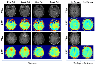
APT-CEST post Gadolinium. Should it be avoided? Comparison of pre- & post-Gadolinium CEST on glioma at 3T.
Francisco Torrealdea, Joe Hearle, Vincent Evans, Moritz Zaiss, Ana Carvalho, Anath Shankar, Harpreet Hyare, David Atkinson, Xavier Golay, Anna Barnes, Marilena Rega
This study compares APT-CEST between pre- and post-gadolinium in patients with gliomas at 3T, and evaluates the feasibility of performing CEST after administration of T1 contrast. The results of the study demonstrate that Gd administration does not significantly affect the quality of the APT-CEST image, encouraging the acquisition of CEST data, even after the administration of T1 contrast agents.
|
|
5110.
 |
30 |
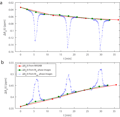
Phase-locked CEST – Introducing dynamic B0-correction to gagCEST
Johannes Windschuh, Moritz Zaiss, Jae-Seung Lee, Alexej Jerschow, Ravinder Regatte
Even a small frequency drift of less than 1Hz/min of the MRI scanner can have a strong impact on gagCEST measurements. We propose a dynamic B0-correction that tracks the frequency shift using the phase images provided by the GRE readout. We show that this correction eliminates the influence of the frequency drift on gagCEST without the need of additional measurement time allowing higher accuracy, reproducibility, and comparability of gagCEST studies.
|
|
5111.
 |
31 |
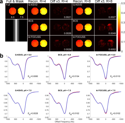
Accelerating CEST with Patch-based Global Orthogonal Dictionary Learning
Huajun She, Xinzeng Wang, Shu Zhang, Ece Ercan, Jochen Keupp, Anath Madhuranthakam, Ivan Dimitrov, Robert Lenkinski, Elena Vinogradov
This work investigates accelerating CEST imaging using patch-based global spatial-temporal dictionary learning (G-KSVD). We extend the dictionary learning for CEST acceleration. CEST data has high spatial-temporal correlation, so we can utilize the global Z-Spectrum information as well as the spatial information to form the global spatial-temporal dictionary. The dictionary is learned iteratively from overlapping patches of the dynamic image sequence along both the spatial and temporal directions. The proposed method performs better than the BCS and k-t FOCUSS methods for both phantom and in vivo brain data at high reduction factor of R=8.
|
|
5112.
 |
32 |
Accelerated CEST Imaging with Parallel Deep Convolutional Neural Networks
Huajun She, Shu Zhang, Xinzeng Wang, Ece Ercan, Jochen Keupp, Anath Madhuranthakam, Ivan Dimitrov, Robert Lenkinski, Elena Vinogradov
CEST is a new contrast mechanism in MRI. However, a successful application of CEST is hampered by its slow acquisition. This work investigates accelerating CEST imaging using parallel convolutional neural networks (PCNN). We extend the Cascade-CNN into a multi-channel model and train the network establish a mapping from the multi-coil input to multi-coil output. This work is the first try to apply deep learning and convolutional neural networks technique in accelerating CEST imaging. The in vivo brain results show that the proposed method demonstrates a high quality reconstruction of the MTRasym maps with different saturation pulses at R=4.
|
|
5113.
 |
33 |
Accelerating 3D CEST Imaging with Low Rank Sparse Reconstruction
Huajun She, Ece Ercan, Shu Zhang, Xinzeng Wang, Jochen Keupp, Anath Madhuranthakam, Ivan Dimitrov, Robert Lenkinski, Elena Vinogradov
Chemical exchange saturation transfer (CEST) is a new contrast mechanism in MRI. However, a successful application of CEST is hampered by its slow acquisition especially in the 3D applications. Compressed sensing (CS) is powerful for reconstruction of highly undersampled data. This work implements the 3D pulsed steady-state CEST acquisition sequence and extended the low rank plus sparse (L+S) method to a 3D version. The phantom and in vivo human brain results demonstrate our design has the potential to accelerate the 3D CEST imaging about 4 times.
|
|
5114.
 |
34 |
Quantitative Magnetization Transfer Imaging in Murine Kidneys with Renal Artery Stenosis
Kai Jiang, Lilach Lerman
Quantitative magnetization transfer (qMT) imaging was used to measure bound water fraction in mouse kidneys with renal artery stenosis (RAS). MT-weighted images at variable offset frequencies and amplitudes, as well as B0, B1, and T1 maps of control and RAS kidneys, were acquired. A two-pool qMT model was used to estimate the bound water fraction as well as other relaxation and exchange parameters. An increased bound water fraction was found in the cortex, outer medulla, and inner medulla of the RAS kidneys. In conclusion, qMT imaging offers potential new biomarkers for assessment of RAS kidneys.
|
|
5115.
 |
35 |
Assessing changes in kidney pH in acute kidney injury model using acidoCEST MRI
Atul Minhas, Jack Sharkey, Edward Randtke, Patricia Murray, Bettina Wilm, Mark Pagel, Harish Poptani
Kidneys are responsible for regulation of pH homeostasis, and cytotoxicity caused by cancer therapeutics can significantly alter renal function and homeostasis. Chemical exchange saturation transfer (acidoCEST) MRI has been proposed to measure tissue pH in-vivo using exogenous contrast agents. In this study, we used the acidoCEST technique to measure changes in kidney pH after acute kidney injury (AKI) in rodents. Typically, CT contrast agents such as Iopamidol (300 mg iodine/mL) are used as CEST contrast agent in acidoCEST MRI. However, the accuracy of acidoCEST using CT contrast agents relies on the delivery of the contrast agent to the target organ. To address this issue, we performed acidoCEST and FAIR-EPI based perfusion imaging to assess pH and blood flow changes in a mouse model of AKI. Results show that perfusion of kidneys affect pH measurements.
|
|
5116.
 |
36 |
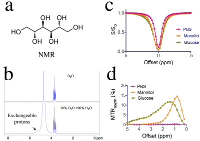
Detection of the accumulation of mannitol in rat brains using CEST MRI
Jing Liu, Chengyan Chu, Lin Chen, Jia Zhang, Rohit Srivatava, Piotr Walczak, Jiadi Xu, Peter van Zijl, Miroslaw Janowski, Guanshu Liu
Mannitol is a clinically widely-used osmotic agent. Accumulation of mannitol in the interstitium of the brain, however, can cause severe adverse effects. Here we used CEST MRI to detect mannitol directly through its inherently carried exchangeable hydroxyl protons. After comprehensively characterizing the CEST properties of mannitol in vitro, we demonstrated that the intra-arterial infusion of mannitol at an excess dose led to a significantly elevated CEST signal at 0.9 ppm, indicating that CEST MRI has great clinical potential to be used as a monitoring tool for mannitol treatment.
|
|
5117.
 |
37 |
Nuclear Overhauser enhancement effect of low $$$B_{1}$$$ power CEST RF in human brain at 3.0 T
Yuki Kanazawa, Masafumi Harada, Mitsuharu Miyoshi, Ikuho Kosaka, Kotaro Baba, Hiroaki Hayashi, Yuki Matsumoto
The purpose of this study is to clarify the relationship between APT and NOE effects derived from CPE fitting of the human brain on a 3 T MR scanner. CEST imaging with different B1 values of the brain was performed in healthy subjects. The mean NOE values of white matter at 0.5 µT were higher than all regions (P < 0.05). CPE-spectrum shows greater sensitivity for both APT and NOE peaks than conventional Z-spectrum and MTRasym. It is found that NOE imaging on a 3.0 T scanner is sensitive on low-B1 power regardless of the CEST fitting process.
|
|
5118.
 |
38 |
GluCEST MRI: Reproducibility, background contribution and source of glutamate changes in the MPTP mouse model of Parkinson’s disease
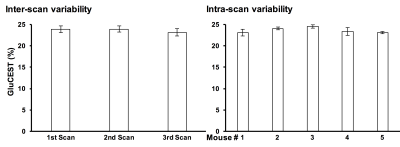
Puneet Bagga, Stephen Pickup, Dan Martinez, Rachelle Crescenzi, Ari Borthakur, Gaurav Verma, Joel Greenberg, John Detre, Hari Hariharan, Ravinder Reddy
Glutamate Chemical Exchange Saturation Transfer (GluCEST) MRI provides indirect detection of glutamate in vivo by measuring the exchange of glutamate amine protons with bulk water. The GluCEST contrast is potentially contaminated by a contribution of other metabolites exhibiting proton chemical exchange. We evaluated the reproducibility and background contamination to the GluCEST and source of the GluCEST changes in MPTP mouse model. Approximately 28% of GluCEST contrast appears to be derived from sources other than glutamate that are also not detectable by MRS. Glial proliferation caused by neuroinflammation was found to be the cause of elevated glutamate in mice exposed to MPTP.
|
|
5119.
 |
39 |
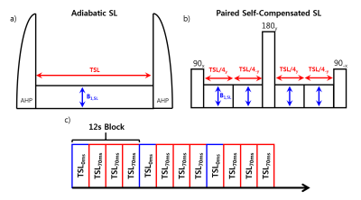
Toward Safer Monitoring of Glucose Transport in a Rat Brain Tumor Necrosis using 3-O-Methyl-Glucose Chemical Exchange-sensitive Spin-Lock Magnetic Resonance Imaging
Julius Chung, Moon-Sun Jang, Geun Ho Im, Wonmin Choi, Tao Jin, Seong-Gi Kim, Jung Hee Lee
Glucose exchange-sensitive spin lock imaging has been shown to have promise in monitoring glucose uptake with reasonable sensitivity. There are design choices that can be made in such an experiment such as whether to use an analog for better sensitivity and how to establish an efficient spin-lock. We examine metabolic uptake of a rat brain tumor with necrosis using 3-O-methyl-glucose, a safer glucose analog than 2DG, and examine differences between using an adiabatic pulse and a paired self-compensated pulse with lower peak power. Both pulses demonstrated delayed uptake in the infarcted tumor region although with higher sensitivity using adiabatic pulses.
|
|
5120.
 |
40 |
Magnetization Transfer in Lipids - Role of Exchangeable Groups and Water Binding
Video Permission Withheld
Weiqi Yang, Jae-Seung Lee, Johannes Windschuh, Maureen Leninger, Nate Traaseth, Alexej Jerschow
We study the magnetization exchange mechanism in lipid systems, with relevance to imaging myelin via MT contrast. Studies of samples with different lipid compositions reveal exchange time scales, and the role of structural features in the contrast mechanism. Insights from molecular dynamics provide estimates of the contribution of the dipolar pool equilibration to the MT amplitude. The effect of lipid head groups, and the contribution of cholesterol and proteins are examined. It is hoped that these findings will help explaining the origin of White Matter MT contrast and will allow better myelin quantification by tailored saturation sequences.
|
|
5121.
 |
41 |
In vivo Kinetic CEST MRI of sodium salicylate(NaSA): Comparison of MTRasym and Subtraction of saturation-weighted images
Yanrong Chen, Chengwang Jin, Yan Luo, Chongxue Bie, Yingcheng Zhao, Xiaowei He, Xiaolei Song
Salicylate analogues feature chemical shift far from water (Δω = 8-10 ppm), however, there are almost no available reports on their in vivo detection of salicylate upon intravenous administration. We aim to optimize the in vivo detection of NaSA, by comparing compared MTRasym and a Dynamic Salicylate Enhancement (DSE). For the mice brain with LPS-induced inflammation, there are ~4% DSE signal which displays a clear kinetic trend. While MTRasm values are very small, oscillating between -2% to 0% due to the unsymetric MTC. In conclusion, our DSE method is able to track the dynamic signal changes following the infusion of NaSA.
|
|
5122.
 |
42 |
Assessment of a clinically feasible Bayesian fitting algorithm using a simplified description of Chemical Exchange Saturation Transfer (CEST) Imaging
Aaron Kujawa, Mina Kim, Eleni Demetriou, Annasofia Anemone, Dario Longo, Moritz Zaiss, Xavier Golay
A Bayesian fitting algorithm was combined with analytical approximations of the Bloch-McConnell (BM) equations with the aim to considerably reduce processing time. The accuracy of the algorithm was assessed with simulated data and data from phantom experiments and compared to fit results obtained with the numerical solution of the BM equations. Continuous-wave and pulsed saturation was considered. The results showed agreement between estimates and ground truth as well as between the approximate analytical and numerical model implementations of the Bayesian algorithm. A considerable reduction of processing time was achieved.
|
|
5123.
 |
43 |
Including water nutation in an analytic solution for pulsed CEST
Christopher Lankford, Zhongliang Zu, Elizabeth Louie, Mark Does, Daniel Gochberg
Pulsed chemical exchange saturation transfer (CEST) MRI lacks an analytical solution, impeding data analysis and optimization efforts. A recently proposed solution has mitigated this problem, but it ignores water pool nutation and is thus inaccurate near the water resonance frequency. This work proposes a solution that accounts for water nutation assuming a known flip angle function which can be numerically estimated with no a priori knowledge about tissue parameters. The nutation-corrected solution closely matches numerical Bloch-McConnell simulation, even near the water resonance frequency.
|
|
5124.
 |
44 |
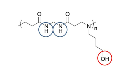
Chemical exchange saturation transfer magnetic resonance imaging of functionalized poly(N, N’-methylene bisacrylamide 4-aminobutanol) gel
Weiqiang Dou, Jos M.J. Paulusse, Heinz Peter Janke, Xiaolei Song, Jiadi Xu, J.W.M Bulte, Arend Heerschap
Poly (amido amine)s like poly(N, N’-methylene bisacrylamide 4-aminobutanol) (MBA-ABOL) are compounds with promising biomedical applications, which, however, require that they can be visualized without contrast application. In this study we investigated if they can be imaged in a “label free fashion” by CEST MRI making use of their exchangeable amide and hydroxyl protons. We systematically determined optimal conditions for CEST in MBA-ABOL in solution and then demonstrated that the material can be imaged both in vitro and ex vivo, implanted in a rat leg, with a strong CEST effect from the amide protons and substantial effect from the hydroxyl protons.
|
|
5125.
 |
45 |
Clinically Feasible Model-based Analysis of Amide Proton Transfer MRI in Acute Ischaemic Stroke
Paula Croal, Yunus Msayib, Kevin Ray, James Larkin, Brad Sutherland, George Harston, Alistair Buchan, Peter Jezzard, James Kennedy, Nicola Sibson, Michael Chappell
Model-based analysis of CEST MRI is a robust quantitative method, however, the lengthy acquisition and processing times make it less clinically feasible. It has recently been proposed that partial acquisition of Z-spectra provides a faster approach, but at the cost of increased variability and large alterations in baseline Amide Proton Transfer (APT) effect. Here we present a refined approach, accounting for magnetisation transfer effects, which reduces acquisition and processing times and also decreases variability in the data. We demonstrate its ability to detect pathological reductions in the APT effect in both preclinical and clinical cohorts of acute ischaemic stroke respectively.
|
|
5126.
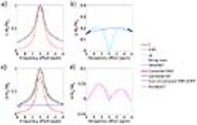 |
46 |
Improved quantification of amide proton transfer effect with direct water saturation- and magnetization transfer-correction in a glioma rat model at 3 Tesla
Yin Wu, Yinsheng Chen, Yiying Zhao, Shasha Yang, Jing Zhao, Zhongping Chen, Phillip Sun
Quantification of in vivo APT effect with routine asymmetry analysis is problematic due to concomitant contributions. Herein, a steady-state CEST signal solution was utilized to estimate direct water saturation (DWS) to improve Z-spectral quantification in a pulsed-RF CEST imaging. Specifically, APT effect was measured from the DWS and magnetization transfer (MT)-corrected CEST signals in glioma rat brains post-chemoradiotherapy at 3 Tesla. The proposed method revealed significant APT signal difference among regions of control (3.13±0.38%), necrosis (3.95±0.31%) and tumor (4.56±0.34%), consistent with histological observations and superior than routine asymmetry analysis.
|
|
5127.
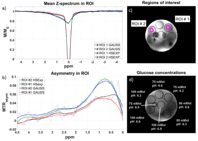 |
47 |
In-vivo Z-spectra acquisition with decreased direct saturation using adiabatic spin-lock pulses at 9.4T
Kai Herz, Chirayu Gandhi, Klaus Scheffler, Moritz Zaiss
Off-resonant spin-lock imaging enables a lot of possibilities for T1ρ and chemical exchange (CE) sensitive applications. For this purpose, a matching amplitude of the tipping and the locking pulse is required, which can be difficult due to the high power requirements of adiabatic pulses. In this work, we present a newly shaped adiabatic half-passage pulse, usable at low power to match the amplitude of the pulses. Off- and on-resonant saturated images acquired at 9.4T are shown. The new pulse shape is able to generate robust images with comparatively low power at ultra-high-field strengths.
|
|
5128.
 |
48 |
Optimization of overlap-resolved CEST for specific mapping of Glutamate and GABA
Frederico Severo, Noam Shemesh
Glutamate-CEST (GluCEST) has been recently emerging as a powerful new technique for mapping Glutamate in the brain. A recent development termed overlap-resolved CEST (orCEST) has demonstrated that other metabolites may contaminate GluCEST contrast, and proposed how to resolve the wanted signals; here we optimize orCEST contrast to reflect Glutamate and GABA with very high sensitivity and good specificity. orCEST experiments are optimized on metabolite phantoms, and then are applied in-vivo in the rat. The results are promising for neurotransmitter mapping in-vivo.
|
|
| Back |
| The International Society for Magnetic Resonance in Medicine is accredited by the Accreditation Council for Continuing Medical Education to provide continuing medical education for physicians. |
