|
Electronic Poster Session
Neuro |
Thursday, 21 June 2018
Electronic PosterNeuro
5247 -5270 Brain Microstructure
5271 -5294 Multiple Sclerosis: Brain & Spinal Cord Applications
5295 -5318 Psychoradiology
5391 -5414 Neuro Outside the Brain
5415 -5437 Neonatal & Pediatric Neuroimaging |
| |
Brain Microstructure
Electronic Poster
Neuro
Thursday, 21 June 2018
| Exhibition Hall |
08:00 - 09:00 |
| |
|
Computer # |
|
5247.
 |
49 |
 White Matter Microstructural Change Following Traumatic Brain Injury Assessed by Simultaneous Multi-Slice Multi-Shell Diffusion MRI - A Preliminary Study White Matter Microstructural Change Following Traumatic Brain Injury Assessed by Simultaneous Multi-Slice Multi-Shell Diffusion MRI - A Preliminary Study
Ping-Hong Yeh, Nicholas Goh, Cheng Guan Koay, Chihwa Song, Wei Liu, Grant Bonavia, John Ollinger, Gerard Riedy
Mild traumatic brain injury (mTBI) is difficult to diagnose and characterize. In this study, we applied simultaneous multi-slice multi-shell diffusion MRI to assess white matter microstructural changes in chronic military mTBI. Preliminary results showed parameters derived from Mean Apparent Propagator MRI method are superior to the parameters derived from diffusion tensor imaging or diffusion kurtosis imaging in differentiating tissues with distinct structural and architectural features, and thus has increased ability to identify microstructural changes in mTBI.
|
|
5248.
 |
50 |
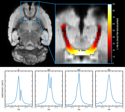
Effects of tissue microstructure on water resonance line-shape in post-mortem rat brain
Sean Foxley, Gregory Karczmar, Kazutaka Takahashi
Many neurodegenerative diseases are characterized by microstructural changes in white matter, including demyelination and cell loss. Such changes have been demonstrated to produce measurable effects on the MR signal. This work examines these effects from post-mortem fixed rat brain on voxel-wise, high-resolution water spectra acquired using a multi-gradient echo pulse sequence. Results demonstrate that components of the spectra are differentially affected by both white matter orientation relative to B0 as well as tissue microstructure. This suggests that water proton spectra may be sensitive to the tissue microenvironment and could serve as potential MRI based biomarkers of neurodegenerative diseases.
|
|
5249.
 |
51 |
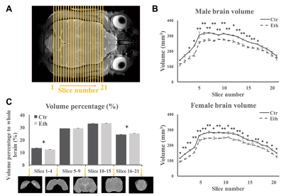
Effects of early alcohol exposure on functional organization and microstructure of a visual-tactile integrative circuit
Shiyu Tang, Su Xu, Alexandre Medina, Rao Gullapalli
Children with fetal alcohol spectrum disorders (FASD) often have deficits associated with multisensory processing. Because ethanol disrupts activity-dependent neuronal plasticity, a process that is essential for refining connections during cortical development, we hypothesize that early alcohol exposure results in alterations in multisensory cortical networks, which could explain the multisensory processing deficits seen in FASD. Here, we use a gyrencephalic animal model to test the prediction that early alcohol exposure alters the functional connectivity and microstructural features of a visual-tactile integrative area with resting-state functional magnetic resonance imaging and diffusion kurtosis imaging.
|
|
5250.
 |
52 |
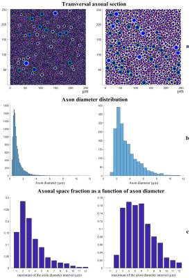
Axonal distributions: a simulation study to estimate Diffusion MRI signal contributions in white matter
Andrea Chiappiniello, Valentina Reggioli, Roberto Tarducci, Marco Catani, Flavio Dell’Acqua
In the last decade, many techniques that use diffusion MRI to obtain an axon diameter estimate as micro-structural integrity index have been developed. However, recent studies showed that diffusion signal may be not sensitive enough to quantify axon diameters. In this study, we simulated a simplified model of white matter to evaluate the contribution of intra-axonal compartment to diffusion MRI signal in white matter. We found that, even in distributions with a small mean diameter, big axons still substantially contribute to the total axonal volume. We conclude that quantifications of human axon diameters from diffusion MRI may still be possible.
|
|
5251.
 |
53 |
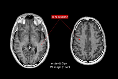
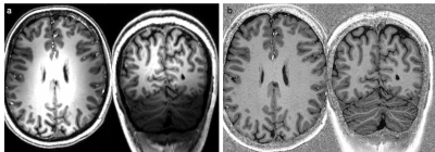
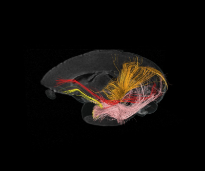
White Matter Fiber Structure Revealed by Synthetic MRI of the Longitudinal Magnetization Relaxation Rate (R1): Effects of Age at 1.5T
Hernan Jara, Stephan Anderson, Osamu Sakai
Purpose: To investigate the potential of R1-weighted Synthetic-MRI for unraveling the microstructure of white matter and for constructing accurate high resolution brain connectomes. Methods: Eighteen research subjects ranging in age from 0.6 to 87years were scanned with multispectral qMRI (T1, R1, T2, and PD) and analyzed with R1-weighted Synthetic MRI. Results: connectome renderings as function of increasing age show the expected increased WM track bundle packing and anatomical distributions evolution as function of age. Conclusion: R1-weighted WM Fibrography is a promising complementary alternative to DTI-WM Tractography for studying the microarchitecture of white matter.
|
|
5252.
 |
54 |
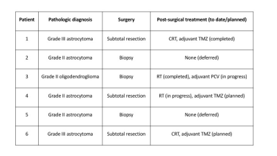
Ultra-High Gradient Diffusion MRI Reveals Distinct Microstructural Changes in Diffuse Gliomas Before and After Radiation Therapy
Ina Ly, Qiuyun Fan, Barbara Wichtmann, Aapo Nummenmaa, Ovidiu Andronesi, Brian Nahed, William Curry, Daniel Cahill, Tracy Batchelor, Jayashree Kalpathy-Cramer, Bruce Rosen, Susie Huang, Elizabeth Gerstner
The lack of a sensitive imaging method capable of capturing the full extent of glioma cell infiltration represents a significant challenge to accurate treatment planning and monitoring of therapeutic response. Here, using a recently developed diffusion MRI method (Linear Multi-Scale Model; LMM), we estimated the changes in restricted, hindered, and free water in six glioma patients pre- and post-treatment. We found scan-to-scan reproducibility of diffusion profiles in normal brain and identified distinct diffusion profiles in the tumor and peritumoral regions at different time points, thus highlighting the robustness of the LMM and its feasibility in the clinical setting.
|
|
5253.
 |
55 |
Brain Microstructure Characterization: Initial Experience and Optimization of protocols for the Siemens Terra 7T System
Video Permission Withheld
Maria Stefanescu, David Lohr, Aleksander Kosmala, Maxim Terekhov, Laura Schreiber
Recent technological advances have brought new ultrahigh-field MRI (UHF) systems to the market, which nourish the expectation of better image quality with vendor-supplied sequences than with older systems. This would be an important factor for a wider distribution of UHF system and, thus, for moving UHF technology forward from a research tool to a clinical application. Therefore, the aim of this study was to perform a pilot study to assess the new Siemens Magnetom Terra 7T system with regard to typical clinical applications of 7T MRI in the brain.
|
|
5254.
 |
56 |
Characterization of white matter structures growth in common marmosets
Fumiko Seki, Keigo Hikishima, Yuji Komaki, Marin Nishio, Junichi Hata, Akiko Uematsu, Norio Okahara, Erika Sasaki, Hideyuki Okano
This study investigated developmental patterns of white matter structures in common marmosets using DTI and MTR. Longitudinal MRI was performed to 23 marmosets at the age of 1-34 months. Tract-based ROIs were created for assessment of major fiber bundles. Population growth trajectories of association fibers estimated using Gompertz function showed different developmental patterns. As previously reported, inferior longitudinal fasciculus (ILF) showed earlier maturation with slow speed, whereas inferior fronto-occipital fasciculus (IFOF) showed slower maturation with fast speed observed in MTR, RD and FA. It indicates ILF might mature compared with IFOF at birth, which was consistent with human studies.
|
|
5255.
 |
57 |
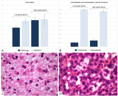
Non-invasive assessment of glioma microstructure using VERDICT MRI with comparison to histopathology
Video Permission Withheld
Fulvio Zaccagna, Frank Riemer, Andrew Priest, Kieren Allinson, Mary McLean, James Grist, Tomasz Matys, Jonathan Gillard, Colin Watts, Stephen Price, Martin Graves, Ferdia Gallagher
Gliomas are characterized by diffuse infiltration, high heterogeneity and poor prognosis. Imaging tumor heterogeneity may improve diagnosis and therapy planning. The Vascular, Extracellular and Restricted Diffusion for Cytometry in Tumors (VERDICT) MRI technique is a multi-compartmental model that exploits tissue microstructure. This preliminary study demonstrated the feasibility of translating VERDICT MRI in human brain imaging to investigate the microstructure of glioma with an abbreviated protocol. We demonstrated that VERDICT-derived cell size does not differ from the measured size on pathological slides and we found clear trends in LGG and HGG that may be useful to better differentiate types of glioma.
|
|
5256.
 |
58 |
Microstructure-mesh projection: Combining shape analysis with diffusion MRI models
Kirsten Lynch, Yonggang Shi, Arthur Toga, Kristi Clark
The hippocampus is a heterogeneous structure consisting of subfields with distinct cytoarchitectonic and connectivity patterns. In order to capture the complexity of hippocampal structure, we propose a framework that combines the localized specificity of shape analysis with the microstructural sensitivity obtained with diffusion MRI models. The microstructure-mesh projection pipeline projects local model parameters within the hippocampus onto the surface to enable visualization and analysis of regional microstructural features. In a pediatric dataset, regional patterns of microstructural maturation within the hippocampus were observed.
|
|
5257.
 |
59 |
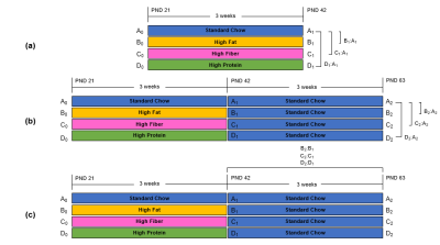
Diffusion Tensor Imaging to Investigate Diet-induced Changes in Neuronal Tissue Microstructure and Organization
Maribel Torres Velazquez, M. Elizabeth Meyerand, John-Paul Yu
Altered gut microbiome populations are associated with a broad range of neurodevelopmental disorders. Disruption of the gut microbiome via dietary intake has been shown to influence brain function and behavior in animal models. Utilizing diffusion tensor imaging we identified global changes in white matter structural integrity occurring in a diet-dependent manner. Subsequent diet-crossover experiments demonstrate the varying permanence of these diet-induced changes and the degree of plasticity associated with these changes. These studies allow us to further explore our understanding of the gut-brain-microbiota axis by revealing possible links between altered and dysbiotic gut microbiome populations and changes in brain structure.
|
|
5258.
 |
60 |
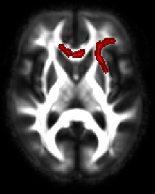
ASSOCIATION OF MID-LIFE VASCULAR RISK FACTORS AND LATE-LIFE WHITE MATTER MICROSTRUCTURE IN COGNITIVELY NORMAL OLDER WOMEN
Vijay Venkatraman, Christopher Steward, Cassandra Szoeke, Rowa Aljondi, Patricia Desmond
In this study, we explored the use of diffusion imaging measures, as possible biomarkers in clinical trials. We examined the association between vascular risk factors and white matter microstructure in normal aging. Consequently, we studied the relationship between composite and individual mid-life vascular risk factors with late-life white matter microstructure in a cohort of cognitively normal women. The results showed no association between composite score and microstructure. However, there was a significant association between systolic blood pressure and white matter microstructure such as the corpus callosum. Future work is needed to understand this relationship and its effect on cognition.
|
|
5259.
 |
61 |
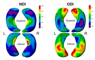
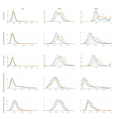
Brain microstructure alterations associated to Alzheimer's disease assessed by diffusion tensor and neurite orientation dispersion and density imaging.
Emma Muñoz-Moreno, Laura Obrado, Raúl Tudela, Xavier López-Gil, Gemma Piella, Guadalupe Soria
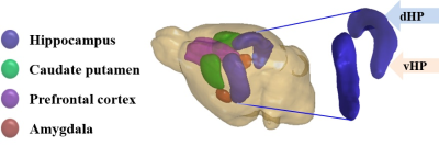
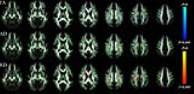
NODDI characterizes neurite orientation dispersion (ODI) and intracellular volume fraction (ICVF), related to neurite density, based on diffusion magnetic resonance imaging. In this study, we have applied NODDI to evaluate excised brains of TgF344-AD, a transgenic rat model of Alzheimer’s disease (AD) and compared them with brains from control rats. Specific brain regions were evaluated: amygdala, caudate putamen, insular cortex and antero-dorsal and posterior hipppocampi. ODI and ICVF shown a different distribution in AD and control rats, with a tendency to higher values in AD, that could not be observed by standard diffusion parameters such as fractional anisotropy (FA).
|
|
5260.
 |
62 |
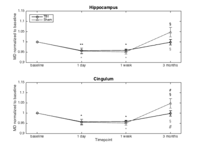
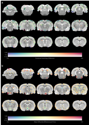
Characterisation of microstructural alterations in a weight drop mTBI rat model: a longitudinal diffusion MRI and histological analysis
Kim Braeckman, Benedicte Descamps, Leen Pieters, Karen Caeyenberghs, Christian Vanhove
TBI is the leading cause of acquired disability of young adults and due to the subtle nature, conventional scans show no evidence of injury. In this multi-shell longitudinal diffusion MRI study of mTBI in rat brain we found that DKI and white matter metrics can be used to follow up recovery in the brain at least until one week after injury. Moreover, histological analysis showed that changes in the metrics could be explained by inflammation and neurofilament compaction. On the other hand, DTI metrics could not differentiate between the sham and TBI group and were comparable in the two groups.
|
|
5261.
 |
63 |
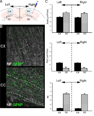
The contribution of astrocytic aquaporin-4 to gray and white matter diffusion ansiotropy
Andre Obenaus, Jacqueline Coats, Andrew Fukada, Wei Sun, Jerome Badaut
We investigated the impact of water channels (aquaporin 4; AQP4) on DTI metrics after silencing expression of AQP4 in the juvenile brain. We observed a significant reduction in AQP4 expression after RNA silencing of AQP4 (siAQP4) along with significantly altered DTI metrics in the cortex but not the corpus callosum (CC). No changes in cellular constituents were found. Histological studies have reported decreased AQP4 expression in acquired brain injuries. Thus, our novel findings suggest that reductions in AQP4 expression may underlie the changes in DTI metrics that are often reported in DTI studies of stroke, traumatic brain injury and others.
|
|
5262.
 |
64 |
Diffusion tensor changes in acute neuroinflammation in rats
Eugene Kim, Camilla Simmons, Karen Randall, Brigida Ranieri, Paula Sureda-Gibert, Tobias Wood, Carmine Pariante, Federico Turkheimer, Diana Cash
Many neurodegenerative and psychiatric disorders feature low level neuroinflammation that is insidious yet difficult to diagnose in vivo. Here we explore the possibility of detecting acute neuroinflammation induced by systemic administration of lipopolysaccharide (LPS) in rats using in vivo diffusion tensor imaging (DTI) at 9.4T. Subtle yet widespread decreases in fractional anisotropy and increases in mean diffusivity were found in LPS-treated rats. Our results confirm the notion that DTI metrics are potential sensitive biomarkers of the dynamic inflammatory response in the brain, raising the possibility of utilizing DTI as a non-invasive in vivo assay for therapeutic interventions.
|
|
5263.
 |
65 |
Combining diffusion and perfusion weighted MRI measurements for disease and treatment monitoring in Multiple Sclerosis
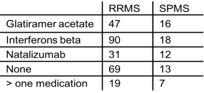
Madalina Tivarus, Xing Qiu, Nicole Zizzi, Giovanni Schifitto
In this large sample retrospective study of MS patients with different diagnoses and treatment regimens we investigated interactions between imaging metrics and age, MS phenotype, and types of treatment, and longitudinal changes associated with disease modifying treatments. We assessed WM injury using DTI metrics (FA, MD), and vascular changes (CBF and CBV) using perfusion DSC, in corpus callosum and in its cortical projections. We found significant correlations of perfusion with age, and DTI metrics with disease type and medication, suggesting that advanced neuroimaging methods such as DTI should become integrated into the clinical evaluation of MS patients for improved management.
|
|
5264.
 |
66 |
Free-Water Elimination Diffusion Tensor Imaging to Assess Nerve Recovery in Excised Rat Nerve
Shashank Manjunath, Isaac Manzanera-Esteve, Wesley Thayer, Mark Does, Richard Dortch
We present our findings using a free water elimination model, which allows for the characterization of edema signal in diffusion measurements. We validate this model on ex vivo rat nerve data after sham surgeries on the sciatic nerve. The free water model effectively accounts for inflammation following sham surgeries, leading to increased accuracy in fractional anisotropy measurements throughout the recovery process. This model will be applied in other injury models (crush, transection/surgical repair) to test its ability to independently monitor inflammation/edema and nerve degeneration/regeneration.
|
|
5265.
 |
67 |
Diffusion tensor imaging in a rat model of brain stem ischemia reveals structural remodeling contralateral to the ischemic lesion
Lydia Wachsmuth, Jens Minnerup, Jan-Kolja Strecker, Kai Diederich, Cornelius Faber
Ischemic stroke of the brain stem affects a considerable number of human patients. However, mechanisms of degeneration and recovery are not well understood and animal models of brain stem ischemia are rare compared to models of cortical stroke. Here we implemented a rat model of brain stem ischemia and applied diffusion tensor MR imaging as a noninvasive means to assess structural connectivity. Probabilistic mapping and histology indicate structural remodeling at the level of thalamus. These results add evidence for a potential compensatory mechanism for the observed partial recovery after brain stem stroke.
|
|
5266.
 |
68 |
Diffusion Changes in Normal-Appearing White Matter in Tracts Affected by White Matter Hyperintensities
Rozanna Meijboom, Susana Muñoz Maniega, Maria Valdés Hernández, Nathalie Royle, Zoe Morris, John Starr, Mark Bastin, Ian Deary, Joanna Wardlaw
White matter hyperintensities (WMH) are common in older brains. We analyzed how WMH affect white matter (WM) tracts and particularly their normal-appearing WM (NAWM). We used MRI of 52 participants (72.2±0.7y) to quantify diffusion parameters of WMH-affected tracts. The intersections of tracts with WMH were identified and volumes quantified. Diffusion parameters were measured for tract-WMH, tract-NAWM, and for tract-NAWM at different distances from the tract-WMH edge, and from the edge of nearby—non-intersecting—WMH. Tract-NAWM showed a gradient of diffusion abnormalities away from tract-WMH, and nearby-WMH. Tract-WMH diffusion, and either tract-WMH volume or whole-brain WMH load, predicted tract-NAWM diffusion.
|
|
5267.
 |
69 |
Evaluating the effect of 3-n-butylphthalide on expression of aquaporin-4 by ultra-high b value diffusion weighted imaging in animal model with focal cerebral ischemia
Baohong Wen, Dandan Zheng, Jingliang Cheng
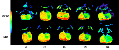
Previous studies have reported that 3-n-butylphthalide (NBP) had beneficial effects on stroke through multiple aspects, including decreasing the area of cerebral infarct, improving energy metabolism , inhibiting the inflammatory response and improving cerebral microvessels . Although the positive effects of NBP on cerebral ischemia and cerebral infarct have been verified in ischemic patients and animal models, the effects of NBP in aquaporin-4 (AQP-4) are still unclear. Recently, ultra-high diffusion weighted imaging was reported to be able to reflect AQP-4 changes . This study would like to evaluate the effect of 3-n-butylphthalide on expression of aquaporin-4 by ultra-high b value diffusion weighted imaging in animal model of focal cerebral ischemia at different time points.
|
|
5268.
 |
70 |
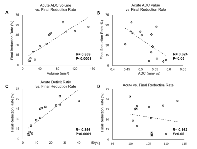
Early Apparent Diffusion Coefficient Deficit Correlates to Final Outcome in Experimental Neonatal Hypoxic Ischemia
Yu-Chieh Jill Kao, Chia-Feng Lu, Chao-Ching Huang, Cheng-Yu Chen
Changes in apparent diffusion coefficient (ADC) at 2 h after hypoxic ischemia (HI) in neonatal rats showed the significant correlation to the final lesion severity. The early ADC deficit appeared within 6 h after HI injury may serve as an index for outcome prediction and the translational evidence to stratify neonates for hypothermia treatment.
|
|
5269.
 |
71 |
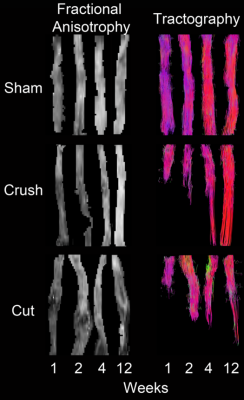
Diffusion Tensor Imaging of Excised Rat Nerve Following Transection and Surgical Repair
Isaac Manzanera Esteve, Angel Farinas Chopite, Marlieke Nussenbaum, Alonda Pollins, Wes Thayer, Mark Does, Dortch Richard
Traumatic peripheral nerve injury (TPNI) from crushing and/or transection can lead to nerve degeneration distal to the site of injury and a temporary loss in sensorimotor function. In this study, we present our findings showing how high-resolution DTI and Tractography measurements of traumatic nerve injury in the sciatic nerve region are capable to identify and characterize nerve injury degeneration/regeneration and injury type in rats. Our findings suggest that DTI and Tractography are viable biomarkers of nerve regeneration and can provide with valuable information in the evaluation of therapeutic interventions
|
|
5270.
 |
72 |
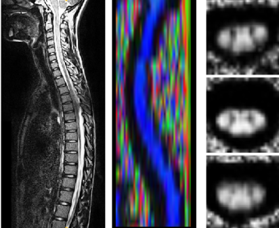
Spinal cord cross section and DTI by vertebral level correlate superior and inferior to injury
Devon Middleton, Shiva Shahrampour, Chris Conklin, Mahdi Alizadeh, Scott Faro, Laura Krisa, MJ Mulcahey, Feroze Mohamed
Examination of diffusion and cord cross section by vertebral level in pediatric subjects has the potential to show useful information in injury diagnosis and prognosis. Correlations between DTI and cord cross section by vertebral level are shown superior and inferior to the injury site.
|
|
Multiple Sclerosis: Brain & Spinal Cord Applications
Electronic Poster
Neuro
Thursday, 21 June 2018
| Exhibition Hall |
08:00 - 09:00 |
| |
|
Computer # |
 |
5271.
 |
73 |
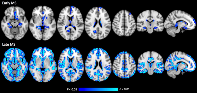
Gray Matter Myelin Alterations in Early and Late Relapsing-Remitting Multiple Sclerosis Evaluated with Quantitative Synthetic Magnetic Resonance Imaging: A Gray-Matter Based Spatial Statistics Analysis
Christina Andica, Akifumi Hagiwara, Keigo Shimoji, Koji Kamagata, Asami Saito, Yuki Takenaka, Tomoko Maekawa, Saori Koshino, Ryusuke Irie, Akihiko Wada, Masaaki Hori, Kanako Kumamaru, Kanako Sato, Kazumasa Yokoyama, Nobutaka Hattori, Shigeki Aoki
Our study demonstrated that myelin volume fraction (MVF) and myelin volume (MyV) obtained by a multi-parametric quantitative synthetic MRI might be useful for evaluating gray matter (GM) myelin alterations and for monitoring disease progression in relapsing-remitting multiple sclerosis (RRMS) patients. GM-based spatial statistics analysis demonstrated decreased MVF in limbic, paralimbic, and deep GM areas in the early-RRMS, and in extensive areas of GM in the late-RRMS. In the meanwhile, MyV was found to be decreased in both RRMS groups compared to healthy subjects, with late-RRMS showing the lowest value, and significantly correlated with disease duration.
|
|
5272.
 |
74 |
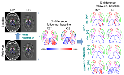
Discriminative Analysis of Regional Evolution of Iron and Myelin/Calcium in Deep Gray Matter of Multiple Sclerosis and Healthy Subjects
Ahmed Elkady, Dana Cobzas, Hongfu Sun, Gregg Blevins, Alan Wilman
We introduce Discriminative Analysis of Regional Evolution (DARE) of iron and myelin/calcium to assess specific changes in Deep Gray Matter (DGM) of Relapsing-Remitting Multiple Sclerosis (RRMS), Progressive MS (PMS) and corresponding age-matched healthy subjects, which we regress with disease severity. DARE enabled discriminative assessment of longitudinal changes in MS, and demonstrated superior performance compared to conventional bulk analysis. Iron decrease and myelin/calcium increase, and myelin/calcium changes, were the primary drivers of observed MRI longitudinal changes in RRMS and PMS DGM, respectively. Specific DARE measures of MS DGM can be used to predict MS Severity Score, and may reflect complex disease pathology.
|
|
5273.
 |
75 |
Surface-based Quantitative Susceptibility Mapping of Cortical Pathology in Multiple Sclerosis
Marco Castellaro, Roberta Magliozzi, Alessandro Palombit, Stefania Montemezzi, Francesca Pizzini, Alessandra Bertoldo, Massimiliano Calabrese
Multiple Sclerosis has been showed to be characterized by extent cortical sub-pial demyelination and iron alterations. Moreover, a “surface-in” gradient of pathology has been showed to be present in MS. In this study, we used QSM to investigate iron and/or myelin changes in the whole cortex. Moreover, we exploit surface-based methods to clarify the presence of a laminar specific changes in cortical susceptibility of Relapsing-Remitting MS patients.
|
|
5274.
 |
76 |
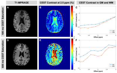
Optimization of CEST MRI at 7 Tesla for Detection of Cortical Gray Matter Pathology in Multiple Sclerosis
Kristin O'Grady, Samantha By, Bailey Box, Quinn Weinberg, Siddharama Pawate, Francesca Bagnato, Seth Smith
Glutamate-sensitive chemical exchange saturation transfer (CEST) MRI has been applied in the human brain and shows promise for detecting pathology related to dysfunctional glutamate regulation. Glutamate abnormalities are linked to cortical gray matter (GM) pathology and cognitive impairment in multiple sclerosis (MS), but quantitative assessment techniques are lacking. We optimized and applied CEST MRI at 7.0T in phantoms and in vivo to evaluate sensitivity to glutamate and the effect of saturation pulse duration. Our results show increased CEST contrast in cortical GM of MS patients relative to controls and demonstrate the potential of CEST in characterizing GM damage in MS.
|
|
5275.
 |
77 |
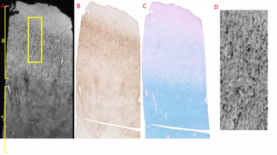
MR Microscopy of Cortical Lesions Reveal Iron Loss in Individual Oligodendrocytes
Stephen Dodd, Govind Nair, Seung-Kwon Ha, Daniel Reich, Alan Koretsky
Imaging of cortical demyelination in chronic multiple sclerosis, has been challenging. Previous MRI studies have detecting signal changes in cortical lesions have been attributed to myelin loss and the presence of iron-laden microglia. Here we demonstrate, in a case study, that MR microscopy may readily identify individual iron-rich cells (primarily oligodendrocytes) and regions. In addition, MR microscopy is shown to allow detailed examination of the central vein signal in white matter lesions.
|
|
5276.
 |
78 |
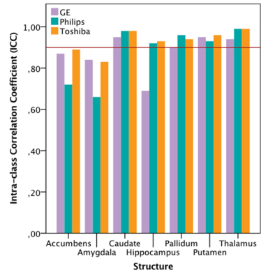
Inconsistency in Grey Matter Volume Estimation of MS Patients: A Multi-Vendor Study at 3 Tesla
Houshang Amiri, Stephanie Bosschaert, Iman Brouwer, Joost Kuijer, Jan de Munck, Marloes Hagens, Joep Killestein, Frederik Barkhof, Hugo Vrenken
MR images are widely used to measure brain atrophy is neurodegenerative diseases. However, reliable evaluation of atrophy is hampered by between- and within-scanner variability and inconsistency. We investigated this in 21 multiple sclerosis patients scanned at three different scanners (twice at each scanner). Volumes of GM, WM and whole brain, as well as deep grey matter structures were assessed using SIENAX and FSL-FIRST, respectively. Voxel-based morphometry was used to localise variabilities in the brain. Our findings suggests that scanner-related factors, and especially between-scanner variability, play a role in inconstancy of brain volume measurements.
|
|
5277.
 |
79 |
Comparison of Methods for Whole-Brain and Grey Matter Atrophy Assessment in Multiple Sclerosis
Video Permission Withheld
Loredana Storelli, Maria Rocca, Elisabetta Pagani, Wim Van Hecke, Mark Horsfield, Nicola De Stefano, Alex Rovira, Jaume Sastre-Garriga, Jacqueline Palace, Diana Sima, Dirk Smeets, Massimo Filippi
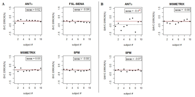
We compared different methods for whole-brain and grey matter (GM) atrophy estimation (ANTs v1.9, CIVET v2.1, FSL-SIENA(X) v5.0.1, Icometrix-MSmetrix v1.7, and SPM v12) in multiple sclerosis (MS). The accuracy and precision were evaluated for cross-sectional and longitudinal whole-brain and GM atrophy measures. All software showed high accuracy and comparable repeatability for cross-sectional measures. However, since there was poor reproducibility and high variability in cross-sectional and longitudinal atrophy measures, changes of MR scanner should be avoided. This study may help in the selection of a suitable pipeline, depending on the requirements of the application (research center, clinical setting or clinical trial).
|
|
5278.
 |
80 |
1H MRS study of glutamate excitotoxicity in hypothalamus of early multiple sclerosis patients
Video Permission Withheld
Petra Hnilicová, Ema Kantorová, Hubert Polácek , Štefan Sivák, Michal Bittšanský, Kamil Zelenák, Egon Kurca, Dušan Dobrota
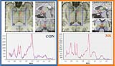
Multiple sclerosis (MS) is considered as an autoimmune disease with expanding axonal and neuronal degeneration in the spinal cord or cerebral cortex during the acute MS phase. The hypothalamus (HYP) is often overlooked yet controls important homeostatic functions. This 1H MRS study performed on 1.5 T MR scanner using 3D CSI with 10×10×12.5 mm3 voxels was focused to altered HYP metabolism in early MS. Considering our results, increased Glx ratios with reduced mIns and tNAA ratios in HYP suggested glutamate excitotoxicity associated with glial activity and neuronal damage. This indicated that HYP plays an important role in early disease evolution.
|
|
5279.
 |
81 |
Examining Cortical Thickness Markers in Relapse Remitting Multiple Sclerosis
Geoffrey Ngo, Ravi Menon
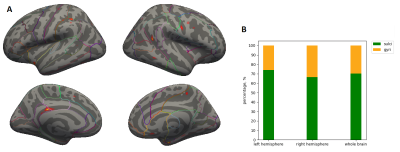
Recent evidence suggests that cortical pathology in Multiple Sclerosis (MS) does not affect all brain regions equally. We aim to investigate regional cortical MS pathology by using cortical thickness measures in a relapsing-remitting group (RRMS). Structural scans from 21 RRMS and 21 controls were processed using Freesurfer to obtain cortical thickness measurements. Group level analysis was performed to investigate the preference of sulcal and gyral thinning. Differences between gyri and sulci thickness between groups were also calculated to see if there was any evidence of cortical-layer specific thinning. Sulcal preference was shown, and no layer specific thinning was observed.
|
|
5280.
 |
82 |
Indirect assessment of cerebral metabolism in deep grey matter areas of progressive multiple sclerosis subjects – preliminary results
Andrea Horvath, Ian Tagge, Manoj Sammi, Katherine Powers, Dennis Bourdette, Xin Li, Rebecca Spain, Charles Springer, William Rooney
Mitochondrial injury and impaired metabolic capacity are hypothesized to drive neurodegeneration in multiple sclerosis (MS). Here, we investigate a novel putative marker of tissue metabolic activity, trans-capillary water flux, derived from dynamic contrast enhanced MRI. In this study we compared 23 subjects with progressive MS to 19 healthy controls. We find significantly reduced measures on capillary water flux in MS thalami compared to controls. Implications for use of this new biomarker are discussed.
|
|
5281.
 |
83 |
Mismatch Between Cerebral Glucose and Oxygen Metabolisms in Young Adults with Relapsing-Remitting Multiple Sclerosis
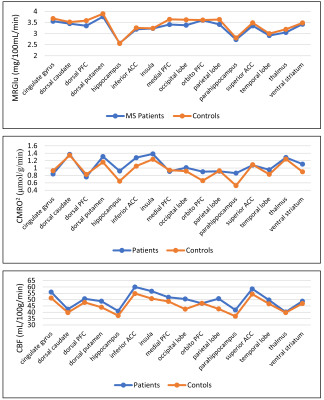
Xiang He, Kenneth Wengler, Elizabeth Bartlett, Leigh Charvet, Tim Duong, Christine DeLorenzo, Lauren Krupp
Oxidative stress has been linked to neuroinflammation that leads to demyelination in multiple sclerosis (MS). While most studies focus on older MS patients, the underlying cause of oxidative stress at this stage of the disease may be obscured. In this study, cerebral metabolic rates of oxygen and glucose in young adult relapsing-remitting MS patients were measured with simultaneous PET/MRI. Several brain regions, most associated with the corticostriatal pathway, exhibited increased oxygen metabolism and decreased glucose metabolism in young MS patients when compared to healthy controls. These observations may elucidate the mechanism for mitochondrial dysfunction and neuroinflammation in MS pathophysiology.
|
|
5282.
 |
84 |
Altered Hippocampal GABA and Glutamate Levels and Uncoupling from Functional Connectivity in Multiple Sclerosis
Did Not Present
fei gao, xuntao yin, weibo chen, guangbin wang
This study offers a novel combination of methods investigating the complex relationships among excitatory/inhibitory neurotransmitters, brain connectivity and cognitive function in health and disease states. Modulation of Glu and GABA neurotransmission may enable the development of new therapeutic strategies for the early stages of MS.
|
|
5283.
 |
85 |
Quantification of white matter tract integrity in primary-progressive multiple sclerosis
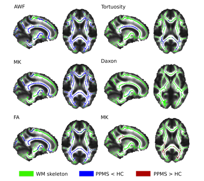
Maria Petracca, Simona Schiavi, Catarina Saiote, Lazar Fleysher, Matilde Inglese
Diffuse white matter (WM) injury is prominent in primary progressive multiple sclerosis (PPMS). Diffusion Kurtosis Imaging (DKI) allows the quantification of non-Gaussian water diffusion, offering the possibility of more detailed characterization of WM damage, in comparison with that provided by diffusion tensor imaging metrics. Here we present application of DKI metrics in PPMS using a Tract-Based Spatial Statistics approach. We observed a diffuse WM microstructural damage, manifested as axonal water fraction, mean kurtosis and fractional anisotropy decrease. In line with histopathological studies, our results suggest the prevalence of axonal damage over demyelination in progressive MS.
|
|
5284.
 |
86 |
Multi-dimensional microstructural imaging offers novel in vivo insights into brain pathology: an application to multiple sclerosis
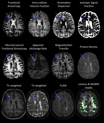
Kasper Andersen, Samo Lasic, Henrik Lundell, Markus Nilsson, Daniel Topgaard, Filip Szczepankiewicz, Lars Hanson, Hartwig Siebner, Morten Blinkenberg, Tim Dyrby
Magnetic resonance imaging is today the most versatile imaging method for characterization of multiple sclerosis (MS) in vivo, but clinical examinations lack sensitivity to capture changes in the tissue microstructure. Using a multi-dimensional microstructural imaging approach, we demonstrate how it is possible to obtain more specific and broader microstructural insights about the underlying pathology of MS. For this we use a comprehensive battery of conventional and novel diffusion weighted imaging and quantitative MRI sequences each capable of explaining different and complementary microstructural properties. This allows us to explore the underlying pathology of MS, which is normally only accessible with histology.
|
|
5285.
 |
87 |
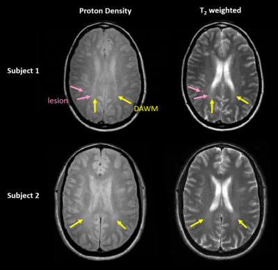
Does the presence of diffusely abnormal white matter in MS affect cognitive function?
James Cairns, Irene Vavasour, Guojun Zhao, Roger Tam, Sarah Morrow, Andrew Riddehough, Alex MacKay, Anthony Traboulsee, Martin McKeown, Wayne Moore, David Li, Cornelia Laule
Multiple sclerosis (MS) subjects with diffusely abnormal white matter (DAWM) typically progress faster on physical disability scores. The impact of DAWM on cognitive measures is unknown. 50 MS participants had conventional MRI and cognitive tests of Trails Making Test, Working Memory, and Processing Speed. The presence of DAWM was not associated with worse cognitive performance. As DAWM is most commonly present in posterior regions of the brain, it may be that these cognitive tests were not sensitive to DAWM-associated pathology.
|
|
5286.
 |
88 |
Advanced diffusion MRI characterization of microstructure changes associated with increasing T1 hypointensity in the white matter lesions of relapsing-remitting multiple sclerosis patients.
Timothy Shepherd, Benjamin Ades-aron, Bettina Conti, Yvonne Lui, Dmitry Novikov, Els Fieremans
Autopsy studies of white matter lesions in relapsing-remitting multiple sclerosis demonstrate that FLAIR-bright lesions represent a juxtaposition of inflammation, demyelination, axonal injury and gliosis, whereas T1 hypointense lesions represent more severe confluent injury and axonal loss. We used a white matter tract integrity (WMTI) previously validated in cuprizone animal models of demyelination to better characterize specific in vivo microstructure changes associated with graded T1 signal intensity changes in multiple sclerosis lesions.
|
|
5287.
 |
89 |
DTI Analysis in FLAIR-positive Lesions and Normal-Appearing White Matter in Young Adult Multiple Sclerosis Patients
Tao Wang, Sindhuja Govindarajan, M. Parra, Patricia Stefancin, Andrew Labella, Kenneth Wengler, Chuan Huang, Xiang He, Leigh Charvet, Lauren Krupp, Tim Duong
Neuroimaging studies showed that there is abundant diffusion tensor imaging (DTI) research that has been done on older multiple sclerosis (MS) patients. By contrast, similar research is relatively sparse in young/pediatric MS patients. Our study is interested in discovering the effects of MS on young adult patients to see whether the same pattern as in older patients appears. Our results suggest that DTI diffusivity data provides insights in the pathophysiology of MS in young adults. Diffusivity data may serve as an imaging biomarker of early disease pathophysiology in MS.
|
|
5288.
 |
90 |
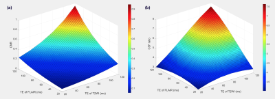
Utility of Combination Image of T2-weighted and FLAIR using Synthetic MRI for Improved Lesion Contrast in Multiple Sclerosis
Yasuhiro Fujiwara, Yumi Inoue, Masayuki Kanamoto, Shota Ishida, Toshiki Adachi, Hirohiko Kimura
To improve multiple sclerosis plaque, we attempted to produce Synthetic FLAIR3 (SyFLAIR3) combined from FLAIR and T2WI using Synthetic MRI. The purpose of this study was to determine optimal contrast weighting for SyFLAIR3 and to evaluate whether the SyFLAIR3 can improve the T2 contrast on WM. To effectively suppress CSF signal for SyFLAIR3, it was necessary to optimize the combination of TE for T2WI and FLAIR. The optimized SyFLAIR3 using Synthetic MRI makes it possible to improve contrast in MS lesion.
|
|
5289.
 |
91 |
Accelerated Isotropic Sub-Millimeter Whole-Brain Susceptibility Imaging at 3T: Application to Multiple Sclerosis
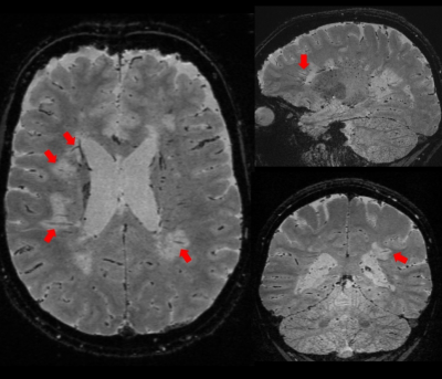
Sunil Patil, Henrik Odéen, J. Andrew Derbyshire, Gunnar Krueger, Dennis Parker, Himanshu Bhat, Daniel Reich, Pascal Sati
High-resolution susceptibility-weighted MRI has recently gained attention as a novel imaging biomarker in multiple sclerosis (MS) such as the ‘central vein sign’ and ‘phase rims’. This preliminary study demonstrates the feasibility of an accelerated volumetric (3D) segmented echo-planar-imaging (EPI) sequence, which allows acquiring whole brain images at 0.65 mm isotropic resolution in approximately 3 minutes. Both magnitude (T2*-weighted) and phase (QSM) information were interpretable and displayed characteristic features of MS lesions. This accelerated 3D EPI anatomical acquisition shows potential to open the door to routine high-resolution susceptibility-weighted imaging to better support diagnosis and therapy monitoring in MS patients.
|
|
5290.
 |
92 |
Quantitative MT (qMT) imaging of the Whole Brain: Conventional 3D MT vs. 3D EP-vfMT methods
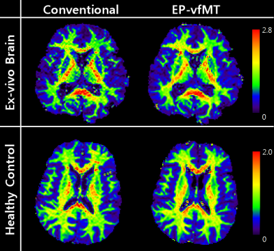
Se-Hong Oh, Dongmyung Shin, Ken Sakaie, Daniel Ontaneda, Mark Lowe
In this study, the whole brain qMT map from conventional MT imaging was compared with that from a newly proposed method, Segmented EPI readout Variable Flip angle Magnetization Transfer (EP-vfMT). The voxel-wise correlation shows a high correlation between the two maps. Compared to the conventional MT method, EP-vfMT provides similar image quality with good reproducibility. It also covers a whole brain volume in a much reduced scan time.
|
|
5291.
 |
93 |
Multicenter assessment of focal and diffuse cervical cord MTR abnormalities in early relapsing-remitting multiple sclerosis
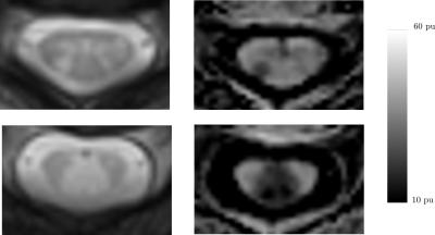
Benoit Combès, Anne Kerbrat, Laureline Monteau, Jean-Christophe Ferré, Emmanuelle Le Page, Josefina Maranzano, Virginie Callot, Pierre Labauge, Xavier Ayrignac, Clarisse Carra Dallière, Nicolas Menjot de Champfleur, Jean Pelletier, Adil Maarouf, David Brassat, Jérome de Seze, Nicolas Collongues, Francoise Durand Dubief , Christian Barillot, Gilles Edan, Elise Bannier
The purpose of this work was to assess the ability of magnetization transfer ratio imaging of the cervical spinal cord to capture relevant differences in the first stage of multiple sclerosis in a multicenter context. For this purpose, we analyzed the MTR values in the lesions, the whole cord and normal-appearing cord from 52 patients in the first 18 months of the disease and from 17 controls. Images were acquired in 5 centers. We showed such measurements were able to capture relevant group differences and displayed slightly higher correlations with patients clinical status than lesion volume.
|
|
5292.
 |
94 |
Assessing the Role of Cord Atrophy Toward Disease Progression in Multiple Sclerosis
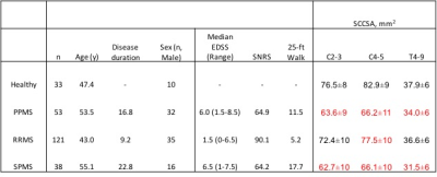
Govind Nair, Shila Azodi, Yoshimi Akahata, Daniel Reich, Steven Jacobson
Plot of spinal cord cross-sectional area (SCCSA) from C1 to T10 reveals distinct patterns in various multiple sclerosis subtypes. While the relapsing remitting subtype revealed thinner cord in the cervical region, progressive subtypes had thinner c- and t-spines compared to healthy subjects. SCCSA measures were related to clinical outcomes, and revealed that up to 10-15% of disability can be explained by differences in SCCSA alone. Furthermore, segregating patients based on the SCCSA seems to give a better stratification of their clinical disability. This study suggests SCCSA can be used as an outcome measure in clinical trials.
|
|
5293.
 |
95 |
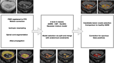
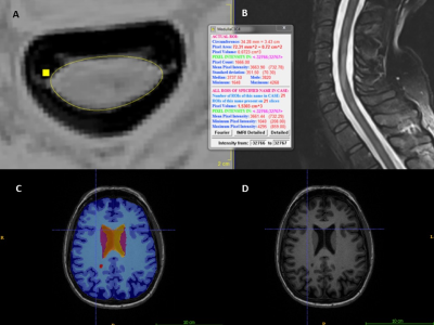
A fully unsupervised method for spinal cord lesion segmentation in Multiple Sclerosis
Carole Sudre, Ferran Prados, Rosanna Cortese, Marios Yiannakas, Hugh Kearney, Olga Ciccarelli, Sébastien Ourselin, Claudia Gandini Wheeler-Kingshott, M. Jorge Cardoso
The presence of focal lesions in the spinal cord is an important diagnostic criteria for Multiple Sclerosis (MS). Accurate estimation of lesion volume is important for monitoring disease progression over time. However, manual and automated lesion segmentation for volume estimation remain challenging, since they rely respectively on the skills of the rater or on the automated criteria set within the algorithms. In this work, we present an adaptation to the spinal cord, of a fully unsupervised hierarchical model selection framework that automatically detects abnormality tissue patterns without any a priori knowledge on pathology location.
|
|
5294.
 |
96 |
Relationship of spinal cord volume, total and regional brain volumes to disability in a large cohort of multiple sclerosis patients
Video Permission Withheld
Michaela Andelova, Jan Krasensky, Lukas Sobisek, Zdenek Seidl, Eliska Kusova, Tomas Uher, Eva Havrdova, Barbora Benova, Bénédicte Maréchal, Tobias Kober, Dana Horakova, Manuela Vaneckova
Identification of MRI biomarkers that predict permanent neurological disability in multiple sclerosis is crucial for assigning patients to correct treatment and for appropriate recruitment of patients for clinical trials. A variety of brain structures and spinal cord have been investigated; however, neither a single structure nor combinations of structures have been routinely used as stable, specific and sensitive biomarkers. Small sample sizes, different MR protocols and segmentation approaches across studies may hamper the identification of such a biomarker. We evaluated global and regional brain volumes and cervical spinal cord volume in a large single-center cohort of multiple sclerosis patients.
|
|
Psychoradiology
Electronic Poster
Neuro
Thursday, 21 June 2018
| Exhibition Hall |
08:00 - 09:00 |
| |
|
Computer # |
|
5295.
 |
97 |
Hypersynchronicity in the default mode-like network and altered NMDA receptor function in a maternal immune activation model
Stephan Missault, Cynthia Anckaerts, Soumaya Ahmadoun, Ines Blockx, Kenny Bielen, Disha Shah, Samir Kumar-Singh, Annemie Van der Linden, Stefanie Dedeurwaerdere, Marleen Verhoye
Maternal immune activation (MIA) is an important risk factor for schizophrenia, which supports the neurodevelopmental hypothesis of this disorder. Two major hypotheses of schizophrenia are the aberrant connectivity hypothesis and the NMDA receptor hypofunction hypothesis. The goal of our study was to investigate functional and structural connectivity, as well as NMDA receptor function in a MIA model using resting-state functional MRI, diffusion tensor imaging and pharmacological MRI. We observed increased functional connectivity in the default mode-like network, as well as a decreased response to the NMDA receptor antagonist in adult rats that were exposed to prenatal immune challenge.
|
|
5296.
 |
98 |
Brain Metabolism in Schizophrenia and First-Degree Relatives: a 7T MRS study
Video Permission Withheld
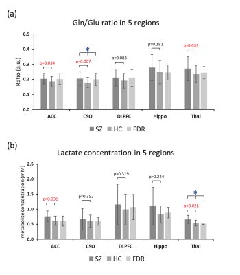
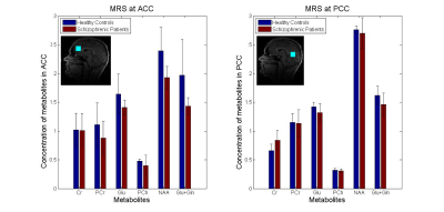
Anna Min Wang, Subechhya Pradhan, Stephanie Korenic, S. Andrea Wijtenburg, Laura Rowland, Peter Barker
Brain metabolism was investigated in 38 patients with schizophrenia (SZ), 38 healthy control (HC) subjects, and 11 first degree relatives of SZ patients using 7T MRS in 5 brain regions. Multiple metabolic abnormalities were found in SZ patients, including increases in the ratio of glutamine to glutamate, increased levels of brain lactate, and decreased levels of γ-aminobuytric acid (GABA) and N-acetylaspartate-glutamate (NAAG). Many of these changes also correlated with measures of cognitive performance and negative symptom severity. 7T MRS is an excellent tool for the non-invasive investigation of brain pathophysiology in SZ.
|
|
5297.
 |
99 |
Ex-vivo diffusion MRI reveals microstructural alterations in stress-sensitive brain regions: A chronic mild stress recovery study
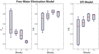
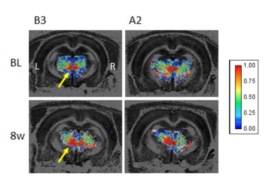
Ahmad Khan, Brian Hansen, Ove Wiborg, Christopher Kroenke, Sune Jespersen
Depression is a leading cause of disability worldwide and causes significant microstructural alterations in stress-sensitive brain regions. However, the potential recovery of these microstructural alterations has not previously been investigated, which we, therefore, set out to do using diffusion MRI (d-MRI) in the chronic mild stress (CMS) rat model of depression. This study reveals significant microstructural alterations after 8 weeks of recovery, in the opposite direction to change induced by stress in the acute phase of the experiment. Such findings may be useful in the prognosis of depression or for monitoring treatment response.
|
|
5298.
 |
100 |
Analysis of resting-state networks alterations of first-episode obsessive–compulsive disorder by ICA -based fMRI
Did Not Present
Junhong Liu, Dandan Zheng, Jingliang Cheng
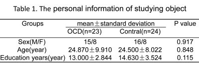
Whole brain resting-state functional connectivity was analyzed using independent component analysis in 47 subjects including 23 first-episode and treatment-naïve patients with obsessive-compulsive disorder(OCD) and 24 health controls. Three abnormal resting-state networks, pDMN, RFP and lVN, have been found in OCD patients. In addition, OCD patients showed increased functional connection in Bilateral cuneus(T=3.8222,P=0.005), Right inferior parietal lobule(T=5.291,P=0.005)and Right middle occipital lobule(T=4.614,P=0.005) compared with controls. It’s considered that changes of abnormal resting-state networks might be associated with emotional and cognitive dysfunction in OCD patients.
|
|
5299.
 |
101 |
Disrupted reward and cognitive control networks contribute to anhedonia in depression
Liang Gong, Chunming Xie, Hongxing Zhang, Zhijun Zhang
In the present study, we investigate the association between intrinsic reward network (β-network) and cognitive control network (δ-network) and anhedonia in depression patient. We found that depression patients showed decreased functional connectivity (FC) in intra- and inter- β- and δ-networks and the FC in both β- and δ-networks were significantly correlated with anhedonic severity in depression patients. Importantly, the integrated neural features of β- and δ-networks would more precisely predict anhedonia symptom. These findings indicated that the neural features in both β- and δ-networks would represent a fundamental mechanism underlying anhedonia in the MDD patients.
|
|
5300.
 |
102 |
Subcortical and medial temporal lobe shape changes following electroconvulsive therapy
Filip Bouckaert, Jurgen Germann, Mallar Chakravarty, Annemieke Dols, François-Laurent De Winter, Lies Van Assche, Jan Van den Stock, Stefan Sunaert, Max Stek, Pascal Sienaert, Mathieu Vandenbulcke, Louise Emsell
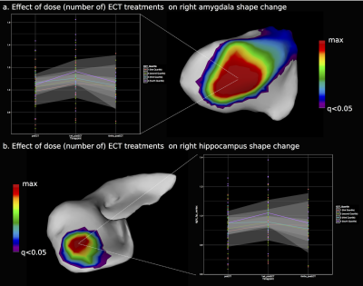
Electroconvulsive therapy is a safe, rapid-acting antidepressant treatment that has consistently been associated with grey matter (GM) volume increase, primarily in the medial temporal lobe (MTL). Here we replicate and extend previous studies by demonstrating substantial surface area and local displacement changes in subcortical and MTL GM one week following the last ECT treatment in 70 patients with late-life depression. We report new data on ECT induced thalamus shape changes, and demonstrate that whilst the number of ECT treatments correlates positively with the degree of medial temporal GM surface area increase, this is not associated with clinical improvement in mood.
|
|
5301.
 |
103 |
Reduced White Matter Integrity Related with Elevated In?ammatory Cytokine Expression and Cognitive Impairments in First-episode Schizophrenia: A Diffusion Tensor Imaging Study Based on Tract-Based Spatial Statistics
Did Not Present
Jie Gao, Yajuan Fan, Lei Wang, Yarong Wang, Feng Zhu, Min Tang, Dongsheng Zhang, Xia Zhe, Xuejiao Yan, Xin Zhang, Zhizheng Zhuo, Xiaoling Zhang
This study aimed to investigate whether elevated in?ammatory cytokine expression induced white matter integrity changes and cognitive impairments in first-episode schizophrenia patients. 27 first-episode schizophrenia patients and 16 healthy controls who underwent diffusion tensor imaging were enrolled. Tract-based spatial statistics analysis exhibited significantly decreased fractional anisotropy and increased radial diffusivity in widespread white matter tracts in patients. Of these tracts, anterior corona radiata (ACR), superior corona radiata, superior longitudinal fasciculus, the body of the corpus callosum, the splenium of the corpus callosum and fornix showed signi?cant correlations with higher in?ammatory cytokine expression. Moreover, ACR and fornix simultaneously showed reduced white matter integrity related to cognitive impairments in working memory and problem solving. These findings provides more evidence for supporting the role of neuroin?ammation in the pathophysiology of schizophrenia.
|
|
5302.
 |
104 |
Disrupted functional connectivity of occipital lobe with frontal and parietal regions in subjects with Cocaine addiction.
Jaspreet Kaur, Divya Gautam, Rahul Garg
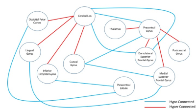
The study aims at exploring a data driven method to analyse connectivity differences between two populations - subjects with cocaine addiction and healthy controls. Sufficient indications of previously undiscovered hypo- and hyper-connectivity was observed in cocaine addicts spanning different lobes of brain suggesting that hypothesis driven approaches are insufficient.
|
|
5303.
 |
105 |
Investigation of Schizophrenia Patients with Clozapine-Induced Obsessive Compulsive Symptoms by Advanced MR Imaging
Arzu Has, Sule Bicakci, Elif Bulut, Aygun Ertugrul, Kader K. Oguz
We aimed to investigate white matter (WM) integrity and structural differences in schizophrenia patients with clozapine-induced obsessive compulsive symptoms (S-OCS) comparing them those without symptoms (S) and healthy controls (HC). Tract-based-spatial-statistic/diffusion-tensor-imaging, voxel-based morphometry, and caudate volume measurements were performed to reveal underlying WM and gray matter (GM) alterations. S-OCS showed less fractional anisotropy (FA) reductions in WM and less areas of reduced GM density than S when compared to HC. FA elevations and increased GM density were observed in S-OCS compared with S. The results may suggest differential effect of clozapine, and/or different baseline pathophysiology in a subgroup of patients.
|
|
5304.
 |
106 |
Whole brain effect of real-time fMRI amygdala neurofeedback emotional training and its association with PTSD symptom reduction
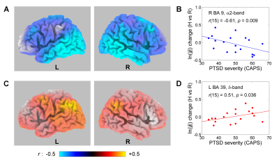
Masaya Misaki, Vadim Zotev, Raquel Phillips, Chung-Ki Wong, Brent Wurfel, Frank Krueger, Matthew Feldner, Jerzy Bodurka
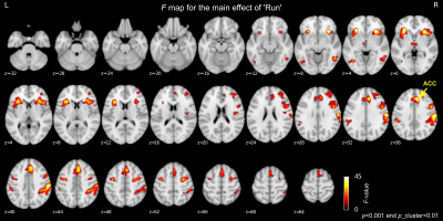
The effect of real-time fMRI neurofeedback (rtfMRI-nf) training with the left amygdala activity on whole brain regions and their association with symptom reduction was investigated in veterans with combat-related PTSD. The main effect of training was seen in the salience network regions including anterior insula and the anterior cingulate cortex (ACC). The decrease in ACC response was significantly correlated with a decrease in PTSD symptoms. These results indicated that the effect of rtfMRI-nf training was not limited to the left amygdala but other emotion-related regions were co-modulated during the training. The treatment response could be meditated by those regions.
|
|
5305.
 |
107 |
Multivariate pattern analysis of DTI reveals differential white matter in depressive patients with and without suicidal ideation.
Did Not Present
Huawei Zhang, Zhiyun Jia
At present there are no objective biological markers that can be used to reliably identify depressive individuals with and without suicidal ideation (SI). DTI data were obtained from 20 depressive patients with SI and 20 depressive patients without SI, scanned using a 3T MRI system. Fractional anisotropy (FA) values of white matter between patients were examined using multivariate support vector machine (SVM). SVM applied to FA images correctly discriminated two groups of patients with a sensitivity of 75% and a specificity of 85% resulting in a statistically significant accuracy of 80% (p≤0.001). The discriminating regions contain the bilateral occipital lobes and parietal lobes, right temporal lobe and splenium of corpus callosum. These results reveal patterns of neuroanatomical alterations that could be used to inform the identification of depressive patients with and without SI at the individual level.
|
|
5306.
 |
108 |
EEG Correlates of Real-time fMRI Neurofeedback of the Amygdala in Combat-related PTSD Evaluated Using eLORETA
Video Permission Withheld
Vadim Zotev, Raquel Phillips, Masaya Misaki, Chung Ki Wong, Brent Wurfel, Matthew Meyer, Frank Krueger, Matthew Feldner, Jerzy Bodurka
We have performed a study of emotion regulation training in veterans with combat-related PTSD using real-time fMRI neurofeedback (rtfMRI-nf) with simultaneous EEG. Eighteen PTSD patients learned to upregulate their left amygdala activity using rtfMRI-nf during a positive emotion induction task based on retrieval of happy autobiographical memories. EEG source analysis with eLORETA revealed task-specific changes in the current source densities in the upper alpha and delta EEG bands that significantly correlated with PTSD severity. These results suggest that the rtfMRI-nf training in combination with EEG source analysis provides new insights into the neurobiology of PTSD.
|
|
5307.
 |
109 |
Multimodal metabolic imaging using single voxel MRS and [11C]ABP688 PET in Schizophrenic Patients
Linda Orth, Ravichandran Rajkumar, Cláudia Régio Brambilla , Ezequiel Farrher, Andreas Matusch, Shukti Ramkiran, Andrej Ruch , Jörg Mauler, Elena Rota Kops, Lutz Tellmann, Jürgen Scheins, Bernd Neumaier, Markus Lang, Johannes Ermert, Hans Herzog, Karl-Josef Langen, Christoph Lerche, N. Jon Shah, Irene Neuner
By utilizing a multimodal imaging approach, the levels of glutamate and other metabolites in the anterior cingulate and the posterior cingulate cortex were assessed by MRS and compared to the metabotropic glutamate receptor 5 (mGluR5) binding potential using [11C]ABP688 PET in schizophrenic patients and healthy controls. Glutamate levels seem to be elevated in schizophrenic patients. PET analysis revealed no differences in binding potential (BPND) in both diagnostic groups. However, adding smoking status to analysis, there is a significant reduction in BPND in smokers compared to non-smokers, suggesting a connection between mGluR5 binding potential and nicotine dependence.
|
|
5308.
 |
110 |
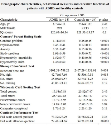
A multi-methodological fMRI resting state voxel-wise analysis to assess brain abnormalities of children with ADHD
Yingxue Gao, Hailong Li, Xuan Bu, Lianqing Zhang, Xinyu Hu, Peiran Hu, Xiaoxiao Hu, Xiaoqi Huang
We used three different measurements including regional homogeneity (ReHo), voxel-mirrored homotopic connectivity (VMHC) and functional connectivity strength (FCS) to explore local and interhemispheric FC in drug naïve ADHD children. And we found lower ReHo and FCS in ADHD located in almost identical region of right middle frontal gyrus and correlated with each other. In addition, we also found lower VMHC in the bilateral occipital lobe, which was related with characteristics of WCST and CPRS-R. This finding may provide new insights into functional connectivity changes in ADHD and promote the exploration of functional network in the future.
|
|
5309.
 |
111 |
Quantative Tractography Reveals Alteration in Corticospinal Tract Associated with Motor Abnormalies in Medication-Naive Attention-Deficit /Hyperactivity Disorder Children
Xuan Bu, Qingxia Lin, Lu Lu, Lianqing Zhang, Xiaoxiao Hu, Hailong Li, Xinyu Hu, Chuang Yang, Xiaoqi Huang
In the current study, we aim to quantify diffusion measures at multiple nodes along the trajectory of corticospinal tract in ADHD children. We found altered FA and RD in distinctive CST regions. Besides, significant correlations between neuropsychological measurements and abnormal white matter microstructure implicated critical role the disturbed CST played in the pathology of motor deficits in ADHD.
|
|
5310.
 |
112 |
The effects of relapse on gray matter volume changes in patients with Major Depression – a longitudinal VBM study
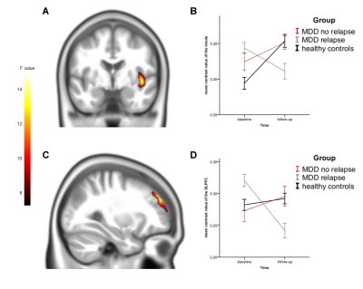
Harald Kugel, Dario Zaremba, Katharina Dohm, Ronny Redlich, Dominik Grotegerd, Robert Strojny, Susanne Meinert, Christian Buerger, Verena Enneking, Katharina Foerster, Jonathan Repple, Nils Opel, Bernhard Baune, Pienie Zwitserlood, Walter Heindel, Volker Arolt, Udo Dannlowski
Structural brain alterations in major depressive disorder (MDD) are associated with patients' course of illness, especially in progressive and recurrent MDD. Here, a longitudinal study investigated the influence of relapse on gray matter volume. As a result, Voxel based morphometry showed a decrease of insular and DLPF gray matter volume in patients with at least one relapse, while volume in patients without relapse was stable. This illustrates the negative effect of relapse on structural brain alterations.
|
|
5311.
 |
113 |
Characteristic Changes of Shape in Subcortical Nuclei in Major Depressive Disorder
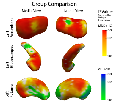
Lianqing Zhang, Naici Liu, Lu Li, Lu Lu, Xiaoxiao Hu, Hailong Li, Xuan Bu, Xinyu Hu, Qiyong Gong, Xiaoqi Huang
We analysis alterations of volume and shape of subcortical nuclei in a relatively large sample of adult patients with Major Depressive Disorder (MDD) using an automatically segmentation and vertex-based shape analysis protocol. We found that hippocampus, putamen and accumbens were impaired in patients with MDD and subregional shape of hippocampus, accumbens and pallidum may have potential predictive value of treatment response in patients with MDD. Shape analysis may provide more evidence of neuropathology related to depression from a different perspective. Future study should consider shape and volume analysis simultaneously.
|
|
5312.
 |
114 |
Alterations in functional brain network topology in Tourette’s syndrome
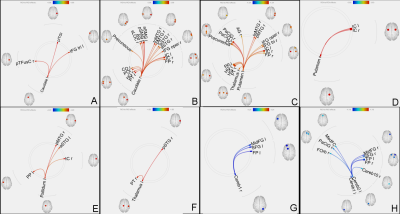
Shukti Ramkiran, Larissa Heidemeyer, Ravichandran Rajkumar, N Jon Shah, Irene Neuner
Tourette syndrome (TS) is a neurodevelopmental disorder with typical onset in childhood. Its characteristic motor tics are said to be attributed to dysfunction in the cortico-striato-thalamo-cortical circuit and cerebellar communication. Brain functional connectivity along with network topology analysis provides a useful tool to understand communication strategies in the brain. Hence we aim to investigate alterations in functional and effective connectivity in brains of patients with TS. Based on prior results1,2, we hypothesize that connectivity of basal ganglia, thalamus and cerebellum with other regions will be altered.
|
|
5313.
 |
115 |
Altered Brain Activity in Jet Lag by Regional Homogeneity (ReHo): A resting state fMRI study
Feifei Zhang, Zhiyun Jia, Qiyong Gong
To identify how jet lag influence brain activity in rest we calculate ReHo values of 23 adult participants who were on a transmeridian flight across eight-time zones from west to east .Participants in ‘Jet Lag’ state compare to recovered state showed decreased ReHo value in the right inferior parietal lobule (BA40, BA7) and the right angular gyrus and increased in the bilateral occipital lobe. Acute circadian disruption caused by jet lag can lead to mild temporary visual cognitive dysfunction.
|
|
5314.
 |
116 |
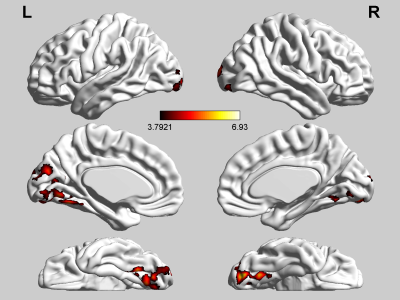
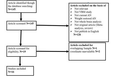
Meta-analytic investigations of grey matter alterations in patients with anorexia nervosa.
Simin Zhang, Weina Wang, Xiaorui Su, Qiyong Gong, Qiang yue
The purpose of the present Meta-analytic study was to summarize the grey matter volumetric alterations and elucidate how the changes were associated with symptoms and pathophysiology in anorexia nervosa(AN). We use effect-size signed differential mapping (ES-SDM) to conduct meta-analytical whole brain volumetric differences between patients AN and healthy controls (HCs). The studies showed volume reduction in bilateral median cingulate cortex(MCC), posterior cingulate cortex (PCC), precuneus, supplementary motor area(SMA) and left cerebellum, which provide evidence for abnormalities in emotion regulation, behavior regulation and sensorimotor area in nervosa anorexia.
|
|
5315.
 |
117 |
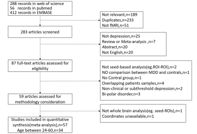
Meta-analysis of Resting-State Functional Connectivity in Major Depressive Disorder
Shi Tang, Xinyu Hu, Lianqing Zhang, Lu Lu, Xuan Bu, Xiaoxiao Hu, Hailong Li, Qiyong Gong, Xiaoqi Huang
To investigate the most reliable resting-state functional connectivity(rsFC) abnormalities in adult dignosed as having major depressive disorder(MDD) with existing studies.After a comprehensive literature search of studies, meta analysis was conducted using Signed Differential Mapping(SDM) software package. We found dysfunction in large-scale brain regions in MDD patients, including hyperconnectivity in fronto-cingulate-parietal area and hypoconnectivity in bilateral superior temporal gyrus(STG). These findings paralleled to the core feature of MDD patients and may underlie the cognitive and affective abnormalities in depressive disorder.
|
|
5316.
 |
118 |
The morphometric brain alteration in current and remitted major depressive disorder: a meta-analysis
Xin Xu, Zhiyun Jia, Qiyong Gong
To investigate the gray matter volume (GMV) alteration in major depressive disorder (MDD) patients at different episode state, here we conducted a meta-analysis which tried to integrate the Voxel-based morphometry (VBM) studies by using Seed-based d Mapping. This study detected that lower GMV in the left insula in both current and remitted MDD patients compared to HC. And the current conjunction meta-analysis indicated that GMV in bilateral anterior cingulate (ACC) were decreased in current MDD patients but increased in remitted MDD patients. Our findings here motivate a morphological alteration pattern of MDD linked to dynamic mood dysfunction state.
|
|
5317.
 |
119 |
The effects of illness duration on white matter connectivity in drug-naive schizophrenia
Fei Li, Su Lui, Li Yao, Wei Liao, Gongjun Ji, Xiaoqi Huang, Qiyong Gong
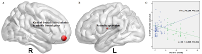
This study investigated the topological alterations of white matter connectivity in schizophrenia patients with a long illness duration by using diffusion tensor imaging and graph theoretical analysis and explored the relationship of such characteristics with the duration. We recruited three groups including the healthy controls, drug-naive schizophrenia patients with a short illness duration (0.1 to 10 months) and a long duration (12 to 36 months), and found that only the patients with a long illness duration exhibited decreased connection strength than the controls and a correlation between the nodal degree of rolandic operculum and the duration, suggesting a neuroprogressive process.
|
|
5318.
 |
120 |
Functional Connectivity as a Potential Predictor of Treatment Response in Patients With Major Depressive Disorder
Hailong Li, Xinyu Hu, Lianqing Zhang, Lu Lu, Xiaoxiao Hu, Xuan Bu, Shi Tang, Qiyong Gong, Xiaoqi Huang
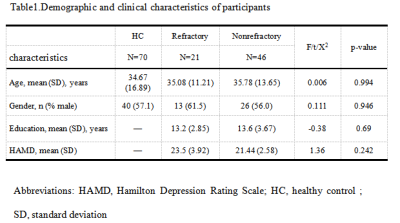
Resting-state functional connectivity(FC) analyses using a subcallosal cingulate cortex(SCC) seed was applied to major depressive disorder(MDD) patients to characterize a potential neuro-imaging biomarker that identifies the treatment outcome. MDD patients were divided into refractory and non-refractory group according to the treatment response. We found distinguished FC alterations between the three groups especially the lack of FC between SCC and Anterior Cingulate Cortex in the refractory MDD patients. In addition, the alterations in FC correlated with clinical symptoms in different ways in two MDD groups.
|
|
Neuro Outside the Brain
Electronic Poster
Neuro
Thursday, 21 June 2018
| Exhibition Hall |
09:00 - 10:00 |
| |
|
Computer # |
|
5391.
 |
73 |
Semi-automatic, Machine Learning Segmentation of Peripheral Nerves in Healthy Volunteers and Patients
Fabian Balsiger, Mirjam Arn, Carolin Steindel, Benedikt Wagner, Marwan El-Koussy, Waldo Valenzuela, Mauricio Reyes, Olivier Scheidegger
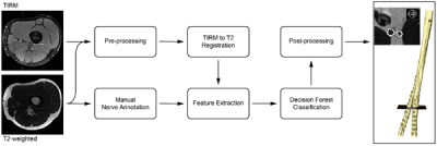
Magnetic resonance neurography (MRN) is increasingly used to diagnose peripheral neuropathy. Here, we propose a semi-automatic multimodal machine learning-based segmentation algorithm to segment peripheral nerves from MRN images. Our algorithm was tested on 9 volunteers and 25 patient cases suffering from sciatic neuropathy. Compared to manual segmentation, Dice coefficients were 0.723 ± 0.202 and 0.443 ± 0.228, respectively, with segmentation times of 5 ± 1 for semi-automatic, and 24 ± 8 minutes for manual segmentation. Our preliminary results suggest that machine learning-based segmentation of the sciatic nerve is possible in healthy and diseased nerves in clinically feasible time.
|
|
5392.
 |
74 |
Improved brachial plexus visualization using an adiabatic iMSDE-prepared STIR 3D TSE
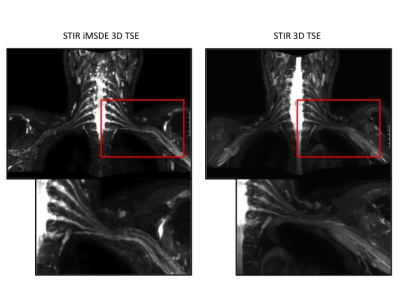
Elisabeth Klupp, Barbara Cervantes, Nico Sollmann, Franziska Treibel, Dominik Weidlich, Thomas Baum, Ernst Rummeny, Claus Zimmer, Jan Kirschke, Dimitrios Karampinos
The close proximity of blood vessels to the brachial plexus nerves can confound nerve visualization in the preferably used fat suppressed 3D T2 weighted sequences. Vessel suppression can be increased by means of an additional motion-sensitizing preparation (e.g. iMSDE). The aim of this work was the evaluation of STIR 3D-TSE in conjunction with an adiabatic T2 preparation incorporating iMSDE-based motion sensitization for MRN of the brachial plexus in a clinical routine-setting quantitatively and qualitatively. The additional motion-sensitizing iMDSE preparation reveals robust blood suppression, leading to higher CNR, increased conspicuity of the nerves, better image quality and less artifacts.
|
|
5393.
 |
75 |
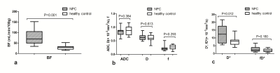
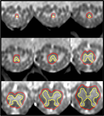
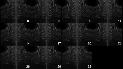
Investigating the value of arterial spin labeling (ASL) and intravoxel incoherent motion (IVIM) imaging on the diagnosis of nasopharyngeal carcinoma (NPC) in T1 stage
Video Permission Withheld
Meng Lin, Xiaoduo Yu, Lizhi xie
T1-stage NPC was difficult to diagnosis using conventional MRI and additional examinations were often needed. This study aimed to investigate ASL and IVIM on diagnosing T1-stage NPC. We measured the BF by ASL and IVIM parameters (D, D*, f, fD*) of T1-stage NPC tumors and nasopharyngeal mucosa of healthy control. The mean BF and D* of T1-stage NPC were higher than those of healthy control. And the BF correlated positively with D* and fD*. Therefore, ASL and IVIM could reflect blood perfusion difference between T1-stage NPC and benign nasopharyngeal mucosa, which is potential to help the early diagnosis of NPC.
|
|
5394.
 |
76 |
Normalised Grey Matter and White Matter Volumes in the Neurologically Intact Conus Medullaris
Marios Yiannakas, Martina Liechti, Patrick Cullinane, Xixi Yang, Ahmed Toosy, Jalesh Panicker, Claudia Gandini Wheeler-Kingshott
Magnetic Resonance Imaging derived measures of spinal cord (SC) grey matter (GM) and white matter (WM) volume are useful for indirectly assessing neurodegeneration over time (i.e. atrophy). However, for the correct interpretation of such morphometric analyses, one must take into consideration the natural variability that exists between subjects, which is unrelated to a disease process. Various normalisation strategies have been proposed for use in the upper SC, but evidence from similar applications in the lower SC is currently lacking. In this work, we present our first approach to normalisation of GM/WM volumes in the neurologically intact conus medullaris.
|
|
5395.
 |
77 |
Development of the fast 3D-MR neurography using the optimized combination of the compressed sensing and parallel imaging
Takuya Aoike, Noriyuki Fujima, Masami Yoneyama, Kinya Ishizaka, Hiroyuki Sugimori, Kohsuke Kudo
We assessed the rapid acquisition design in 3D-MR neurography (3D-MRN) using compressed sensing (CS) with the combination of the parallel imaging technique. High sparsity in 3D-MRN raw data was considered to be compatible with high CS acceleration factor. This result will be make patients comfortable in daily clinical MRN scanning.
|
|
5396.
 |
78 |
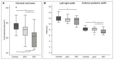
Structural and quantitative MRI to identify lesion level dependant neurodegeneration after SCI
Video Permission Withheld
Michela Azzarito, Patrick Grabher, Maryam Seif, Patrick Freund
Patients with spinal cord injury (SCI) undergo neurodegeneration affecting the spinal cord and the brain. However, the volumetric and microstructural patterns of degeneration, their relation to lesion level and clinical outcomes are uncertain. In this study, both structural and quantitative MRI approaches are used in order to identify lesion dependent neurodegeneration following SCI. It was found that lesion level drives structural changes in the spinal cord but not brain.
|
|
5397.
 |
79 |
Spinal Cord Perfusion is Associated with Diffusion and clinical mJOA score in Preoperative Patients with Cervical Spondylotic Myelopathy
Chunyao Wang, Xiao Han, Wen Jiang, Guangqi Li, Jinchao Wang, Hua Guo, Huijun Chen
Cervical Spondylotic Myelopathy (CSM) is a chronic progressive disorder of spinal cord with a relatively ill-defined onset of pathogenesis. A series of state-of-art quantitative and functional MR imaging techniques have been proposed aiming to find out specific indicators in prediction and diagnosis of CSM at early phase, but lack of sufficient evidences. Spinal cord blood supply change was recognized as one of the crucial pathophysiological process in CSM. Hence, we investigate the relationship between spinal cord blood perfusion assessed by MR DSC with DTI metrics and clinical mJOA score. Finally, we find spinal cord blood flow is significantly correlated with diffusion metrics and mJOA.
|
|
5398.
 |
80 |
Diffusion weighted T2-mapping for the determination of tissue characteristics in patients with head and neck squamous cell carcinoma
Noriyuki Fujima, Masami Yoneyama, Eunju Kim, Takuya Aoike, Suzuko Aoike, Kohsuke Kudo
We investigated the utility of T2 mapping with the pre-pulse of diffusion gradient (DW T2-map) for the determination of tissue characteristics in head and neck squamous cell carcinoma. Significant difference in T2-value of tumor tissue between that with and without diffusion gradient was observed. In addition, DW T2-map was suggested to be one of the diagnostic tool for the prediction of tumor histological grade. DW T2-map can be useful tool for the assessment of tumor tissue characteristics with greater detail.
|
|
5399.
 |
81 |
Simultaneous Diffusion Tensor Imaging and T2 relaxometry in Lumbar Nerve Roots using Dual-Echo Single-Shot DW-EPI
Masami Yoneyama, Takayuki Sakai, Eunju Kim, Tetsuo Ogino, Atsuya Watanabe, Marc Van Cauteren
Diffusion tensor imaging (DTI) is promising for evaluation of lumbar nerve root compression in the extraforaminal area. A quantitative assessment using T2 relaxometry is also promising to evaluate nerve injury. Hence, nerve root quantification using both DTI and T2 relaxation properties may improve the diagnosis of nerve roots in patients with lower back pain, but it requires a long scan time. In this study, we developed a new sequence to simultaneously obtain both diffusion parameters and T2 value in one single scan (Diffusion-Relaxation Matrix: DRM). DRM simultaneously provides diffusion tensor imaging and T2 map without prolongation of acquisition time. This quantitative combination may be helpful to further assess the lumbar nerve root pathology.
|
|
5400.
 |
82 |
Quantitative MR Neurography with Robust Fat Suppression
Masami Yoneyama, Akio Hiwatashi, Xinzeng Wang, Osamu Togao, Ivan Dimitrov, Ananth Madhuranthakam, Iain Ball, Marc Van Cauteren
MR neurography plays a major role in the diagnostic work-up of peripheral nerve pathologies and a quantitative evaluation based on T2 values can be clinically useful in estimating treatment effects and determining prognosis. Recently, we proposed a new sequence (SHINKEI-Quant) to add a quantitative information to MR neurography. To solve some issues caused by the current fat suppression techniques (SPAIR and STIR), we propose to combine SHINKEI-Quant with a new two-point dual-echo 3D DIXON-TSE (DE-mDIXON) technique. SHINKEI-Quant with DE-mDIXON simultaneously provides both MR neurography with high SNR, uniform fat suppression, and T2 maps with T2 values similar to conventional method. This sequence may be helpful to quantitatively assess nerve pathology.
|
|
5401.
 |
83 |
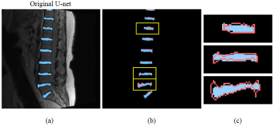
Automatic delicate segmentation of the intervertebral discs from MR spine images using deep convolutional neural networks: ICU-net
Sewon Kim, Won Bae, Dosik Hwang
The segmentation method using Deep Convolutional Neural Networks shows good performance in medical imaging. In particular, U-net is a well-known and successful model. However, U-net based on classification network shows weakness in fine segmentation. We developed a new model by changing layers and structure of U-net. Our model enables more detailed segmentation of the intervertebral discs in spine MR images.
|
|
5402.
 |
84 |
A longitudinal study of APT CEST contrast in the spinal cord of patients with multiple sclerosis at 3T
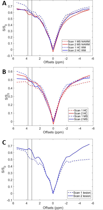
Richard Lawless, Quinn Weinberg, Bailey Box, Samantha By, Francesca Bagnato, Seth Smith
Current clinical MRI sequences cannot characterize biochemical tissue changes within the spinal cord. Therefore, MRI biomarkers sensitive to biochemical tissue changes are needed. Amide proton transfer chemical exchange saturation transfer (APT CEST) is an emerging MRI contrast method sensitive to the exchange rate and concentration of amide proton moiety. In this work, we sought to assess the reproducibility and longitudinal change of spinal cord APT CEST in patients with MS. Our results suggest that APT CEST in the spinal cord is capable of reproducibly identifying underlying changes in spinal cord tissue pathology.
|
|
5403.
 |
85 |
Low Apparent Diffusion Coefficient Value Predicts Early Progression of Skull Base Meningiomas
Ching Chung Ko, Sher Wei Lim, Tai Yuan Chen, Jeon Hor Chen, Chien Feng Li, Yow Ling Shiue
A recent study described the extent of tumor resection and ADC values could offer better prediction of progression/recurrence (P/R) in meningiomas than histopathological grading. Although complete resection of tumor is a key determining factor of recurrence in meningiomas, it is often difficulty to achieve for the skull base meningiomas (SBM) due to complex neurovascular structures. In this study, we investigated the preoperative CT and MR imaging features for the prediction of early P/R in SBM, with emphasis on ADC values. Our results found that low ADC value and adjacent bone invasion could predict high risk of early P/R in SBM, and therefore, offer clinically vital information for the planning of treatment.
|
|
5404.
 |
86 |
In vivo 31P magnetic resonance spectroscopy of human parotid gland
Toshiyuki Sato, Hiroyoshi Isoda, Hirotsugu Nakai, Shigeshi Kohno, Koji Tokunaga, Hironori Shimizu, Seiya Kawahara, Kaori Togashi
This is the first in vivo study to investigate major phosphorus metabolites of human parotid gland with 31P magnetic resonance spectroscopy (MRS) using 3D chemical shift imaging (CSI) technique. Five healthy volunteers were measured on a clinical 3.0 T MRI scanner, and three of them were remeasured immediately after intake of tablets containing vitamin C. The spectra reveal large adenosine triphosphate (ATP) and phosphocreatine (PCr) intensities. Following intake of tablets containing vitamin C, decrease of β-ATP can be observed. In vivo 31P MRS can be used to assess bioenergetics of the human parotid gland within a reasonable scan time.
|
|
5405.
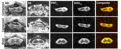 |
87 |
Detection of Spinal Cord Compression Injury in the Rat using Filter-Probe Diffusion Encoding
Matthew Budde, Natasha Beucher, Seung Yi Lee, Brian Schmit, Shekar Kurpad
A rat model of chronic spinal cord compression was evaluated with reduced field of view (rFOV) diffusion tensor imaging and filter-probe diffusion encoding. The results demonstrate the sensitivity of diffusion MRI for detecting injury.
|
|
5406.
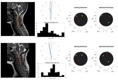 |
88 |
Anatomically-informed and patient-specific diffusion tensor sampling schemes for the cervical spinal cord
Kurt Schilling, Samantha By, Quinn Weinberg, Bailey Box, Bennett Landman, Adam Anderson, Seth Smith
In this study, we minimize uncertainty in Diffusion Tensor Imaging (DTI) parameters by optimally selecting diffusion gradient directions utilizing prior structural knowledge. By measuring the anatomy of the spinal cord for each subject, we created patient-specific, anatomically-informed DTI encoding schemes for 256 subjects, utilizing just 6 diffusion directions. In addition, we create an optimal encoding 6-direction scheme for the anatomically-average person. Through simulations and experimental validation on a physical fiber phantom, we show improvements in FA estimation, even when compared to conventional 30 direction schemes. This technique results in minimal scan time and increased accuracy in order to maximize clinical value.
|
|
5407.
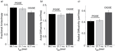 |
89 |
Oscillating Gradient Spin Echo (OGSE) Diffusion Tensor Imaging of the Human Spinal Cord: Application to Multiple Sclerosis
Samantha By, Seth Smith, Kurt Schilling, Quinn Weinberg, Sean Devan, Francesca Bagnato, Junzhong Xu
Compared to conventional pulsed gradient spin echo (PGSE) techniques, oscillating gradient spin echo (OGSE) provides access to shorter diffusion times, thereby enabling greater sensitivity to microstructure on smaller scales. Here, we report initial results of OGSE in the human spinal cord. Unlike in the brain, axial diffusivity in the spinal cord appears to be negligibly affected with diffusion time. Relative to the PGSE sequence (tdiff= 66 ms), the OGSE sequence (tdiff=8.77 ms) shows a 37% mean increase in radial diffusivity (RD) in healthy controls. When applied to MS, OGSE shows a larger difference in RD in comparison to healthy controls.
|
|
5408.
 |
90 |
MRI-guided Focused Ultrasound to Improve Drug Delivery in Spinal Cord Injury
Donna Cross, Allison Payne, Amanda Stump, Henrik Odéen , Megan Ostlie, Ethan Reichert , Chloe Cross, Yoshimi Anzai, Gregory Hawryluk
Spinal cord injury (SCI) affects over 17,000 individuals each year in the United States, and most patients are left with some permanent paralysis. MRI-guided focused ultrasound (MRgFUS), when applied to the spinal cord with microbubbles to generate sonoporation, can transiently open the blood spinal cord barrier for effective drug delivery by breaking the glial scar. We used MRgFUS to increase permeability in the blood spinal cord barrier in a SCI rat model. Rats that underwent an MRgFUS procedure showed increased contrast caudal to the injury site, indicating drugs could potentially permeate these regions and assist with axonal regrowth.
|
|
5409.
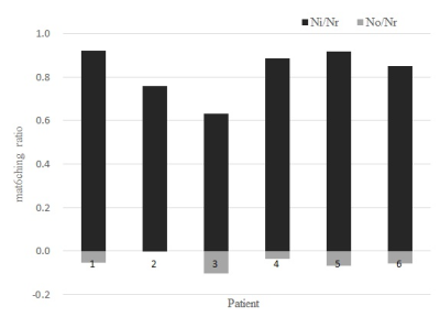 |
91 |
Evaluation of a new semi-automatic delineation method for quantitative analysis of tumors: application to head and neck squamous cell carcinoma
Kanae Moriyama, Hiroyuki Sugimori, Noriyuki Fujima, Toru Yamamoto
Objective determination of tumor region of interest (ROI) on MR images is expected for quantitative analysis of tumors. We developed a semi-automatic method of ROI delineation. We applied this method to head and neck squamous cell carcinoma and evaluated the matching of the ROI delineation by our method and an experienced radiologist. The ADC values were compared for different ROI delineation methods. Averagely, 80% of the area of the ROI matched and the difference of the ADC value was within 2% (sd), showing that our new method of tumor delineation can be used to analyze the tumor characteristics quantitatively.
|
|
5410.
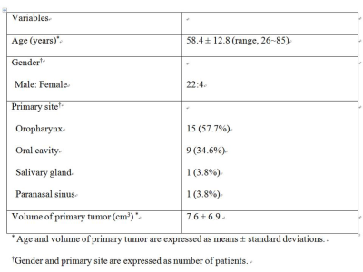 |
92 |
Correlation of tumor blood flow in head and neck squamous cell carcinoma by pseudo-continuous arterial spin labeling with parameters of dynamic contrast-enhanced MRI
Young Jun Choi, Jeong Hyun Lee, Josef Pfeuffer
The aim of this prospective study was to evaluate the correlation between tumor blood flow (TBF) measurement using pseudo-continuous arterial spin labeling (pCASL) and parameters of DCE-MRI in patients with head and neck squamous cell carcinoma (HNSCC). We scanned 26 patients with HNSCC using 3T MRI with both pCASL and DCE-MRI. There were significant correlation between TBF of pCASL and wash-in, signal enhancement ratio, and Vp of DCE-MRI, with a correlation coefficient of 0.649, 0.642, and 0.507, respectively (P<0.01). The pCASL can be a useful tool for noninvasive assessments of the TBF in patients with HNSCC.
|
|
5411.
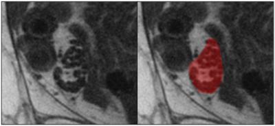 |
93 |
Texture analysis applied to MRI of nerves affected with chronic inflammatory demyelinating neuropathy.
Paolo Felisaz, Andrea Poli, Giovanni Vitale, Raimondo Vitale, Laura Piccolo, Andrea Cortese, Niels Bergsland, Anna Pichiecchio, Stefano Bastianello
Introduction. Texture analysis was applied to MR images of peripheral nerves obtained with MR micro-neurography. Materials and Methods. Ankle tibial nerves were imaged at 3T in 10 patients affected with chronic inflammatory demyelinating neuropathy and 10 healthy subjects. Multiple subsets of textural features were compared, using different extraction methods and statistical analyses. The most discriminating features were selected and compared to the automatically extracted subsets. Results. Feature subset extracted from the whole pool of features performed better than the ones obtained by specific groups of features. Conclusion. Texture analysis may have a role in discriminating between pathologic and normal nerves.
|
|
5412.
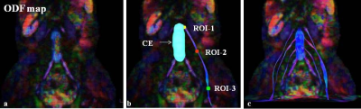 |
94 |
How to separate peripheral nerves from muscles
Ibrahim Ibrahim, Jaroslav Tintera, Vít Herynek, Antonín Škoch, Ivan Humhej, Milan Hájek
MR tractography of the peripheral nerves (PN) is challenging due to the difficulty to acquire high quality DWI data for peripheral nerve bundles reconstruction. The aim of this study was to propose an algorithm for separation LSP bundles from muscles using segmentaion of cauda equina and normalized quantitative anisotropy.
|
|
5413.
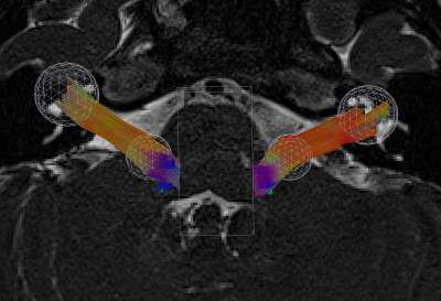 |
95 |
Feasibility of Quantitative Diffusion MR Tractography of the Vestibulocochlear Nerve in Children with Unilateral Profound Sensorineural Hearing Loss
Video Permission Withheld
Ryan Cabeen, Elina Kari, Courtney Voelkner, Marta Kulich, Laurel Fisher, Yonggang Shi, Meng Law, Arthur Toga
Diffusion MR tractography modeling of the vestibulocochlear nerve (VCN) has clinical value for characterizing hearing loss and surgical planning for cochlear implants; however, the neuroanatomy of the VCN presents technical challenges to image-based modeling. We conducted a feasibility study to evaluate VCN tractography in children with unilateral sensorineural hearing loss using advanced diffusion MRI acquisitions and modeling techniques. The results indicate tractography of the VCN is clinically feasible and quantitative tractography metrics reflect lateralization of hearing loss. We further found significant improvements from a reverse-phase encoding acquisition and multi-shell multi-compartment diffusion modeling.
|
|
5414.
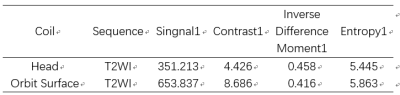 |
96 |
Phantom Validation Study for Custom-made Orbit Surface Coil
Did Not Present
ZONG RUI ZHANG, QING HUA CHEN, XIAO QI WANG, YAN TAO NIU, JUN FANG XIAN
Previously introduced the new custom-made orbit imaging surface coil was tested in this research. The study gives a brief overview of the quantitative information that can be derived from it. Combining information from normal standard 15 channel head coil can aid in answering the typical advantages and future clinical application of the new surface coil. This phantom study is to verify the orbit surface coil’s feasibility in clinical use.
|
|
Neonatal & Pediatric Neuroimaging
Electronic Poster
Neuro
Thursday, 21 June 2018
| Exhibition Hall |
09:00 - 10:00 |
| |
|
Computer # |
|
5415.
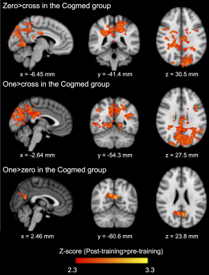 |
97 |
Working memory training and brain structural and functional correlates in children born preterm
Claire Kelly, Deanne Thompson, Jian Chen, Elisha Josev, Leona Pascoe, Megan Spencer-Smith, Chris Adamson, Chiara Nosarti, Lex Doyle, Marc Seal, Peter Anderson
In a randomised controlled trial, we investigated if adaptive, computerised working memory training using Cogmed was associated with greater neural changes compared with a placebo training program. Participants were a population-based cohort of 91 school-age children born <28 weeks’ gestation or <1000 g birthweight. Children had structural, diffusion and task-based functional MRI before and two weeks following five weeks of Cogmed or placebo. There was little evidence for larger changes in cortical morphometry, white matter microstructure, or brain functional activity following Cogmed compared with placebo. In our study, Cogmed did not benefit brain structure or function in preterm-born children.
|
|
5416.
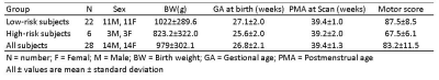 |
98 |
Early Identification of Reduced Brain Functional Connectivity in Very Preterm Infants with Motor Impairments
Lili He, Hailong Li, Nehal Parikh
Very preterm infants (<32 weeks gestational age) are at high risk for motor impairments. Investigation of brain network connectivity will improve our understanding of how brain organizational changes influence motor function and can result in improved individual risk stratification. In this work, we found reduction in functional connectivity in multiple motor and sensory regions, soon after birth, in very preterm infants at high risk of motor impairments assessed at 2 years of age. Our findings may enable mechanistic understanding and facilitate early, more accurate prediction of motor impairments.
|
|
5417.
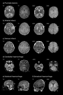 |
99 |
Prevalence of brain lesions in a contemporary cohort of newborns with complex congenital heart disease prior to surgery
Christopher Kelly, Catarina Tristão Pereira, Sophie Arulkumaran, Lucilio Cordero Grande, Emer Hughes, Rui Pedro Teixeira, Johannes Steinweg, Joseph Hajnal, John Simpson, A. David Edwards, Mary Rutherford, Serena Counsell
Infants born with congenital heart disease (CHD) are known to experience a distinct pattern of neurodevelopmental and behavioural impairment in later life. In this study, we scanned 64 infants with complex CHD prior to surgery, and used high-resolution structural, diffusion and susceptibility-weighted magnetic resonance imaging to understand the burden of brain lesions in a contemporary cohort. We characterised lesions and produced quantitative lesion maps, registered to a population template. We found a lower incidence of infarction than has been reported in previous comparable cohorts, and we explore the clinical reasons that may explain such variation.
|
|
5418.
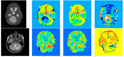 |
100 |
Multicentre pediatric brain tumour dynamic susceptibility contrast (DSC-) MRI with contrast agent leakage correction
Stephanie Withey, Jan Novak, Lesley MacPherson, Laurence Abernethy, Barry Pizer, Richard Grundy, Simon Bailey, Dipayan Mitra, Theodoros Arvanitis, Dorothee Auer, Shivaram Avula, Andrew Peet
Dynamic susceptibility contrast (DSC-) MRI provides measures of relative cerebral blood volume (rCBV) in pediatric brain tumors. Correction of rCBV for the effects of contrast agent leakage can be done using post-processing techniques. Forty pediatric patients with high and low grade tumors underwent DSC-MRI scans pre-treatment at four centers. DSC-data was analyzed with and without leakage correction to calculate rCBV and the leakage parameter, K2. There were significant differences between all parameters when comparing the high and low grade tumor groups. Low grade tumors tended to show T1-dominant leakage effects while high grade tumors showed predominantly T2* dominance.
|
|
5419.
 |
101 |
Brain growth over the first 13 years of life in typically developing and very preterm children
Deanne Thompson, Lillian Matthews, Bonnie Alexander, Katherine Lee, Claire Kelly, Rod Hunt, Jeanie Cheong, Megan Spencer-Smith, Marc Seal, Jeffrey Neil, Terrie Inder, Lex Doyle, Peter Anderson
Few longitudinal cohort studies exist characterizing regional brain volumes from birth to adolescence. This study derives brain volumes in approximately 100 regions at term-equivalent, 7 and 13 years of age for 102 very preterm and 20 full-term children. The trajectory of brain development in many regions differed between very preterm and full-term children over the first 7 years of life. From 7 to 13 years brain growth slowed, ceased or regressed in both groups in a region-specific manner, apart from subcortical regions that continued to increase in volume. This study provides novel insights in typical and atypical regional brain volumetry.
|
|
5420.
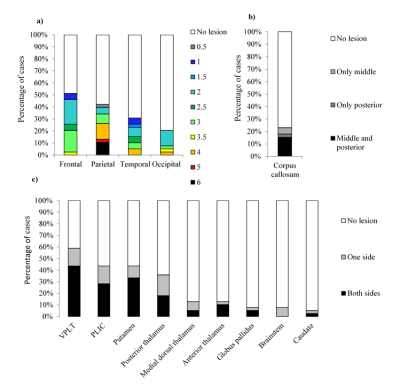 |
102 |
A simple semi-quantitative scale for brain MR imaging is associated with motor function, communication and cognition in dyskinetic cerebral palsy
Olga Laporta-Hoyos, Simona Fiori, Kerstin Pannek, Julia Ballester-Plane, David Leiva, Lee B Reid, Alex M Pagnozzi, Elida Vázquez, Ignacio Delgado, Alfons Macaya, Roser Pueyo, Roslyn N Boyd
Previous neuroimaging studies of DCP have been based on categorical descriptors or advanced neuroimaging protocols that cannot be easily utilised in a clinical context. To enable clinical translation, we characterized brain lesions, and their association with clinical outcomes in 39 participants with DCP, using a semi-quantitative scale for brain MRI that is clinically accessible due to its relative simplicity and reliance only on standard clinical images. Our results indicate that (A) ventral posterior lateral thalamus and frontal lobe are the most frequent locations of observable lesions in DCP and that (B) sqMRI can index motor, communication and cognitive functioning.
|
|
5421.
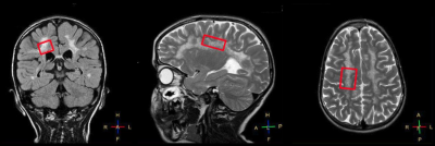 |
103 |
Quantitative 1H Single Voxel Spectroscopy In Metachromatic Leukodystrophy Patients Treated With Haematopoietic Stem Cell Gene Therapy: Preliminary Results
Video Permission Withheld
Paola Scifo, Pasquale Anthony Della Rosa, Matteo Canini, Francesca Fumagalli, Valeria Calbi, Andrea Falini, Alessandro Aiuti, Cristina Baldoli
The efficacy of Haematopoietic Stem Cell-Gene Therapy for Metachromatic Leukodystrophy is under evaluation. CNS damage is currently rated by means of visual MR severity scores. The aim of this study was to investigate whether quantitative spectroscopy measurements correlate with MR severity scores. This preliminary analysis shows that both NAA/Cr and NAA/Cho ratios are associated with progression in total MR Severity Score, Atrophy and Demyelination Load. This suggests that MR spectroscopy could be useful to longitudinally and quantitatively evaluate the disease burden in MLD patients.
|
|
5422.
 |
104 |
FETAL CORTICAL PARCELLATION BASED ON GROWTH PATTERNS
Jing Xia, Caiming Zhang, Fan Wang, Oualid M. Benkarim, Gerard Sanroma , Gemma Piella, Miguel A. Gonzalez Ballester, Nadine Hahner, Elisenda Eixarch, Dinggang Shen, Gang Li
The available cortical parcellations created for adults are inapplicable for fetuses, due to dramatic differences in brain appearances and dynamic growth of fetal brains. Considering sulcal-gyral landmarks are not stably established in fetal brains, we propose to use the growth patterns of cortical properties to parcellate fetal cortical surfaces. Growth patterns, which reflect underlying changes of microstructures, enable an accurate definition of distinct regions in development and function, as microstructures determine the molecular organization and functional principles of the cortex. We apply the growth-pattern-based method on 25 normal fetuses from 26 to 29 gestational weeks and generated biologically meaningful parcellations.
|
|
5423.
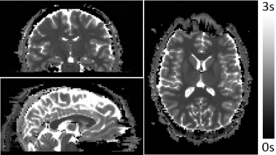 |
105 |
Fetal T1 mapping using multi-inversion EPI at 3T
Borjan Gagoski, Daniel Park, Ville Renvall, Esra Abaci-Turk, Elfar Adalsteinsson, Thomas Witzel, Lawrence Wald, Patricia Grant, Jonathan Polimeni
T1 mapping of the fetal brain is hindered by substantial, frequent, and random fetal motion, making many current quantitative techniques impractical in the fetus. We have applied to the fetal brain (and adult brain for validation purposes) a recently introduced T1 mapping technique based on EPI readout that is preceded by a non-selective inversion pulse, and where the order of the acquired slices is permuted from one inversion recovery to the next, allowing efficient, high temporal sampling of the T1 relaxation curve. We believe that this method is capable of providing accurate T1 maps of the fetal brain.
|
|
5424.
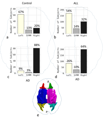 |
106 |
Chemotherapy reduces microstructural asymmetry in the brain
Junyu Guo, Yuanyuan Han, Yimei Li, Wilburn Reddick
Previously, we discovered two types of brain microstructural asymmetry: myelin-related asymmetry and axon-related asymmetry in healthy volunteers. In this study, we found that chemotherapy with methotrexate significantly reduced the microstructural asymmetries in patients with acute lymphoblastic leukemia, having a greater impact on asymmetries in younger patients than in older ones. Our results indicate that chemotherapy may lead to atypical brain development long before patients become symptomatic. These results may help to provide insights into neurocognitive deficits caused by chemotherapy.
|
|
5425.
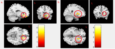 |
107 |
The Impact of Prenatal Endocrine-Disrupting-Chemical Exposure on Brain Function in Teenagers
Chi-Ieong Hong, Shu-Li Wang, Chao-Yu Shen, Jeng-Dau Tasi, Tou-Rong Chen, Jun-Cheng Weng
The products made by endocrine disrupting chemicals (EDCs) widely exists in our daily life, which could be a risk of our health. We tried to find the relationship between prenatal exposure to EDCs’ concentrations and teenager’s brain function using resting-state functional MRI (rs-fMRI). Our results showed the correlation between prenatal perfluorononanoic acid (PFNA)/MeHg exposure and functional alterations of the teenage brain in caudate gyrus and putamen.
|
|
5426.
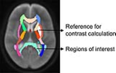 |
108 |
Assessment of myelination in developing brains by using the inhomogeneous magnetization transfer MRI
Xianjun Li, Yuli Zhang, Fan Wu, Xiaoqian Jia, Xiaoyu Wang, Chao Jin, Qinli Sun, Heng Liu, Xiaocheng Wei, Jian Yang
In vivo assessment of myelination is critical for understanding the typical and pathological brain development. Inhomogeneous magnetization transfer (ihMT) has been proposed recently and may be useful to quantify myelin contents. This study tried to evaluate performances of magnetization transfer ratio (MTR) and ihMT ratio (ihMTR) in assessing the myelination of toddler and adolescent brains. The ihMTR maps hold larger image contrasts than MTR maps. Moreover, ihMTR demonstrates larger relative changes during the period from the toddler to adolescent. Results in this study suggested that ihMTR was specific for characterizing white matter and sensitive to myelination during the brain development.
|
|
5427.
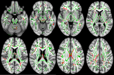 |
109 |
White Matter Integrity in Adolescents with Attention Deficit Hyperactivity Disorder with Comorbid Bipolar Disorder assessed by Tract Based Spatial Statistics
Arzu Has, Mahmut Mujdeci, Gokce Say, Hediye Gunbey, Nilufer Okumus, Yusuf Karaer, Halime Cak Esen, Ozlem Yildiz Gundogdu, Rahsan Gocmen, Yonca Anik
White matter (WM) integrity is an important factor for the pathophysiology of attention-deficit/hyperactivity disorder (ADHD) and bipolar disorder (BD) in adolescents. In this study, we hypothesized that adolescents with attention deficit hyperactivity disorder with comorbid bipolar disorder (ADHD+BD) would yield different pattern and extent of WM abnormalities than ADHD and BD. Sixty adolescents were categorized into four groups as ADHD, BD, ADHD+BD and healthy controls. The comparative diffusion tensor imaging analyses were performed between each patient group and healthy controls. Adolescents with ADHD+BD displayed different pattern and extent of WM abnormalities than ADHD and BD revealed by tract-based spatial statistics.
|
|
5428.
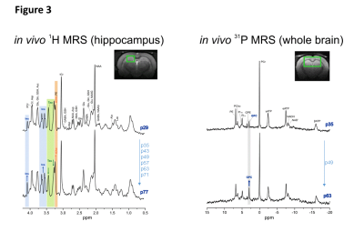 |
110 |
Late post-natal brain metabolic changes in healthy rats, a longitudinal in vivo 1H and 31P MRS study
Veronika Rackayova, Olivier Braissant, Rolf Gruetter, Valerie McLin, Cristina Cudalbu
New-born rats are known to undergo important brain metabolic changes during first 4-5 weeks of their lives. However precise description of metabolic changes at latter stages is missing. At this period rats might still undergo changes due to final brain maturation what may impact data interpretation of experiments during this window. This study aimed to explore possible metabolic changes in the rat brain during the late post-natal period (post-natal days 29 - 77), using in-vivo 1H and 31P-MRS and a temporal resolution of 1 week. We report significant changes in Ins, tCho (mainly due to GPC increase), Tau and Glu in rats considered as young adults.
|
|
5429.
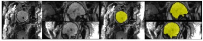 |
111 |
Automated Extraction of the Fetal Brain from Functional MRI Data
Saige Rutherford, Mike Angstad, Jasmine Hect, Andre Zapico, Moriah Thomason, Chandra Sripada
In this study, we present a novel application of a Convolution Neural Network algorithm to a challenging image segmentation problem: fetal brain segmentation. Resting-state fMRI data was obtained from 192 fetuses (gestational age 20-40 weeks, M=31.9, SD=4.28). The output from automated extractions are compared with the ground truth of manually drawn brain masks. We report that automated fetal brain localization and extraction is achievable at the same integrity of manual methods, in a fraction of the time.
|
|
5430.
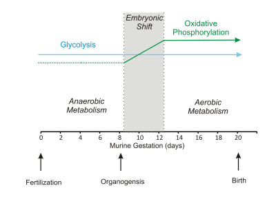 |
112 |
In vivo 1H NMR spectroscopy study of mitochondrial pyruvate carrier 1 (MPC1) deficient mouse reveals energy shift in the development of embryonic brain
Video Permission Withheld
Hongxia Lei, Zeinab Ammar, Corina Berset, Jean-Claude Martinou
Pyruvate degradation is an important step for oxidative phosphorylation. We studied brains of murine embryos devoid of mitochondrial pyruvate carrier 1 (MPC1) using 1H MRS. We demonstrates that after the embryonic energy shift, lactate accumulation can explain embryonic lethality in embryo devoid of MPC1.
|
|
5431.
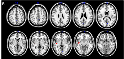 |
113 |
Differential effects of hunger and depression on cerebral blood flow in healthy adolescents
Céline Charroud, Emmanuelle Le Bars, Emily Sanrey, Jérémy Deverdun, Josef Pfeuffer, Nicolas Menjot de Champfleur, Philippe Coubes
This study aims to explore the appetite effect on taste and depression on healthy adolescents using Arterial Spin Labeling. Fifteen participants complete the Multiscore Depression Inventory for Children test and two MRI sessions: pre-lunch (hunger) and post-lunch (satiety). We found an increased CBF – cerebral blood flow – during hunger in the posterior insula (anticipation and motivation of feeding) and during satiation in the precuneus, lingual gyrus and cuneus (inhibition pattern of food intake). We show that the correlations between depression and CBF are modulated by appetite in the precuneus, operculum, lingual, cuneus, middle frontal gyrus and inferior parietal lobule.
|
|
5432.
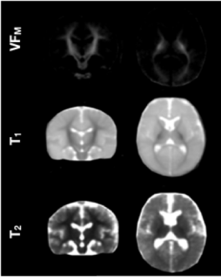 |
114 |
Influences of the Gut Microbiome on Early Brain Development
Douglas Dean, Elizabeth Planalp, Nicholas Vogt, Kristin Dowe, Alysha Rameshk, Kristine Mclaughlin, Abigail Freeman, Andrew Alexander
Increasing evidence from animal studies suggests the gut microbiome has a significant role on early brain development and function. However, little is known about this role on human brain development and in particular, on myelination. Using quantitative multicomponent relaxometry and 16S rRNA sequencing, we examined measures of myelin content and the gut microbiome from a cohort of typical developing infants. Infant brain measures were found to be differentially associated with the relative abundancies of certain bacteria phylum, suggesting that microbial communities may have a significant influence on processes of early brain development.
|
|
5433.
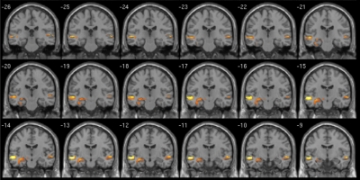 |
115 |
Establishing a developmentally appropriate fMRI paradigm for presurgical mapping of memory in children
Amanda Wood, Elaine Foley, Parnpreet Virk, Helen Ruddock, Paras Joshee, Kelly Murphy, Stefano Seri
The use of functional MRI to evaluate risk to memory function following temporal lobe surgery in children cannot rely on adult tools. We present a novel fMRI paradigm that is brief, independent of reading ability, and therefore a candidate for presurgical evaluation. Data from 36 adults and 19 children (all healthy controls) show that the paradigm captures the expected leftward asymmetry of mesial temporal activation in adults. A more symmetrical pattern is observed in children, consistent with the emergence of hemispheric specialisation across childhood. These data have important implications for the interpretation of presurgical memory fMRI in the paediatric setting.
|
|
5434.
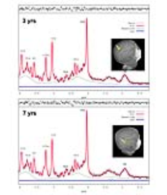 |
116 |
Trajectories of brain lactate and energy metabolite concentrations do not contribute to elevated aerobic glycolysis across childhood
Helene Benveniste, Gerald Dienel, Zvi Jacob, Hedok Lee, Rany Makaryus, Albert Gjedde, Fahmeed Hyder, Douglas Rothman
Lactate is produced in normal brain even when O2 levels are ample, and high lactate production may explain aerobic glycolysis (AG) in developing brain. We evaluated steady state lactate concentrations ([Lac]) in brains of 87 children using 1HMRS while they underwent routine MRI examination. The trajectory of [Lac] in cerebral cortex across childhood was below the 0.5-0.7 mM range in normal adult brain. Thus, lactate accumulation and efflux are unlikely to underlie excessive AG in children.
|
|
5435.
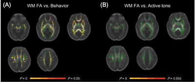 |
117 |
Potential role of diffusion tensor imaging in assessing neonatal neurobehavior development in the newborn stage
Chao Jin, Xianjun Li, Congcong Liu, Yannan Cheng, Miaomiao Wang, Jian Yang
Maturation of brain structure underpins its functionality. From birth, brain structure and neurobehavior abilities progressively mature. However, specific link between these two entities remains unclear. This study therefore aims to investigate such correlation between neonatal neurobehavior and brain white matter (WM) microstructural development measured by diffusion tensor imaging (DTI)-fractional anisotropy (FA). Results indicated that neonatal behavior abilities significantly correlated with sensorimotor WM FAs; while active-tone just correlated with motor-associated WM. These findings demonstrate the specialized correlations of specific neurobehavior abilities with microstructural maturation in corresponding WMs, suggesting the potential role of DTI in assessing the neonatal neurodevelopment in clinical practice.
|
|
5436.
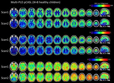 |
118 |
Quantifying cerebral hemodynamics in developing children using multiple post-labeling delay pseudo-continuous arterial spin labeling
Nick Todd, Helen Santoro, Haley McEvoy, Danny Wang, Lino Becerra, David Borsook, Duncan Hodkinson
Arterial Spin Labeling (ASL) is an attractive option for studies in pediatric populations due to its ability to acquire quantitative cerebral perfusion measurements without ionizing radiation. Brain vascular dynamics in children are different from adults and rapidly changing with age. ASL sequence parameters must therefore be carefully chosen and longitudinal studies must account for normally occurring neurodevelopment. Here we present a multiple-post label delay pseudo-continuous ASL approach to quantifying cerebral blood flow, arterial transit time, and cerebral blood volume in children. We show analysis of the PLD selections with respect to ATT values and test-retest reliability of the CBF measurements.
|
|
5437.
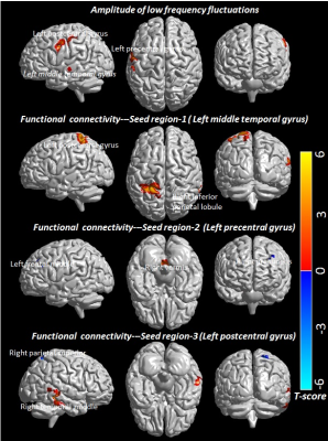 |
119 |
Changes in Resting-State Functional Brain Activity are Correlated with Waning Cognitive Functions in Pediatric HIV
Santosh Yadav, Rakesh Gupta, Sheema Hashem, Ajaz Bhat, Ravindra Garg, Vimala Venkatesh, Pradeep Gupta, Alok Singh, Muhammad Azeem, Deepak Kaura, Mohammad Haris
We evaluated the functional brain changes in HIV-infected children by mapping the amplitude-of-low-frequency-fluctuations (ALFF) and functional connectivity (FC) using rs-fMRI. Association of these changes with cognitive measures was also explored. ALFF and FC were significantly altered in multiple brain regions involved in auditory, visual, language, motor and sensory activity. The waning cognitive functions in HIV-infected children were associated with the changes in ALFF and FC. These two imaging parameters in association with the cognitive evaluation may provide better understanding of the functional brain activity in HIV-infected children.
|
|
| Back |
| The International Society for Magnetic Resonance in Medicine is accredited by the Accreditation Council for Continuing Medical Education to provide continuing medical education for physicians. |
