|
Electronic Poster Session
Diffusion |
Thursday, 21 June 2018
Electronic PosterDiffusion
5200 -5222 Fiber Orientation & Fiber Tracking
5223 -5246 Microstructure: Modeling
5319 -5342 Diffusion MRI: Acquisition & Reconstruction
5343 -5366 Diffusion MRI: Post-Processing
5367 -5390 Detecting Microstructural Changes Using Diffusion |
| |
Fiber Orientation & Fiber Tracking
Electronic Poster
Diffusion
Thursday, 21 June 2018
| Exhibition Hall |
08:00 - 09:00 |
| |
|
Computer # |
|
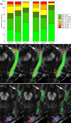
5200.
 |
1 |
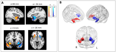
 The uncinate fasciculus: hemispheric asymmetries of DTI metrics and curvature mapping The uncinate fasciculus: hemispheric asymmetries of DTI metrics and curvature mapping
Lia Talozzi, Claudia Testa, Stefania Evangelisti, Lorenzo Cirignotta, Claudio Bianchini, Micaela Mitolo, Paola Fantazzini, Caterina Tonon, David Manners, Raffaele Lodi
We reconstructed the uncinate fasciculus bilaterally by probabilistic tractography in a group of 29 healthy subjects, in order to quantitatively investigating hemispheric asymmetries. We evaluated tract volume and DTI metrics. Our tract Laplacian parameterization successfully described curving bundles and allowed the calculation of along-tract measures and curvature mapping. Group variability maps showed a more dorsal route in the left hemisphere, towards the lateral fronto-orbital cortex. We also found a higher fractional anisotropy on the right compared to the left and different tract curvature. These asymmetries could be associated to specific tract functions as semantic and emotional processing, selectively affected in pathologies.
|
|
5201.
 |
2 |
Ischemic optic neuropathy: Using Track-Weighted Imaging to detect changes in the neuroretina
Arnaud Attyé, Felix Renard, Clement Jean, Laurent Lamalle, Alexandre Krainik
Track-Weighted Imaging was recently introduced as a qualitative tractography-based method with super-resolution properties and high anatomical contrast. We report the use of this method to explore the human retina. In healthy volunteers, we showed that reconstructions of neuroretinal fascicles were influenced by diffusion acquisition parameters, ocular laterality, and ocular dominance. We have raised the hypothesis that patients with anterior ischaemic optic neuropathy, a disease that leads to degeneration of neuroretinal cells, could present with specific neuroretinal fascicle injuries. We demonstrated that presence of the temporal fascicle in the affected eye provides an objective outcome radiological sign correlated with visual recovery.
|
|
5202.
 |
3 |
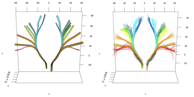
Tractography-Based Atlas of the Healthy Cortico-Spinal Tract
Aymeric Stamm, Alessandro Zito, Valeria Callioni, Ilaria Sartori, Luca Torriani, Simone Vantini
The corticospinal tract is a critical white matter pathway as it connects the primary motor cortex to the spinal cord and handles voluntary motion. Atlases of major brain connections do exist, but, surprisingly, atlases that depict the actual localisation of a specific pathway are missing. In this work, we propose a comprehensive statistical methodology for generating an atlas of the healthy CST. This should help designing efficient statistical tests for detecting damaged tissue along the CST and consequently improving patient outcome in a number of brain pathologies (tumors, strokes, Parkinson and related disorders, etc.).
|
|
5203.
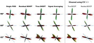 |
4 |
Bootstrapping Fibre Orientation Distributions: Accuracy advantages and other benefits of estimating shape uncertainty
Video Permission Withheld
Marina Rakic, Robert Dallyn, Flavio Dell'Acqua
Inherently low signal-to-noise ratio in diffusion-MRI results in high uncertainty in the estimated underlying fibre orientation distributions. True bootstrapping and model- based residual bootstrapping approaches have been proposed to estimate the uncertainty in the estimated peak directions. Here we compare the two methods using simulations and observe that more information can be extracted by looking at the variation in the entire shape of the FOD, as opposed to just the peak direction. Results also suggest that, in this case, residual bootstrapping might not provide a representative estimation of directional uncertainty due to non-negligible model bias.
|
|
5204.
 |
5 |
Biases in classical structural parcellation
Michael Jensen, Henrik Thomsen, Sune Darkner, Matthew Liptrot, Niklas Kasenburg, Karl-Anton Dorph-Petersen, Aasa Feragen
The classical structural parcellation algorithm by Behrens et al1 remains widely used in clinical research, largely thanks to its simplicity and availability in standard MRI processing software. However, its construction and dependency on tractography leads to several biases that can severely affect the conclusions drawn from it, but which are not well known. We illustrate these biases via the original thalamus parcellation experiment on Human Connectome Project (HCP) data3. Based on our experiments, we outline open problems for future structural parcellation algorithms, and possible directions for overcoming them.
|
|
5205.
 |
6 |
Exploring peritumoral neural tracts by using Neurite Orientation Dispersion and Density Imaging
Video Permission Withheld
Shin Tai Chong, Hung-Wen Kao, Chien-Yuan Lin, Chiao-Chi Chen, Chun-Yi Lo, Ching-Po Lin, Chen Chang
We hypothesized that the Neurite Orientation Dispersion and Density Imaging (NODDI)-based tractography could improve the reconstruction of the fiber tracts that by tracking through regions of peritumoral edema. In visual comparison with diffusion tensor imaging (DTI), this model provided similar performance in healthy side and better performance in the corticospinal tract (CST) of the affected side. This technique may help neurosurgeons to define an optimal safe margin for total resection.
|
|
5206.
 |
7 |
The impact of structure tensor informed fibre tractography (STIFT) on structural connectivity
Kwok-Shing Chan, David Norris, José Marques
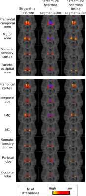
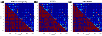
Structure tensor informed fibre tractography(STIFT) incorporating white matter morphology from gradient echo data to diffusion data is beneficial in the presence of kissing and highly curved fibres. However, previous demonstrations of STIFT provide only qualitative results and its performance beyond the initial targets (optic radiation and cingulum) is still unknown. This study investigated the quantitative effects of STIFT through structural connectivity comparison between diffusion tractography and STIFT. We found that there is no change of connectivity with STIFT in major structural connections.
|
|
5207.
 |
8 |
DTI-based Network Analysis of APP/PS1 Mouse Brains with Age and Sex
Samuel Grant, David Hike, Abdol Ismail, Victor Wong, Scott Boebinger, Tara Palin
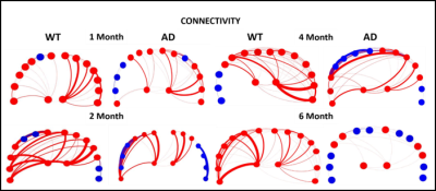
This study utilizes DTI and graph theory as a novel way for identifying early pathology and connectivity changes related to Alzheimer’s Disease. As a function of phenotype, age and sex, DTI studies were performed on APP/PS1 mouse brains and age-matched wild type controls at 11.75 T. Current data shows a drop in FA and a decrease in connectivity in the temporal region of the brain. High resolution 3D images acquired at 21.1 T display the presence of amyloid plaques, which temporally correlate with the progression of structural connectivity alterations in this transgenic preclinical model.
|
|
5208.
 |
9 |
The unresolved problem of synaptic connectivity in the context of the cerebro-cerebellar loop
Fulvia Palesi, Fernando Calamante, Giovanni Savini, Egidio D'Angelo, Claudia Gandini Wheeler-Kingshott
Recently advanced tractography has been used for assessing the feasibility of characterizing cerebro-cerebellar tracts, acknowledging the issue of how tractography deals with polysynaptic connectivity in particular through the thalamus. In this work, four different synaptic connectivity configurations at thalamic level were hypothesized and each one of them was reconstructed using high-quality HCP data. Our findings revealed the key role of thalamic connectivity for characterizing the cerebro-cerebellar connections. We can hypothesize that the relation between streamlines entering/outing the thalamic synapses is multiplicative and our findings using polysynaptc thalamic tracts support the fact that cognitive/associative areas are the mainly involved in the cerebro-cerebellar connections.
|
|
5209.
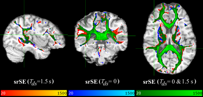 |
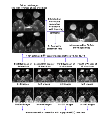
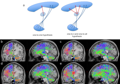
10 |
Improved fibre tracking with optimized diffusion-weighted single-refocused spin echo EPI
Manoj Shrestha, Pavel Hok, Ulrike Nöth, Bianca Lienerth, Ralf Deichmann
This study investigates the suitability of data acquired with an optimized diffusion-weighted (DW) single-refocused spin-echo EPI sequence for fibre tracking. The proposed scheme uses dummy scans of 1.5s duration prior to the acquisition of each multi-slice data volume, thus driving eddy-currents into a steady-state, thus allowing to use an optimized parameter setting of the eddy-current correction tool “EDDY”. Results show that the proposed sequence yields better fibre tracking results than conventional DW sequences with a more precise estimation of the subsidiary fibre orientations, showing additional connections to the lateral frontal, parietal and temporal cortices and to the thalamus.
|
|
5210.
 |
11 |
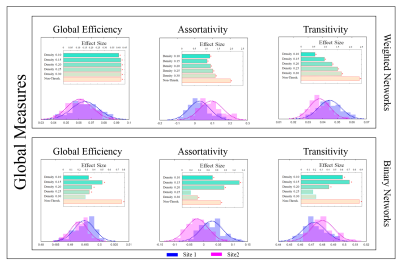
Are Brain Networks Reproducible across Sites?
Abdol Aziz Ould Ismail, Drew Parker, Sriram Moparthy, Birkan Tunç, Ragini Verma
The field of connectomics has introduced new computational tools to the system neuroscience domain, facilitating novel investigations into the mechanisms of the human brain. While methodological advancements in connectomics have found numerous applications with exciting interpretations, a systematic analysis on the reliability and reproducibility of the results and inferences drawn from the findings is still missing. We aim to fill this gap by studying the reproducibility of connectomics findings on diffusion data that has been collected at different sites with varying acquisition protocols. Structural network properties at local, meso, and global scales were investigated to determine what is different and what is preserved in the presence of site related variation. Our comprehensive investigation revealed that despite significant differences between local and global properties, the topological features were mostly preserved.
|
|
5211.
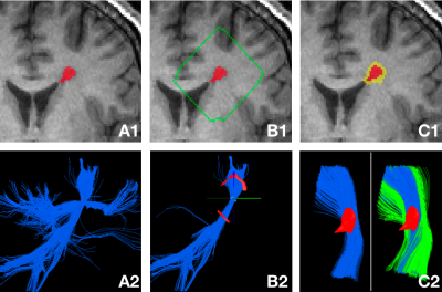 |
12 |
Personalized Lesion Profiling in Multiple Sclerosis
Sidong Liu, Alexander Klistorner, Chenyu Wang, Yang Gao, Brittany Gilchrist, Yang Song, Yuyi You, Junen Yao, Weidong Cai, Michael Barnett
The pathology of multiple sclerosis (MS) lesions is highly subject-specific, therefore personalized lesion analysis is important in assessing the degree of disease-related brain damage and the patient’s response to therapy. Separating and comparing lesional and non-lesional tissue provides an “internal control” and, therefore, increased precision of measurement of the degree of lesional damage in the brain. Here we present a novel and fully automatic method to create a personalized lesion profile of the subject based on DTI tractography, which can be used to assess whole brain lesional damage. This technique can be applied to measure microstructural change using any MRI modality, and may have high translational impact in clinical trials and neurological research.
|
|
5212.
 |
13 |
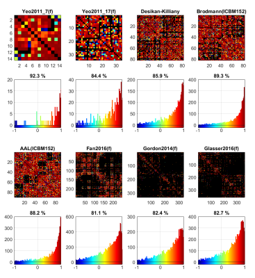
Reproducibility of Human Diffusion Connectomes Generated from Different Atlases: A Multicenter Study
Qiqi Tong, Hongjian He, Peipeng Liang, Ting Gong, Tianyi Qian, Yi Sun, Qiuping Ding, Chen Li, Kuncheng Li, Jianhui Zhong
In a multicenter study, high reproducibility among centers is essential. However, the construction of diffusion connectomes requires multiple complex steps. Errors may be easily hidden in the connectome matrix and possible deteriorate the intrinsic neural network analysis. The registration and parcellation based on the brain template are the final steps and are vital to the connectome matrix. A different anatomical and functional image-based atlas may produce variable reproducibility in diffusion connectome matrices. In this work, seven frequently used atlases were compared to test the reproducibility of different templates.
|
|
5213.
 |
14 |
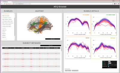
AFQ-Browser: Supporting reproducible human neuroscience research through browser-based visualization tools
Jason Yeatman, Adam Richie-Halford, Josh Smith, Anisha Keshavan, Ariel Rokem
MRI research faces various challenges with regards to reproducibility: scientists are generally aware that data sharing is an important component of reproducible research, but it is not always clear how to share data in a manner that allows other researchers to understand and reproduce published findings. Here we describe AFQ-Browser, a software tool that builds an interactive website as a companion to a diffusion MRI study and leverages web-visualization technologies to create linked views between different aspects of a diffusion MRI dataset (anatomy, diffusion metrics, subject metadata). This facilitates exploratory data analysis, fueling new scientific discoveries based on previously published datasets.
|
|
5214.
 |
15 |
Adaptive linear discriminant analysis for complex networks to study extreme prematurity and intrauterine growth restriction effects at school age
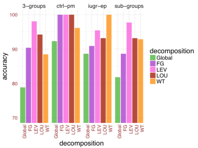
Serafeim Loukas, Djalel Meskaldji, Elda Gomez, Lana Vasung, Dimitri Van De Ville, Petra Huppi
In this study, we combined complex network theory with machine learning in order to grasp potential biomarkers of brain development. The data consists of brain connectomes (brain connectivity matrices) of 53 children aged six years old. For each subject, we estimated brain network-based measures at four different levels: connection, node, module and global levels. Then we applied linear discriminant analysis and support vector machine in order to extract features and we compared their performances. We showed that node and module levels are the best choices to extract relevant and interpretable biomarkers in order to distinguish between different brain development conditions.
|
|
5215.
 |
16 |
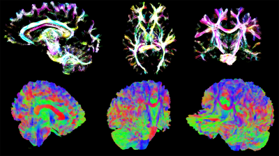
Advances in structural and functional connectivity visualization using the Fibernavigator
Maxime Chamberland, Maxime Descoteaux, Derek Jones
Scientific data visualization is constantly challenged by the continuously growing diffusion MRI (dMRI) field. Exploring and interacting with high-dimensional datasets is central to every analysis pipeline, allowing us to better understand the behavior of a certain tracking algorithm, for example. In this abstract, we present a brief overview of the recent advances in data analysis & visualization available inside the Fibernavigator package.
|
|
5216.
 |
17 |
Enhancing bundle topology for tractography visualization using silhouette rendering
Maxime Chamberland, Derek Jones
Global illumination rendering techniques are often considered the gold standard for displaying 3D structures. They are however computationally expensive and most neuroimaging software packages do not support such advanced methods. Here, we applied cartoon-like shading to tractography-derived bundles to enhance visualization of their topological features. Combined with glass-brain rendering, we believe that this non-realistic rendering technique can help both new comers to the field and experienced scientists to better perceive shape specific features of brain bundles, enhancing both neuroanatomical understanding and neurosurgical planning.
|
|
5217.
 |
18 |
Generation of a muscle fibre orientation atlas of the in vivo tongue
Luuk Voskuilen, Ludi Smeele, Alfons Balm, Ferdinand van der Heijden, Gustav Strijkers, Aart Nederveen
While diffusion tensor imaging based atlases in the brain are common, no muscle fibre atlases for the tongue have been created. An unbiased atlas was generated from ten healthy volunteers, by iteratively registering fibre orientation distributions calculated via constrained spherical deconvolution to a template. The atlas shows good reproducibility and better performance than an image intensity based atlas. Various tongue muscles can be detected in tractography of the atlas. This atlas may facilitate automatic segmentation of tongue muscles after tumour resection to evaluate impaired tongue function.
|
|
5218.
 |
19 |
Functional- and Structural-Connectivity Connectome Fingerprints Correlate With Cognitive Behavior Traits in HCP Datasets
Ying-Chia Lin, Steven H. Baete, Xiuyuan Wang, Fernando E. Boada
Connectome analysis of the human brains structural and functional architecture provides a unique opportunity to understand the organization of brain networks. Recently, connectome fingerprinting using brain functional connectivity profiles as the unique traits was able to identify individuals from the group in the Human Connectome Project (HCP) datasets. In HCP s900 datasets, we extend connectome fingerprinting from functional to structural connectivity, identifying multiple relationships between behavioral traits and brain connectivity.
|
|
5219.
 |
20 |
The Benefits Of Cognitive Training Depend On The Wiring Of The Brain
Karen Caeyenberghs, Claudia Metzler-Baddeley, Peter Wilson, Derek Jones
There is strong evidence that task-specific training leads to changes in brain structure, as assessed using MRI-based techniques that probe microstructure or morphology. In the present study, we want to understand the specific mechanisms of action of task-specific training and identify other critical ingredients. Here, we used a well-established working memory training program and state-of-the art neuroimaging methods in 40 healthy adults. Further research on dose, timing, and duration of training is necessary to generalize the training protocols to the field of structural neuroplasticity.
|
|
5220.
 |
21 |
Automated fibre tractography of the optic radiation for epilepsy surgery planning
Sjoerd Vos, Matteo Mancini, Vejay Vakharia, Rachel Sparks, Mei Chiang, M Cardoso, John Duncan, Gavin Winston, Sebastien Ourselin
Fibre tractography of the optic radiation can be helpful in epilepsy surgery, but its widespread clinical use is hampered by the need for time-consuming expert manual ROI delineations and interrater variability. Our automated approach using anatomically constrained geodesic shape-based averaging is evaluated on two diffusion MRI datasets from different scanners. Automatically generated tracts are within the interrater agreement of two expert raters when evaluating for distance between temporal pole and Meyer’s loop for both datasets, with a tendency towards a more inclusive Meyer’s loop in the automated method.
|
|
5221.
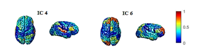 |
22 |
Decomposition of brain structural connectivity using Independent component analysis
Farzaneh Keyvanfard, Abbas Moghaddam, Alessandra Griffa, Patric Hagmann
Diffusion imaging provides the capability of investigating brain white matter non-invasively. There is an increasing interest in studying whole brain structural connectivity (SC) as a complex network, but examining multiple structural sub-networks has yet to be investigated. In this study we have proposed a specific pipeline to decompose whole brain structural connectivity into different sub-networks using Independent Component Analysis (ICA). Obtaining two structural gender related sub-networks in line with previous findings confirms the feasibility of the approach.
|
|
5222.
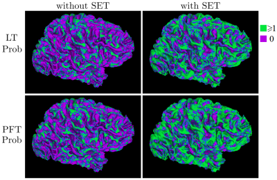 |
23 |
Reducing Tractogram Endpoint Biases with Surface-Enhanced Tractography
Etienne St-Onge, Maxime Descoteaux
In this work, we highlight the importance and advantages of integrating cortical surfaces with dMRI tractography. Doing so facilitates the integration of gray matter (GM) features in white matter (WM) connectivity analysis. Extending streamlines to cortical meshes allows the study of WM structural features from tractography along the cortex. This combined approach also enables the measurement of streamline connections cortical coverage and density bias with different tractography algorithms.
|
|
Microstructure: Modeling
Electronic Poster
Diffusion
Thursday, 21 June 2018
| Exhibition Hall |
08:00 - 09:00 |
| |
|
Computer # |
|
5223.
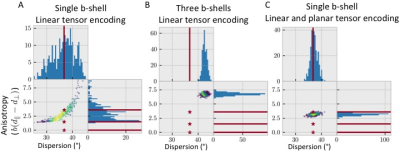 |
25 |
Accurate fibre dispersions from varying the b-tensor shape
Michiel Cottaar, Filip Szczepankiewicz, Matteo Bastiani, Stamatios Sotiropoulos, Markus Nilsson, Saad Jbabdi
Accurate measures of fibre dispersion might improve tractography through voxels with bending and/or fanning configurations and also provide more accurate tissue microstructure indices. When estimated from regular diffusion MRI, however, the accuracy of the dispersion estimate depends on the accuracy of the underlying microstructural model. In this work, we find that encoding water diffusion using multiple shapes of the b-tensor leads to an accurate measurement of fibre dispersion within a single b-shell. We show that this holds for complicated sub-voxel geometries using both simulations and in vivo data.
|
|
5224.
 |
26 |
Intra-axonal Fractional Anisotropy from Fiber Ball Imaging
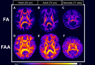
Hunter Moss, Emilie McKinnon, Andreana Benitez, Dorothea Jenkins, G. Glenn, Jens Jensen, Joseph Helpern
Using fiber ball imaging (FBI), we isolate the intra-axonal compartment of the white matter with strong diffusion weighting in order to suppress the extra-axonal water fraction and then calculate its associated fractional anisotropy (FA). We call this parameter the fractional anisotropy axonal (FAA) in contrast to the conventional FA, and we compare these two measures in three subjects: a healthy young adult, a cognitively intact older adult with severe white matter abnormalities, and a neonate with acute hypoxic ischemic injury. Our results indicate that FAA reveals diffusion anisotropy that is not apparent using the conventional FA.
|
|
5225.
 |
27 |
Deep Learning Captures More Accurate Diffusion Fiber Orientations Distributions than Constrained Spherical Deconvolution
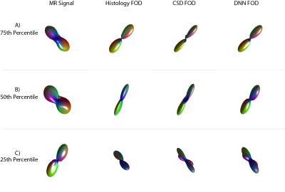
Vishwesh Nath, Kurt Schilling, Prasanna Parvathaneni, Allison Hainline, Colin Hansen, Camilo Bermudez, Andrew Plassard, Justin Blaber, Vaibhav Janve, Yurui Gao, Iwona Stepniewska, Adam Anderson, Bennett Landman
Confocal histology provides an opportunity to establish intra-voxel fiber orientation distributions that can be used to quantitatively assess the biological relevance of diffusion-weighted MRI models, e.g., constrained spherical deconvolution (CSD). Here, we apply deep learning to investigate the potential of single shell diffusion-weighted MRI to explain histologically observed fiber orientation distributions (FOD) and compare the derived deep learning model with a leading CSD approach. This study (1) demonstrates that there exists additional information in the diffusion signal that is not currently exploited by CSD, and (2) provides an illustrative data-driven model that makes use of this information.
|
|
5226.
 |
28 |
Influence of the size and curvedness of neural projections on the orientationally averaged diffusion MR signal
Evren Özarslan, Cem Yolcu, Magnus Herberthson, Hans Knutsson, Carl-Fredrik Westin
We studied the orientationally averaged diffusion weighted MR signal for diffusion along general curves at all three temporal regimes of the traditional pulsed field gradient measurements. We found that long fibers as well as short fibers that are straight could yield the $$$q^{-1}$$$ decay. The absence of such a decay suggests fibers that are short and curvy. We note that the true asymptotic behavior of the signal decay is characterized by the Debye-Porod law, which suggests $$$\bar{E}(q)\propto q^{-4}$$$ at very large $$$q$$$-values. This study is expected to provide insights for interpreting the diffusion-weighted images of the central nervous system.
|
|
5227.
 |
29 |
Estimation of the brain microstructure in the presence of crossing fascicles from a dictionary of Monte Carlo signals
Gaëtan Rensonnet, Benoit Scherrer, Gabriel Girard, Jonathan Rafael-Patino, Simon Warfield, Benoit Macq, Jean-Philippe Thiran, Maxime Taquet
We estimate microstructural features of crossing fascicles using Monte Carlo simulations to represent the diffusion attenuation of each fascicle. The measured diffusion-weighted MRI signal is reconstructed as a sparse weighted sum of pre-computed signals and microstructural properties are estimated from the selected fascicle configurations. The consistent results obtained on various synthetic phantoms as well as on in vivo brain data suggest the potential of our method for the quantitative estimation of microstructural features in fascicle crossings.
|
|
5228.
 |
30 |
Advanced quantification of dispersion and directionality in three-dimensional electron microscopy images and diffusion MR imaging of injured rat brain
Raimo Salo, Ilya Belevich, Eppu Manninen, Eija Jokitalo, Olli Gröhn, Alejandra Sierra
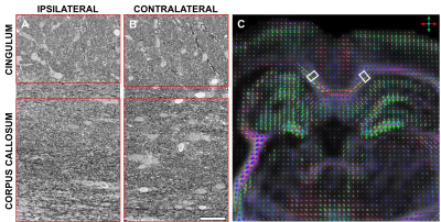
The relationship between orientation distribution functions (ODFs) from advanced dMRI and the real tissue microstructure is under active research. We utilized three-dimensional (3D) serial block-face scanning electron microscopy in the corpus callosum and cingulum, and implemented 3D structure tensor (ST) analysis to produce ODFs comparable to dMRI ODFs from the same locations. The ODFs were parametrized using Watson distribution. Our results showed a clear correspondence in orientation estimates, but dispersion estimates were not as clearly correlated. ST analysis combined with 3D SBEM has great potential to unveil the complexity of the underlying microstructure in biological tissues.
|
|
5229.
 |
31 |
Sensing Von Economo Neurons in the Insula with Multi-shell Diffusion MRI
Did Not Present
Demian Wassermann, Van-Dang Nguyen, Guillermo Gallardo-Diez, Jing-Rebecca Li, Weidong Cai, Vinod Menon
Sensing microstructural characteristics of human brain tissue with clinical scanners has been an area of heated debate in the diffusion MRI (dMRI) community . In this work we propose that diffusion MRI on clinical scanners is sensitive to the presence of Von Economo neurons.
Von Economo neurons, located in the insular and anterior cingular cortices, are large neurons present only in mammals with high cognitive functions. Albeit these neurons' role is not yet known, evidence suggests they facilitate rapid long-range information integration.
In this work, we provide theoretical and in-silico evidence that the dMRI signal is sensitive to the presence of Von Economo neurons as well as preliminary evidence on human dMRI images.
|
|
5230.
 |
32 |
Prediction of Neurite Indices From Diffusion Tensor Imaging in the human cerebral cortex.
Hikaru Fukutomi, Thai Akasaka, Koji Fujimoto, Takayuki Yamamoto, Tomohisa Okada, Kaori Togashi, Takuya Hayashi
Recent studies suggest that Neurite Orientation Dispersion and Density Imaging (NODDI) provides valuable information about cortical neurites. However, it requires lengthy time to acquire multi-shell data of diffusion weighted imaging (DWI), as well as to calculate parameters, neurite density index (NDI) and orientation dispersion index (ODI). We propose a method to estimate cortical NDI and ODI from diffusion tensor imaging (DTI), which is based on a mathematical relationship between NODDI and DTI assuming negligible cerebrospinal fluid (CSF) in the cortex. We also show the accuracy and time for scanning and computation.
|
|
5231.
 |
33 |
Stick, ball, and plane phantoms for validation of diffusion MRI
Karin Bryskhe, Johan Larsson, Dan Lundberg, Greta Eklund, Hong Jiang, Filip Szczepankiewicz, Daniel Topgaard
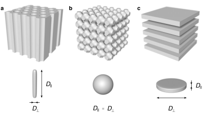
When using quantitative measurements, sensitive to subtle errors, access to a stable reference object is crucial to validate parameter accuracy, and system fluctuations. Here, we propose three complementary phantoms providing ideal systems for validation of metrics of diffusivity, microscopic anisotropy, and compartment orientation. We have used liquid crystals to design phantoms exhibiting local diffusion tensors with normalized anisotropy (DΔ) equal to the theoretical values of +1 (sticks), 0 (balls), as well as –½ (planes). We also confirm that our proposed phantoms have the desired properties with regards to voxel-average diffusion tensors and microscopic diffusion tensors.
|
|
5232.
 |
34 |
Microscopic diffusion anisotropy imaging in the kidneys
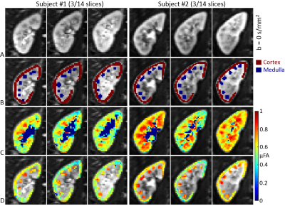
Fabio Nery, Matt Hall, David Thomas, Enrico Kaden, Filip Szczepankiewicz, Isky Gordon, Chris Clark
Diffusion tensor imaging (DTI) is a widely used diffusion weighted imaging (DWI) approach, successful owing to its sensitivity to changes in tissue microstructure. Recent advances in diffusion MRI acquisition using arbitrary b-tensor shapes allow the in-vivo estimation of tissue microscopic fractional anisotropy (μFA), which unlike conventional fractional anisotropy (FA) is not confounded by orientation dispersion. The aim of this work is to investigate, for the first time, the feasibility of μFA quantification in the kidneys of healthy volunteers.
|
|
5233.
 |
35 |
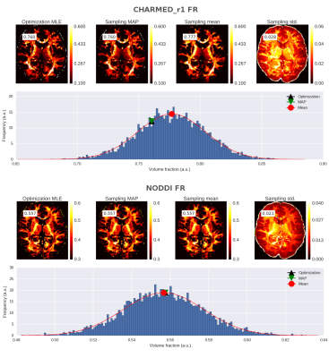
Robust and fast MCMC sampling of diffusion MRI microstructure models
Robbert Harms, Alard Roebroeck
In this work we extend the Maastricht Diffusion Toolbox (MDT) software package with MCMC capabilities, thereby introducing robust and fast sampling of diffusion MRI microstructure models. MDT's object oriented modular design allows arbitrary specification and combination of models, likelihood functions, prior functions and proposal distributions. GPU based computations allow for ~80x faster MCMC sampling; e.g. the 81 volume example NODDI dataset can be sampled with 15000 samples in 1.5 hour, making MDT suitable for probabilistic inference of all implemented models, both dMRI microstructure as quantitative MRI models. The software is open source and freely available at https://github.com/cbclab.
|
|
5234.
 |
36 |
Unified multi-modal characterization of microstructural parameters of brain tissue using diffusion MRI and multi-echo T2 data
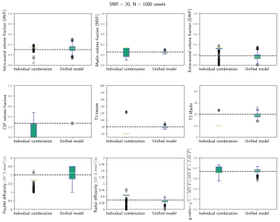
Erick Canales-Rodríguez, Marco Pizzolato, Yasser Alemán-Gómez, Nicolas Kunz, Caroline Pot, Jean-Philippe Thiran, Alessandro Daducci
In this study we propose a theoretical model to estimate different microstructure indices from diffusion MRI and multi-echo T2 (MET2) data. The proposed estimation framework takes into account the common and complementary information provided by both modalities. While the MET2 data allow us to model the myelin compartment, the diffusion data enable us to better characterize the intra-axonal and extra-axonal compartments. Results from numerical experiments support the hypothesis that the new unified estimation is more accurate than the alternative approach based on the individual sequential fitting of both image modalities. The performance was stable for noise levels commonly found in clinical protocols.
|
|
5235.
 |
37 |
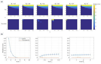
Estimating axon diameter distributions beyond the physical limits of acquisition capabilities
Mark Drakesmith, Suryanarayana Umesh Rudrapatna, Silvia de Santis, Derek Jones
Estimation of axon diameter distributions (ADDs) is hindered by the fact that axon diameters below a certain limit, which are prevalent throughout the nervous system, are invisible to a diffusion MRI protocol. Here we propose a simple modification to the AxCaliber protocol where only the portions of the ADD that can feasibly generate a signal are modelled, by fitting to a truncated distribution. We show in simulations and human data acquired on a high-gradient (300mT/m) system that using this approach produces ADD estimates much closer to those observed in histology.
|
|
5236.
 |
38 |
Microstructure characterisation of fixed prostate tissue using ultra high-resolution diffusion-weighted MRI
Maira Tariq, Andrada Ianus, Nyoman Kurniawan, Gary Cowin, Daniel Alexander , Roger Bourne, Eleftheria Panagiotaki
This work explores ultra-high field microimaging of prostate tissue using diffusion compartment modelling. The acquired ultra-high-resolution data provides a unique opportunity to investigate the accuracy of the diffusion estimates as well as the contribution of various tissue components to the diffusion signal. We compare several multi-compartment models to explore the best microstructure model for prostate tissue. We find plausible results for the data and models used, which will be compared to corresponding histology. Future work will use more samples and extend the models compared to get a comprehensive analysis of the data.
|
|
5237.
 |
39 |
Voxel size matters: big voxels are required to generate realistic extra-axonal dMRI signals from Monte Carlo simulations
David Romascano, Jonathan Rafael-Patino, Ileana Jelescu, Muhamed Barakovic, Tim Dyrby, Jean-Philippe Thiran, Alessandro Daducci
Monte Carlo simulations provide diffusion MRI signals that can be used to evaluate microstructure models, but that can also be incorporated into microstructure reconstruction methods. It is therefore important for the generated signals to be as realistic as possible. This work shows preliminary evidence that, in the case of white matter models, the symmetry of the perpendicular extra-axonal signal generated with Monte Carlo simulations depends on the voxel size. Simulations corresponding to millimeter-sized voxels should therefore be computed using substrates of equivalent size, or by averaging signals generated from multiple small voxels.
|
|
5238.
 |
40 |
Deep learning based segmentation of cardiomyocytes to aid numerical simulations of diffusion cardiovascular magnetic resonance
Jan Rose, Wee Zhao Chua Khoo, Sonia Nielles-Vallespin, Pedro Ferreira, David Firmin, Andrew Scott, Denis Doorly
To better understand how the underlying microstructure and pathology affect the DT-CMR signal in vivo, more realistic numerical models that account for irregular myocyte configurations such as sheetlets are necessary. We manually segmented cardiomyocytes from pig histology and confirmed that the resulting substrate is representative of the local microstructure through automatic segmentation of the surrounding tissue with a convolutional neural network. Monte Carlo random walk simulations, covering short and long mixing times and varying compartment diffusivities, show a mismatch between the results for the histology-based substrate and a simple cuboid model with comparable ECV and mean cell size.
|
|
5239.
 |
41 |
Diffusion Weighting with Linear and Planar Encoding Solves Degeneracy in Parameter Estimation
Bibek Dhital, Marco Reisert, Elias Kellner, Valerij Kiselev
While estimating parameters of tissue microstructure models has never been simple, the recently discovered bi-modality (degeneration) of objective function in parameter space render it highly challenging. This study proposes a response to this challenge based on combining the commonly used linear encoding (the b-matrix with the diagonal 0,0,b) with the planar encoding (b,b,0). The signal was simulated in the whole parameter space of the standard model of brain white matter. Exhaustive search in this space demonstrates the absence of bi-modality when results for both encoding schemes are treated together.
|
|
5240.
 |
42 |
Constrained analysis of b-tensor encoding diffusion data with multiple echo times allows estimation of compartment-specific T2 relaxation times in white matter.
Björn Lampinen, Filip Szczepankiewicz, Daniel Topgaard, Oskar Hansson, Danielle van Westen, Markus Nilsson
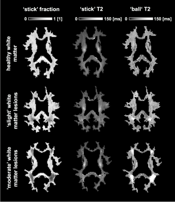
We used b-tensor encoding and multiple echo times to estimate the separate T2 relaxation times and apparent fractions of white matter compartments. Nineteen elderly subjects were imaged, and data were analyzed using a constrained ‘ball-and-stick’ diffusion-relaxation model. Results show that the ‘ball’ T2 relaxation time is inversely related to the fraction of ‘sticks’ in white matter lesions, and that the ‘stick’ T2 relaxation time may be sensitive to the axonal diameter. The approach could be useful to characterize white matter damage.
|
|
5241.
 |
43 |
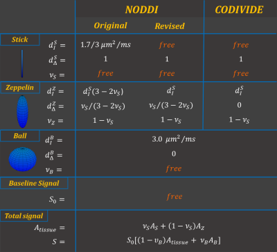
Revised NODDI model for diffusion MRI data with multiple b-tensor encodings
Michele Guerreri, Filip Szczepankiewicz, Björn Lampinen, Markus Nilsson, Marco Palombo, Silvia Capuani, Hui Zhang
This work proposes a revision of the NODDI model to relate brain tissue microstructure to the new generation of diffusion MRI data with multiple b-tensor encodings. NODDI was developed originally for conventional multi-shell diffusion data acquired with linear tensor encoding (LTE). While adequate for LTE data, it has been shown to be incompatible with data using spherical tensor encoding (STE). We embed a different set of assumptions in NODDI, while retaining the tortuosity constraint, to accommodate both LTE and STE data. Experiments with human data with multiple b-tensor encodings confirm the efficacy of the revision.
|
|
5242.
 |
44 |
Isotropic Diffusometry MRI of the human brain
Alexandru Avram, Joelle Sarlls, Peter Basser
From diffusion MRI data acquired with isotropic diffusion encoding over a wide range of b-values, we estimate intravoxel distributions of mean diffusivities using a regularized Inverse Laplace Transform analysis. In vivo spectra of intra-voxel mean diffusivities measured in healthy subjects show consistent single-peak distributions in most brain regions, with subtle differences between white matter, cortical gray matter, subcortical gray matter, corpus callosum, and cerebrospinal fluid. This non-invasive, model-free, whole-brain approach to quantifying spectra of intrinsic tissue water mobilities could improve the clinical diagnosis and characterization of stroke, cancer, and other diseases.
|
|
5243.
 |
45 |
Quantification of white matter pathologies by a diffusion MRI technique modeling both intra- and extra-axonal compartments
Video Permission Withheld
Chunyu Song, Tsen-Hsuan Lin, Peng Sun, Sheng-Kwei Song
A new diffusion MRI histology (D-Histo) is proposed to model both intra- and extra-axonal diffusion as well as isotropic diffusion components within an image voxel. Both Monte-Carlo simulation, in vivo MRI of EAE mouse optic nerve, and post-MRI histology were used to validate this approach. The intra-axonal fraction derived from D-Histo more accurately assessed fiber fraction, and the intra-axonal axial diffusivities (AD) it derives more sensitively reflected axonal injury.
|
|
5244.
 |
46 |
Axon Diameter Mapping Independent of Crossing Structures using Spherical Mean Technique
Qiuyun Fan, Aapo Nummenmaa, Thomas Witzel, Ned Ohringer, Qiyuan Tian, Lawrence Wald, Susie Huang, Eric Klawiter
Current approaches to axonal size estimation by diffusion MRI assume a single fiber bundle, which limits its application to a few white matter tracts in the healthy human brain. We introduce a new approach to per-voxel axon diameter and volume fraction estimation inspired by the spherical mean framework that is robust to fiber crossings and orientation dispersion. We use this technique to estimate whole brain axon diameter and volume fraction in 6 healthy subjects scanned on the 3T Connectome scanner and demonstrate the utility of this approach to characterize white matter pathology in a patient with multiple sclerosis.
|
|
5245.
 |
47 |
Assessing feasibility and reproducibility of a bundle-specific framework on in vivo axon diameter estimates at 300mT/m
Muhamed Barakovic, Gabriel Girard, David Romascano, Jonathan Rafael-Patino, Maxime Descoteaux, Giorgio Innocenti, Derek Jones, Jean-Philippe Thiran, Alessandro Daducci
In vivo quantitative estimation of axon diameter in the white matter is a potential new tool for studying the structural and functional architecture of the brain. Recently, the feasibility of axon diameter estimation with diffusion-weighted MRI (DW-MRI) has been questioned. In this work, we explore the feasibility of bundle-specific axon diameter mapping in the context of a reproducibility study using the Convex Optimization Modeling for Microstructure informed Tractography (COMMIT) framework. Our results show that DW-MRI axon diameter estimatesof the corpus callosum and of the corticospinal tract are comparable to histological reports in previous studies.
|
|
5246.
 |
48 |
Mapping Normative Mean Apparent Propagator Derived Microstructural Parameters
Adam Bernstein, Alexandru Avram, Amber Simmons, Martin Cota, Neville Gai, Neekita Jikaria, Anita Moses, Christine Turtzo, Lawrence Latour, Dzung Pham, John Butman, Peter Basser
This work establishes preliminary data as well as a processing pipeline to generate normative MAP-MRI parameter maps. This normative data can be used to better understand the values and variation of different diffusion-derived microstructural parameters found in healthy humans. This information can then be used as a baseline for future research or clinical projects.
|
|
Diffusion MRI: Acquisition & Reconstruction
Electronic Poster
Diffusion
Thursday, 21 June 2018
| Exhibition Hall |
09:00 - 10:00 |
| |
|
Computer # |
|
5319.
 |
1 |
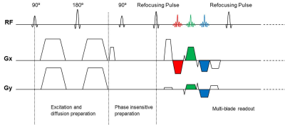
Distortion-free diffusion imaging with SMS PROPELLER DUO
Ali Ersoz, Graeme McKinnon
Although fast spin echo based diffusion-weighted imaging methods provide distortion-free images, they suffer from prolonged scan times. In this work, we incorporated simultaneous multi-slice (SMS) techniques into PROPELLER DUO for accelerated distortion-free diffusion imaging. Results show that SMS PROPELLER DUO can reduce scan time dramatically which can make it a feasible option for clinical applications.
|
|
5320.
 |
2 |
Non-CPMG PROPELLER diffusion imaging: comparison of phase insensitive preparation with split acquisition
Kun Zhou, Wei Liu, Shi Cheng
Turbo spin echo (TSE) based diffusion weighted (DW) PROPELLER imaging has the advantage of reduced sensitivity to B0 inhomogeneity and T2 decay caused image blurring, comparing to DW-EPI sequence. However, the violation of CPMG condition is a challenge for DW-PROPELLER sequences. Phase insensitive preparation and split acquisition methods can be used to handle the non-CPMG problem. A comparison study of these two methods was performed and the results show that the phase insensitive method has superior image quality while split acquisition method has the advantage of speed and lower SAR.
|
|
5321.
 |
3 |
Multiband Diffusion-Weighted MRI of the Eye and Orbit Free of Geometric Distortions Using a RARE-EPI Hybrid
Katharina Paul, Till Huelnhagen, Sebastian Schmitter, Oliver Stachs, Thoralf Niendorf
This study shows that multiband diffusion-weighted RARE-EPI has the capability to acquire distortion-free images of the eye and orbit with ample diffusion contrast for slices in close proximity. The results underpin the challenges of ocular imaging at 3.0 T and 7.0 T for echo planar imaging and demonstrate that these issues can be offset by using accelerated RARE based approaches. This benefit can be exploited for the assessment of spatial arrangements of the eye segments and their masses with the goal to provide guidance in diagnostic assessment and treatment of ophthalmological diseases.
|
|
5322.
 |
4 |
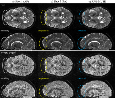
Distortion-Free High-Resolution Diffusion MRI with RPG-MUSE
Iain Bruce, Christopher Petty, Allen Song
Diffusion-weighted images acquired with multi-shot EPI at high-resolution are susceptible to inter-shot motion artifacts and geometric distortions resulting from magnetic-field inhomogeneities. This study presents an efficient means of inherently accounting for both motion and distortion artifacts by alternating the phase encoding gradient polarities in odd/even shots of multi-shot EPI. When acquired in this fashion, the proposed RPG-MUSE model simultaneously accounts for both shot-to-shot motion induced artifacts and geometric distortions during the reconstruction of multi-shot diffusion weighted images. This technique requires no additional scan time, and accounts for B0 and eddy current induced distortions specific to each diffusion-weighted volume.
|
|
5323.
 |
5 |
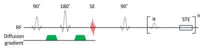
Diffusion-weighted MRI of the prostate without susceptibility artifacts: Multi-shot STEAM with radial undersampling
Andreas Merrem, Klaus-Dietmar Merboldt, Jakob Klosowski, Jens Frahm
Undersampled radial STEAM MRI allows for diffusion-weighed imaging without susceptibility artifacts. Here, this technique was developed for applications to the prostate by moving to a multi-shot acquisition and reconstruction method to optimally process data with limited SNR. Numerical simulations defined the conditions for accurate ADC measurements. In vivo studies of healthy subjects resulted in ADC values in the central gland of the prostate which are consistent and in agreement with previous literature values. A comparison of DW STEAM and DW EPI of the prostate confirmed the major benefit of STEAM sequences which are free of susceptibility-induced distortions.
|
|
5324.
 |
6 |
Improved signal-to-noise ratio with highly asymmetric STEAM-EPI (HASTEAM-EPI)
Manoj Shrestha, Ulrike Nöth, Ralf Deichmann
Several methods exist for increasing the signal-to-noise ratio (SNR) in high spatial resolution imaging, e.g., by using reduced field-of-view techniques or reduced echo-times (TE). Here, a novel module dubbed “highly asymmetric STEAM" (HASTEAM) is proposed, where the stimulated echo (STE) occurs prior to the EPI readout and thus maintains a constant duration of STE preparation, independent of the acquisition matrix-size. This shortens TE and allows for higher SNR, in particular for high spatial resolutions. SNR gain of HASTEAM over the standard STEAM is simulated and compared to in vivo results for different resolutions in structural and diffusion-weighted imaging.
|
|
5325.
 |
7 |
Comparison of Different Methods for Motion-induced Data Corruption Detection Using k-space Information in Diffusion Imaging
Zhe Zhang, Hua Guo, Zhangxuan Hu, Yaou Liu, Yilong Wang, Chun Yuan
In diffusion imaging, subject motion together with diffusion encoding gradient may introduce data corruption. Several methods for corrupted data detection using k-space information such as DC peak amplitude, entropy and signal distribution metric have been proposed, and the detection directly from acquired k-space enables instant data rejection and re-acquisition. This work compared and evaluated these methods using the same single-shot data set. The results show that all methods can successfully detect corrupted shots, and demonstrate good detection consistency with each other and also with ADC measurement.
|
|
5326.
 |
8 |
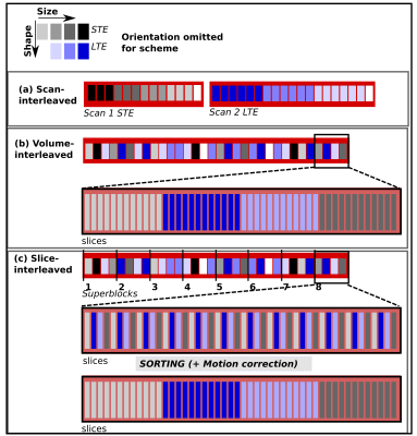
Highly efficient diffusion MRI by Slice-interleaved Free-waveform Imaging (SIFI)
Jana Hutter, Markus Nilsson, Daan Christiaens, Torben Schneider, Anthony Price, Joseph Hajnal, Filip Szczepankiewicz
Diffusion encoding along multiple directions in a single shot facilitates probing of tissue microstructure that is not accessible with conventional (linear) tensor encoding. However, it tends to engage gradients on multiple axes in a pattern that yields higher energy consumption, which can become a critical limiting factor for gradient system performance. Here, we show that Slice Interleaved Free-waveform Imaging (SIFI) of b-tensor size, orientation, and shape reduces peak power consumption and heating, which translates to markedly reduced repetition time, shorter examination times and higher temporal SNR.
|
|
5327.
 |
9 |
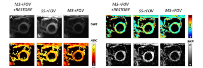
Efficient mutli-slice reduced field of view cardiac T2-ADC mapping with a restore pulse
Kévin Moulin, Eric Aliotta, Daniel Ennis
Slice-following significantly reduces the scan time in cardiac diffusion weighting imaging by enabling free-breathing acquisition compatible with multi-slice coverage. However reduced field of view techniques result in a significant SNR penalty when combined with this multi-slice strategy. The objective of this work was to develop and evaluate a reduce field of view T2+ADC protocol compatible with multi-slice acquisition by using a “restore” pulse to improve higher SNR-efficiency.
|
|
5328.
 |
10 |
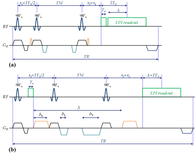
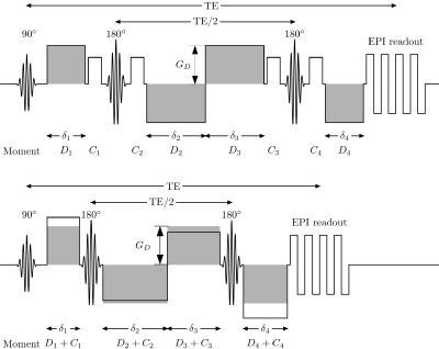
Combined Crushers: Omitting Separate Crusher Gradients in the Twice-Refocused Spin Echo Diffusion MRI Sequence
Richard Buschbeck, Seong Dae Yun, Markus Zimmermann, Ezequiel Farrher, N. Jon Shah
A new method is proposed that allows for the omission of crusher gradients in the Twice-Refocused Spin Echo (TRSE) sequence. The proper spoiling of the unwanted signal pathways is achieved by modifying the amplitudes and durations of the diffusion gradients. Both the crusher moment as well as the desired diffusion weighting is eventually provided only by the diffusion gradients. This results in several advantages like shorter minimum TEs and higher SNRs, all of which can be beneficial in diffusion MRI.
|
|
5329.
 |
11 |
Flow-compensated IVIM model: accuracy and precision as function of SNR, b-values and perfusion fraction
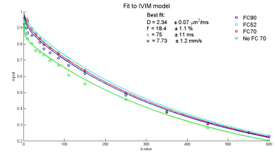
Susanne Rauh, Oliver Gurney-Champion, Uwe Oelfke, Frederik Laun, Andreas Wetscherek
To use intravoxel incoherent motion (IVIM) modelling for clinical applications, the model parameters need to be determined accurately and precisely, which was found challenging. We studied the influence of signal-to-noise ratio (SNR), number of b-values and perfusion fraction on the accuracy of the recently introduced flow-compensated IVIM model parameters (tissue diffusivity, perfusion fraction, characteristic timescale and blood flow velocity). Simulations were performed for typical parameters obtained in healthy volunteers for pancreas and kidney and revealed that for an SNR of 20, 9 b-values and a perfusion fraction of more than 15% are needed for reliable parameter estimation in flow-compensated IVIM.
|
|
5330.
 |
12 |
Simultaneous IVIM and kurtosis acquisition in brain tumours: a trace-based protocol validated in a hybrid MR-PET system
Ricardo Loução, Ana-Maria Oros-Peusquens, Karl-Josef Langen, Hugo Ferreira, N. Jon Shah
Diffusion weighted imaging such as intravoxel incoherent motion (IVIM) and non-Gaussian diffusion (NG-diff) allow the non-invasive study of tissue perfusion and are able to infer the organisation of the microstructure, respectively. Both are highly relevant in pathology characterisation, but are often acquired separately. Here we propose a unified IVIM/NG-diff protocol and apply it to brain tumour patients undergoing preoperative or follow-up scans in a hybrid MR-PET environment. The results agree well with previously reported values using standard techniques. Furthermore, indication for tumour grading power of IVIM parameters was found.
|
|
5331.
 |
13 |
Randomized-Slice Simultaneous Multi-Slice for Diffusion Kurtosis Imaging
Daniel Olson, Volkan Emre Arpinar, Andrew Nencka, L. Tugan Muftuler
Simultaneous Multi-slice (SMS) techniques excite multiple slices simultaneously to accelerate MRI data acquisition. However, slice separation during image reconstruction is not exact and results in coupling between separated voxels. While this may not be critical for most anatomic imaging methods, small but consistent leakage of information from another slice in a DKI dataset will cause bias in diffusion parameter estimates. Here, we implement a randomized-slice pairing technique to alleviate this problem in diffusion MRI acquisitions.
|
|
5332.
 |
14 |
Multi-Shot Diffusion-Weighted EPI Reconstruction Based on Locally Low-Rank Constraints
Xin Ma, Mengye Lyu, Yilong Liu, Ed Wu
Based on the locally low rank (LLR) property of multi-shot diffusion-weighted imaging (DWI), a novel reconstruction scheme is proposed to achieve high signal-to-noise ratio (SNR) without explicit estimation of inter-shot phase variation. The results from in vivo DWI data indicate that the proposed method can effectively improve the image quality at various shot numbers when compared with traditional shot-by-shot reconstruction approaches.
|
|
5333.
 |
15 |
SPIRiT-Based Reconstruction of Interleaved Multi-shot EPI (SPECTRA) for Navigator-Free Diffusion Weighted MRI
Gaojie Zhu, Hai Luo, Xiang Zhou, Meining Chen, Chao Wang, Wei Bian, Ziyue Wu
Multi-shot EPI can achieve a high imaging resolution for Diffusion weighted MRI (DWI) by sampling a large k-space without increasing the degree of T2* blurring. However, its shot-to-shot phase error has to be corrected. Recently, a navigator-free correction method based on SENSE technique has been developed, assuming the phase error is spatially smooth among different DWI shots. In this work, we propose a SPIRiT-based method to correct the phase error and iteratively reconstruct navigator-free multi-shot EPI. Our results suggest it has the advantage of insensitivity to coil map bias and better SNR compared to the SENSE-based method.
|
 |
5334.
 |
16 |
Eddy current characterization and correction using field monitoring for diffusion weighted imaging with maximal strength of 250 mT/m
Yi-Cheng Hsu, Ying-Hua Chu, Thomas Witzel, Qiuyun Fan, Fa-Hsuan Lin
We used a 24-channel field probe array to characterize the eddy current in a connectome scanner during diffusion weighted imaging with the maximal strength of 250 mT/m. Image distortion can be reduced by field monitoring but noticeable artifact remained after characterizing the eddy by up the 3rd-order spherical harmonics. We also found that eddy current lasted longer than 1 s. Our results suggested that the connectome scanner is not a linear time-invariant system, where concurrent field monitoring and high-order spherical harmonics characterization are desired.
|
|
5335.
 |
17 |
The importance of constraints and spherical sampling in diffusion MRI
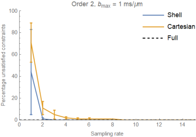
Tom Dela Haije, Aasa Feragen
In this work we analyze the incidence of voxels with physically impossible model parameters, reconstructed from diffusion-weighted data that is acquired using different sampling schemes. Our results show that for cumulants up to order $$$4$$$ constrained least squares can be used to compute a reliable reconstruction of the cumulant expansion of the signal from realistic acquisitions, with spherical sampling producing fewer unsatisfied model constraints compared to space-filling sampling. Voxels where reconstruction is likely to fail are shown to be consistently localized near the white matter-gray matter interface and in deep brain structures.
|
|
5336.
 |
18 |
Phase Inconsistency Compensated Multi-shot Diffusion Image Reconstruction with Locally Low-Rank Constraint
Ziwu Zhou, Fei Han, Yu Gao, Yingli Yang, Peng Hu
Multi-shot diffusion imaging is a promising technique to achieve high-resolution visualization of the microstructure of the issue. However, phase variations induced by physiological motion among different shots remains to be a challenge. To improve the reconstruction accuracy, we propose a novel reconstruction method that integrates the information of phase variation within a compressed sensing framework, and exploits the data redundancy using locally low-rank constraint after phase inconsistency is taken care of.
|
|
5337.
 |
19 |
Model-Based Iterative Reconstruction for in vivo Diffusion Quantification
Video Permission Withheld
Jannik Arbogast, Anna-Katinka Bracher, Meinrad Beer, Henning Neubauer, Volker Rasche
A model-based iterative reconstruction of the apparent diffusion coefficient (ADC) is introduced enabling the quantification of diffusion properties from undersampled DWI data of human knee joints. The approach uses an underlying model function to synthesize k-space data containing crucial phase information. A NLCG is implemented for ADC reconstruction comparing synthetic and measured k-space data. In vivo data of nine subjects show up to 3.5-fold acceleration of the DWI sequence without losing substantial accuracy of the ADC estimates.
|
|
5338.
 |
20 |
A POCS-Enhanced k-Space Reconstruction for 3D Multi-Slab Diffusion Imaging
Erpeng Dai, Xiaodong Ma, Zhe Zhang, Yu-hsuan Wu, Wenchuan Wu, Karla Miller, Chun Yuan, Hua Guo
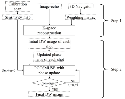
3D multi-slab acquisition is a new technique for high-resolution isotropic diffusion imaging with sufficient SNR maintained. To fill the whole 3D k-space, multi-shot acquisitions are usually needed. However, this can cause the inter-shot phase variations. In this study, we propose a new k-space-based phase correction method for multi-slab diffusion imaging. This proposed method is based on a previous method termed GRAPPA with compact kernel (GRAPPA-CK). Furthermore, POCSMUSE algorithm is subsequently used to enhance the reconstruction of GRAPPA-CK. Finally, the newly proposed method is demonstrated to be compatible with in-plane acceleration in 3D multi-slab imaging.
|
|
5339.
 |
21 |
Novel Image-based Nyquist Ghost Correction of Diffusion-Weighted Echo Planar Imaging using Ghost/Object Minimization
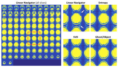
Jessica McKay, Steen Moeller, Sudhir Ramanna, Edward Auerbach, Gregory Metzger, Justin Ryder, Kamil Ugurbil, Essa Yacoub, Patrick Bolan
Diffusion weighted imaging (DWI) acquired with SE-EPI is subject to Nyquist ghosts, which are commonly corrected using a three-line navigator. However, several alternative ghost correction strategies do not require any reference acquisition. These methods define a cost function that is minimized when the image is ghost-free. We propose a novel referenceless method called ghost/object minimization by defining the cost function as the summation over the image divided by its ½-FOV-shifted counterpart. In this work, we test noise sensitivity and demonstrate the feasibility of the ghost/object minimization using simulated ghosts and in vivo acquisitions including brain, prostate, breast, and liver DWI.
|
|
5340.
 |
22 |
Accelerating Intravoxel Incoherent Motion Mapping in the Brain using k-bpp PCA
Georg Spinner, Johannes Schmidt, Sebastian Kozerke
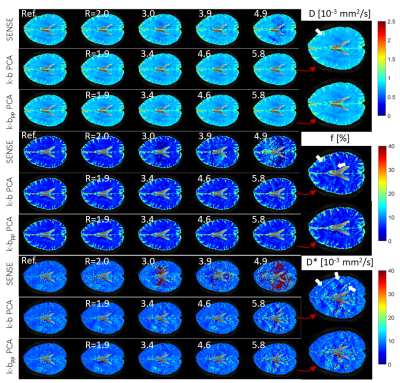
Intravoxel Incoherent Motion (IVIM) parameter mapping yields quantitative information about diffusion and pseudo-perfusion properties. In brain IVIM, parallel imaging with two-fold undersampling has been reported. In order to address noise amplification and unfolding artifacts at undersampling factors greater than two with parallel imaging, reconstruction in spatio-principal component space with data reshuffling (k-bpp PCA) is presented here. Using in-vivo brain data, it is demonstrated that k-bpp PCA allows for significantly increased undersampling factors relative to parallel imaging while maintaining comparable image quality and parameter estimation errors.
|
|
5341.
 |
23 |
Comparison of Navigator-free Multi-shot Diffusion-Weighted Imaging Reconstruction Algorithms for EPI
Malte Steinhoff, Kay Nehrke, Peter Börnert
Multi-shot diffusion-weighted MRI allows for higher spatial resolution than single-shot approaches, but suffers from image artifacts due to motion-induced shot-specific phase variations. Advanced parallel imaging reconstructions can mitigate these effects, but a comparison of these approaches is still missing, especially for the widespread EPI. Following the concept of reproducible research, a number of algorithms were implemented, adapted and refined for EPI, and compared in simulations and in-vivo. Results show different performance and applicability. The iterative feedback and the sharing of joint global information in the reconstruction and appropriate constraints were found to be crucial for high-quality high-resolution DWI.
|
|
5342.
 |
24 |
A diffusion-matched principal component analysis (DM-PCA) based denoising procedure for high-resolution diffusion-weighted MRI
Nan-kuei Chen, Hing-Chiu Chang, Ali Bilgin, Adam Bernstein, Theodore Trouard
A concern with high-resolution DWI and DTI is the limited SNR. Here we report a new denoising procedure, termed diffusion-matched principal component analysis (DM-PCA), which comprises 1) identifying a group of voxels with very similar signal variation patterns along the diffusion dimension, 2) performing PCA along the diffusion dimension for those voxels, and 3) suppressing noisy PCA components. The DM-PCA method performs reliably for input data with a range of SNR and different numbers of diffusion encoding scans, without compromising anatomic resolvability, and should prove highly valuable for imaging studies in research and clinical uses.
|
|
Diffusion MRI: Post-Processing
Electronic Poster
Diffusion
Thursday, 21 June 2018
| Exhibition Hall |
09:00 - 10:00 |
| |
|
Computer # |
|
5343.
 |
25 |
A framework for b-value optimization in DWI
Rick Keesman, Leon ter Beek, Uulke van der Heide
Diffusion-weighted imaging is a valuable asset in the diagnosis of a plethora of diseases. General guidelines are available when measuring (mono-exponential) diffusion in several organs of interest, but there is less consensus on optimal acquisition parameters for more complex models (e.g., intravoxel incoherent motion, kurtosis). We provide a general, easy to implement, solution scheme (similar to the Cramér–Rao lower bound approach) that can be used to optimize the acquisition protocol with respect to the $$$b$$$-values. We present mathematical proof, and phantom as well as in-vivo measurements that corroborate the viability of our method.
|
|
5344.
 |
26 |
Investigation of the Effect of Body Posture on Dynamic ADC Change during Cardiac Cycle in Human Brain
Video Permission Withheld
Naoki Ohno, Tosiaki Miyati, Masatomo Uehara, Yuki Hiramatsu, Riho Okamoto, Mitsuhito Mase, Satoshi Kobayashi, Toshifumi Gabata
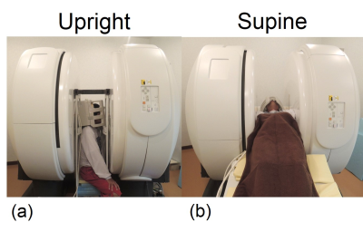
Intracranial conditions are strongly affected by body posture. Apparent diffusion coefficient (ADC) of the brain significantly changes during the cardiac cycle, and this change (ΔADC) reflects intracranial condition. However, the effect of body posture on ΔADC has yet to be confirmed. Therefore, we evaluated the ΔADC and ADC of the brain in upright and supine postures using a multi-posture MRI. ΔADC and ADC of the white matter in the upright posture were significantly higher than those in the supine posture. Multi-posture MRI facilitates the noninvasive evaluation of the effect of gravity on intracranial conditions.
|
|
5345.
 |
27 |
GPU Accelerated Maximum Likelihood Estimation of Diffusion and Kurtosis Tensors with the Rician Noise Model
Viljami Sairanen, Jia Liu, Dario Gasbarra
Diffusion- and kurtosis tensor estimators generally assume independent and a Gaussian distributed noise. While with adequate signal-to-noise ratios (SNR) this assumption can be made without significant bias in the estimated models, this is not true when utilizing high diffusion weighting thus low SNR. In such case, the Rician noise should be considered with maximum likelihood (ML) estimators for example. Moreover, the mathematical properties of ML estimators could unveil novel details of the underlying diffusion process within the brain. In this work, we present super-fast, GPU accelerated ML estimator with Matlab interface to provide a practical tool for such improved estimation.
|
|
5346.
 |
28 |
Rebooting diffusion MRI uncertainty distributions in the presence of outliers with ROBOOT
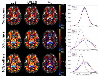
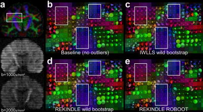
Viljami Sairanen, Derek Jones, Alexander Leemans, Chantal Tax
Characterizing uncertainty distributions in diffusion MRI derived metrics such as fractional anisotropy or kurtosis anisotropy requires non-parametric approaches, since the correct form of the distribution is rarely known a priori. Previously suggested wild bootstrapping methods, however, have not considered the impact of outliers in the data. In this work, we updated the existing wild bootstrap methodology to consider outliers detected by a robust model estimator, adopting a strategy similar to the rejection of the outliers prior to the model estimate. Additionally, we used simulations based on real human data to demonstrate the benefits of our pipeline for recovering uncertainty distributions.
|
|
5347.
 |
29 |
Automated Pattern Recognition Analysis of Intravoxel Incoherent Motion (IVIM)-diffusion to Segment Hypo-perfused Stroke Region
MinJung Jang, SoHyun Han, HyungJoon Cho
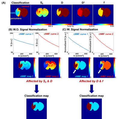
The variation in fitted perfusion factors (f) from different fitting models and the necessary parameter thresholdings are one of the challenges in the robust interpretation of intravoxel incoherent motion (IVIM) diffusion data [1-3]. The purpose of this study is to investigate the feasibility of applying constrained nonnegative matrix factorization (cNMF) to IVIM-diffusion data for the automatic segmentation of hypo-perfused stroke region of rat brain without any model-fittings nor thresholdings.
|
|
5348.
 |
30 |
Digital DWI phantoms for non-Gaussian diffusion with Rician noise
Dariya Malyarenko, Yuxi Pang, Thomas Chenevert
Multi-parametric diffusion models describe non-Gaussian diffusivity of tissue observed by quantitative diffusion weighted imaging (qDWI). The ground-truth values for assumed model parameters are useful for both testing the analysis algorithms and optimizing qDWI acquisition. These values can be provided by virtual phantoms, termed digital reference objects (DROs), based on forward modeling. Here we describe a tool to generate DWI DICOM DROs with varying diffusivity model parameters and b-values including physical noise. Application examples are shown for Kurtosis, Stretched Exponential and Perfusion-fraction Intra-Voxel Incoherent-Motion DWI DROs to quantify noise-induced bias and variation in derived qDWI parametric maps.
|
|
5349.
 |
31 |
Impact of scan and fitting parameters on the accuracy of parameter maps in IVIM-diffusion-MRI of the knee joint
Anna-Katinka Bracher, Meinrad Beer, Volker Rasche, Henning Neubauer
Intravoxel Incoherent Motion (IVIM) imaging has proven its potential for combined diffusion and perfusion imaging of synovitis in the knee joint. In this abstract the impact of the maximum B-value Bmax and the threshold B-value Bcutoff - which separates the B-values for the fitting function in perfusion and non-perfusion part - were investigated. The parameter maps show high dependency on Bcutoff and only slight variations on Bmax.
|
|
5350.
 |
32 |
Diffusion and Perfusion Analysis using Intravoxel Incoherent Motion (IVIM): A Twenty Patient Study with Osteosarcoma
Video Permission Withheld
Esha Baidya Kayal, Devasenathipathy Kandasamy, Kedar Khare, Raju Sharma, Sameer Bakhshi, Amit Mehndiratta
Quantitative analysis of Intravoxel incoherent motion (IVIM) effect of diffusion weighted imaging can assess both diffusion and perfusion component of tissue separately. To the best of our knowledge, IVIM analysis in primary bone tumors has not been reported yet and no reference for IVIM parameter values in bone tumors is available for simulation study. We estimated quantitative IVIM parameters using state-of-the-art IVIM analysis method – Bi-exponential model with adaptive Total Variation penalty function (BE+TV) in a cohort of twenty patients with Osteosarcoma that is useful for IVIM analysis in bone tumors for prognosis and can serve as standard reference for future studies.
|
|
5351.
 |
33 |
Features extracted from diffusion-driven tensor based morphometry can serve as a specific imaging marker for Moebius Syndrome
Neda Sadeghi, Irini Manoli, Timothy Wood, Francis Collins, Ethylin Jabs, Elizabeth Engle, Moebius Syndrome Research Consortium, Carlo Pierpaoli
Quantitative diffusion derived metrics such as fractional anisotropy (FA), and Trace of diffusion tensor (TR) have been used in many studies to assess differences between a subject group and a control group. In this study, in addition to FA and TR, we also look at morphological differences measured by diffusion-driven tensor based morphometry (DTBM). We use DTBM to extract features for use in classification of Moebius syndrome subjects, a rare birth defect characterized by paralysis or weakness of facial muscles and impairment of ocular abduction.
|
|
5352.
 |
34 |
Brain q-space imaging: Mean displacement measurement by SMS, grid sampling, and multi-shell QSI
Koji Sakai, Hiroyasu Ikeno, Hiroshi Imai, Kentaro Akazawa, Masashi Yasuike, Hitomi Nagano, Kei Yamada
The comparisons of mean displacement (MD) from multi-shell QSI (msQSI) and grid sampling QSI (gsQSI) were executed by anisotropic diffusion phantom and human brain. In addition, the effect of the number of slices excited and acquired simultaneously (SMS factor) we also investigated. The MDs of msQSI and gsQSI were different. The shorter diffusion time on gsQSI might effect these differences. SMS factors on msQSI showed difference among the acceleration. On contrary, SMS factors on gsQSI did not show any difference among the acceleration.
|
|
5353.
 |
35 |
Physiological noise at low diffusion weighting reduces repeatability of apparent diffusion coefficient independent of underlying diffusion curve characteristics
Neil Jerome, Igor Vidic, Liv Egnell, Torill Sjøbakk, Agnes Østlie, Hans Fjøsne, Tone Bathen, Pål Erik Goa
The apparent diffusion coefficient is a powerful imaging biomarker, sensitive to microstructure properties, and possessing excellent repeatability. Exclusion of perfusion influence (b<150 s.mm-2) reflects true diffusivity, although fewer data points reduces precision, and thus repeatability. We investigate repeatability of experimental breast diffusion data, and show an unexpected increased repeatability with low b-value data inclusion, in contrast to simulated data. This indicates that experimentally-acquired low b-values contains additional noise, perhaps modulated by the non-Gaussianity of the underlying diffusion processes, that decreases diffusion modelling repeatability independent of the true diffusion curve, and should be considered as part of the analysis strategy.
|
|
5354.
 |
36 |
EDDY QC: Automated quality control for diffusion MRI
Matteo Bastiani, Jesper Andersson, Michiel Cottaar, Fidel Alfaro-Almagro, Sean Fitzgibbon, Sana Suri, Stamatios Sotiropoulos, Saad Jbabdi
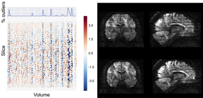
Given the very large number of individual datasets acquired in recent population imaging studies, it is becoming essential to automate data quality control (QC). Here we present an automated QC framework to assess diffusion MRI data both at the single subject and group levels. The QC metrics are derived through different stages of FSL’s pre-processing tools (TOPUP and EDDY). We show that using this framework, it is possible to distinguish between good and bad quality datasets and, importantly, identify subsets of the data that may need careful visual inspection. We hope this QC tool will help harmonisation efforts across sites/studies.
|
|
5355.
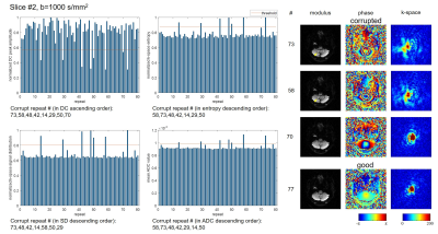
 |
37 |
An open-source framework for analysis of multidimensional diffusion MRI data implemented in MATLAB
Markus Nilsson, Filip Szczepankiewicz, Björn Lampinen, André Ahlgren, João P. de Almeida Martins, Samo Lasic, Carl-Fredrik Westin, Daniel Topgaard
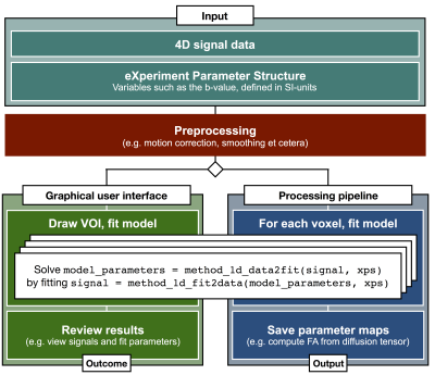
Reproducible research requires robust methods for data management and analysis. We have developed a comprehensive analysis framework for diffusion MRI data based on a robust but flexible management of data and metadata, which enables rapid prototyping and sharing of code. The framework is tailored to deal with multidimensional diffusion MRI data featuring novel types of diffusion encoding strategies, such as b-tensor encoding. It also features routines for higher order tensor mathematics as well as adapted volume registration for motion correction, an analysis GUI and other tools.
|
|
5356.
 |
38 |
Different Models of Ultra-high b-Value DWI for Better Detection of Restricted Diffusion
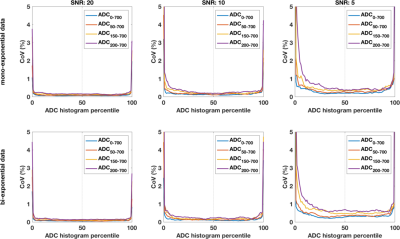
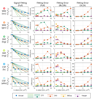
Qiqi Tong, Hongjian He, Tianyi Qian, Yi Sun, Ting Gong, Jianhui Zhong
Diffusion-weighted imaging with high b-value can potentially capture the restricted diffusion in tissues. With high-b-value diffusion weighting, the signal in tissue decays non-exponentially, thus the expansion of DWI signal usually be interpreted at second order or higher. However, in the ultra-high b-value (b>4k s/mm2), the cumulant expansion of higher order would fail to fit the signal. In this study, DTI, DKI, biexponential and Kärger models were used for approximating DWI signals with ultra-high b-values.
|
|
5357.
 |
39 |
Intravoxel Incoherent Motion Imaging of Thermoregulation in Skeletal Muscle
Kemal Sümser, Juan Hernandez-Tamames, Gerard van Rhoon, Margarethus Paulides
Accurate temperature dependent perfusion maps are needed for accurate thermal modelling. In this study, perfusion maps were created for human leg muscle at rest and under cold stress using Intravoxel Incoherent Motion (IVIM) Diffusion Weighted Imaging. IVIM parameters decreasesignificantly under cold stress. Consistency of inter-subject measurements proved IVIM method is suitable for imaging temperature dependent perfusion maps.
|
|
5358.
 |
40 |
Comparative study of DKI model and traditional DWI model in diagnosis of breast cancer
Did Not Present
Ting Li, Siying Wang, Yun Xiong, Kangan Li
The aim of this study is to compare the value of diffusion kurtosis imaging model with traditional single-index diffuse weighted imaging model parameters in the differential diagnosis of benign and malignant breast lesions. The rusults showed that the parameters of DKI model and traditional DWI model can be used to differentiate benign and malignant breast lesions. The diagnostic value of MK value in DKI model is the largest. There is a certain correlation between DKI model parameters and prognostic factors.
|
|
5359.
 |
41 |
Differential Diagnosis of Intrahepatic Cholangiocarcinoma and Hepatocellular Carcinoma by Using Diffusion-tensor Imaging
Video Permission Withheld
Lihua Chen, Ailian Liu, Qingwei Song, Lizhi Xie
The advent of functional MR imaging has facilitated an increased role for imaging in risk stratification and treatment planning. In this study, DTI and DWI MR measurements were performed to investigate the correlation of the FA and ADC values in ROIs of the intrahepatic cholangiocarcinoma (ICC) and hepatocellular carcinoma (HCC), and in further the sensitivity, specificity and accuracy of the parameters for the diagnosis. DTI working at present scanning hardware are more capable to detect the pathophysiological changes unattainable compare to conventional MRI techniques.
|
|
5360.
 |
42 |
The diagnostic capability of diffusion Kurtosis Imaging in distinguishing cervical lymphoma from nasopharyngeal carcinoma metastases
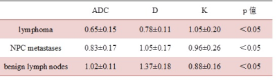
Cuifang Chen, Jing Zhong, Yunbin Chen, Weibo Chen
In our prospective study, We compared DKI and DWI related parameters in order to investigate the diagnostic capability of DKI in distinguishing cervical lymphoma from nasopharyngeal carcinoma(NPC) metastases. The ROC curve showed that K value of DKI had no significantly diagnostic capability, whlie D value of DKI had a better diagnostic performance than the ADC value of DWI in distinguishing the cervical lymphoma from the NPC metastases. Therefore, DKI may be a better noninvasive imaging biomarker for distinguishing the lymphoma and NPC metastases involving the head and neck.
|
|
5361.
 |
43 |
An Analytical Segmented (AS) Approach for Extracting IntraVoxel Incoherent Motion (IVIM) Model Parameters
Erick Buko, Jiming Zhang, Afis Ajala, Pei-Herng Hor, Raja Muthupillai
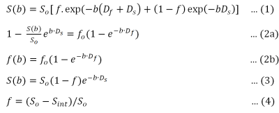
Although IVIM model can provide valuable insight into tissue perfusion and microstructure without the need for contrast, the estimation of model parameters with sufficient accuracy continues to be a challenge. In this work, we propose a analytical segmented (AS) approach for estimating IVIM model parameters describing tissue perfusion (Df, and f), and tissue diffusion (Ds), and compare the performance of this work against conventional approaches using numerical simulations. Results from our work suggest that AS approach can minimize errors in the estimation of metrics characterizing perfusion, i.e., f, and Df compared to conventional approaches (full fitting , segmented and oversegmented methods) even for tissues with low Df/Ds ratios.
|
|
5362.
 |
44 |
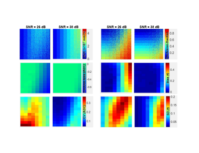
Inadequate modeling of diffusion anisotropy can lead to artefactual IVIM effects: evidence from numerical simulations
Gabrielle Fournet, Luisa Ciobanu, Denis Le Bihan
The interest in studying perfusion anisotropy using IVIM imaging is growing. However, due to the small amount of perfusion in some tissues, IVIM imaging is highly sensitive to imperfections in the analysis. In this work, we investigate the impact of improperly modeling diffusion anisotropy on the IVIM effect. Performing numerical simulations of a perfusion-free tissue using different sets of diffusion directions and tissue orientations, we show that fitting anisotropic diffusion to the Kurtosis model creates a direction- and orientation-dependent IVIM effect, which can be minimized by taking the geometric mean of the signals with at least 6 properly chosen directions.
|
|
5363.
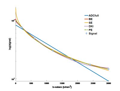 |
45 |
Non-Gaussian DWI of breast lesions at high b-values
Igor Vidic, Liv Egnell, Neil Jerome, Torill Sjøbakk, Agnes Østlie, Hans Fjøsne, Roshan Karunamuni, Nathan White, Rebecca Rakow-Penner, Anders Dale, Tone Bathen, Pål Erik Goa
In this work, we fit multiple non-Gaussian models to high b-value DW-MRI (b= 200-3000 s/mm2) in benign and malignant breast lesions. Models included bi-exponential, stretched exponential, diffusion kurtosis, Padé exponent and ADC (for comparison). We evaluated the quality of fit for each model and investigated the lesion differentiation accuracy for all extracted model parameters. The bi-exponential model provided statistically significant better quality of fit than all the other models, and without systematic bias. This suggests the existence of two effective diffusion compartments in breast lesions. Several of the extracted model parameters performed equally well in terms of lesion differentiation (AUC>0.97).
|
|
5364.
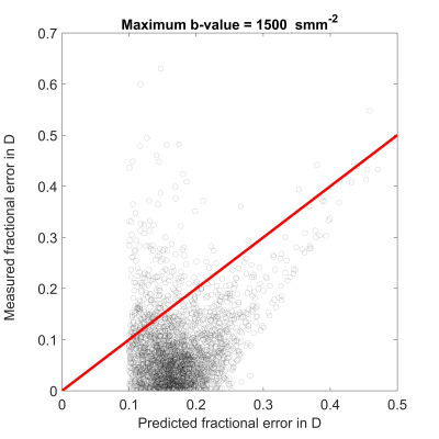 |
46 |
Measurement biases in diffusion and diffusion kurtosis MRI
Nima Gilani, Hamidreza Saligheh Rad, Glyn Johnson
Water diffusion in tissue is non-Gaussian and the expressions used to calculate diffusion parameters are approximations which introduce systematic errors dependent on the maximum b-value employed. The purpose of this study was to characterize biases in estimates of both apparent diffusion coefficient, and kurtosis and to determine their dependence on maximum b-value. Hence, equations were derived to predict biases in measured diffusion parameters and to explain much of the discrepancy between measurements obtained with different b-values. The equations may also be used to choose appropriate maximum b-values for diffusion-weighted, tensor, and kurtosis imaging.
|
|
5365.
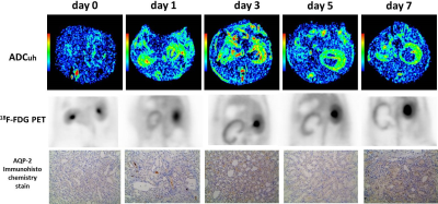 |
47 |
Can Apparent Diffusion Coefficient from Ultra-high b-Values be Used to Assess Renal Function with Murine Model of Unilateral Ureteral Obstruction?
Anqin Li, Zhen Li, Jiali Li, Yao Hu, Daoyu Hu
To evaluate the utility of apparent diffusion coefficient (ADC) data obtained from ultra-high b-values in renal function using with rat model of unilateral ureteral obstruction (UUO). We compared the differences of ADCuh among time points and between the sides, and determined the correlation of ADCuh with positron emission tomography (PET) renal function and expression of aquaporin 2 (AQP-2). ADCuh of the UUO sides on day 0 was significantly lower than that of on day 1, 3, 5 and 7. ADCuh was negatively correlated with SUV both on the UUO side and contralateral side. Therefore, ADCuh may have a certain value to help evaluate renal function.
|
|
5366.
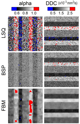 |
48 |
Bayesian estimation applied to stretched exponential modelling of diffusion-weighted imaging data
Peter While
The stretched exponential model provides a means of assessing non-monoexponential signal attenuation in diffusion-weighted imaging data. However, standard least squares approaches to parameter estimation remain susceptible to noise. In this work, we apply two distinct Bayesian approaches to stretched exponential modelling, and assess their performance at low-medium b-values using simulations. The use of a spatial homogeneity prior is found to yield parameter maps with higher precision and accuracy than those obtained with the use of a Gaussian shrinkage prior or nonlinear least squares.
|
|
Detecting Microstructural Changes Using Diffusion
Electronic Poster
Diffusion
Thursday, 21 June 2018
| Exhibition Hall |
09:00 - 10:00 |
| |
|
Computer # |
|
5367.
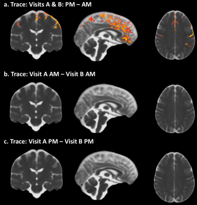 |
49 |
Impact of time-of-day on diffusivity measures of brain tissue derived from diffusion tensor imaging
Cibu Thomas, Neda Sadeghi, Aaron Trefler, Joelle Sarlls, Chris Baker, Carlo Pierpaoli
Although diurnal fluctuations in DTI-based measures of brain tissue properties have been reported, the underlying mechanism has not been investigated. Here, we processed multi-shell diffusion data using the conventional monoexponential tensor model as well as with a dual-compartment tensor model and assessed whether, time-of-day (TOD) fluctuations in DTI measures are due to changes in the magnitude of a CSF-like water compartment, or due to changes in the diffusion properties of parenchymal water. Our results suggest that TOD effects are due to fluctuations in the magnitude of a CSF-like water compartment rather than changes in the diffusion properties of parenchymal water.
|
|
5368.
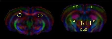 |
50 |
Longitudinal Changes in Gray Matter Regions after Cranial Radiation and Comparative Analysis with Whole Body Radiation: A DTI Study
Did Not Present
Poonam Rana, Apurva Watve, Mamta Gupta, Richa Trivedi, Subash Khushu
Radiation induced white matter changes are well known and vastly studied. However, radiation induced gray matter alterations are still a research question. In the present study, DTI based gray matter changes in C57BL/6 mice were studied following cranial and whole body irradiation at 5 Gy during sub acute and early delayed time point. Longitudinal changes in FA and MD were observed till 8 months post radiation. Comparative analysis depicted differential response after cranial and whole body radiation exposure with prominent alterations in cranially irradiated animals. with most changes at 8 months post irradiation
|
|
5369.
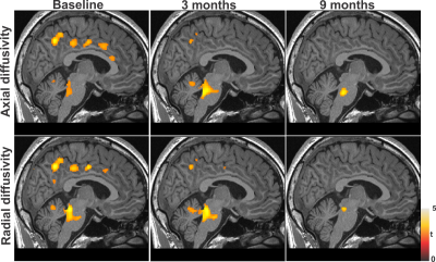 |
51 |
Brain axonal and myelin changes after positive airway pressure treatment in patients with obstructive sleep apnea
Ashish Sahib, Bhaswati Roy, Xiaopeng Song, Sadhana Singh, Luke Ehlert, Ravi Aysola, Daniel Kang, Mary Woo, Rajesh Kumar
Newly-diagnosed, treatment-naïve obstructive sleep apnea subjects show predominately acute tissue changes in gray and white matter, in addition to autonomic, mood, and cognitive deficits, but the extent of brain tissue recovery after PAP treatment and required minimum treatment time is unclear. We examined brain axonal and myelin changes at baseline and tissue recovery after 3 and 9 months’ positive airway pressure treatment in OSA compared to control subjects. Our findings indicate that brain structural changes found in newly-diagnosed OSA subjects can be reversible with long-term PAP treatment.
|
|
5370.
 |
52 |
Investigating the spatial pattern of structural changes of the Optic Radiation in early blind individuals.
Lisa Novello, Chiara Maffei, Hicret Atilgan, Stefania Mattioni, Ceren Battal, Mohamed Rezk, Olivier Collignon, Jorge Jovicich
The study of sensory deprived individuals shows the capability of the brain to massively reorganize. Previous studies show that the optic tract of early blind individuals undergoes structural changes, but little is known about their spatial distribution. We investigated the spatial profile of the structural changes occurring along tractography reconstruction from diffusion MRI data of the optic radiations in early blind individuals compared to age- and gender-matched healthy sighted controls.
|
|
5371.
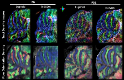 |
53 |
Abnormal cerebellar wiring in a mouse model of Down syndrome revealed by HARDI-based tractography
Dan Wu, Feng Gao, Roger Reeves
We investigated the cerebellar microstructure and wiring diagram in a mouse model of Down syndrome (DS), using HARDI-based tractography analysis. Our results showed an absence of cerebellar climbing fibers (CF) and/or mossy fibers (MF) in the intra-granule layer of the DS mice at neonatal stage, when those fiber tracts started to form in the control neonatal brains. In the adult mice, two groups of crossing fibers—the CF/MF fibers versus the parallel fibers (PF) were identified, but the number of crossing fibers and apparent fiber density was significantly reduced in the granule layer of the DS mice, especially for the CF/MF group.
|
|
5372.
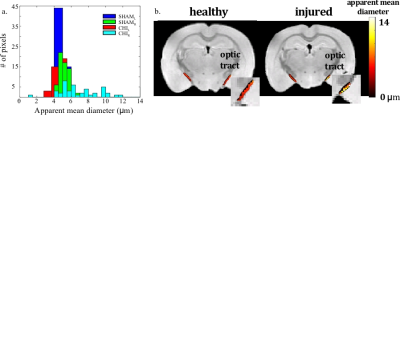 |
54 |
Using Double Diffusion Encoding (DDE) MRI to study tissue microstructure in traumatic brain injury (TBI)
Michal Komlosh, Dan Benjamini, Elizabeth Hutchinson, Sarah King, Margalit Haber, Alexandru Avram, Lynne Holtzclaw, Abhishek Desai, Carlo Pierpaoli , Peter Basser
A double diffusion encoding (DDE) MRI method was used to estimate apparent mean axon diameter (AMD) in a mild traumatic brain injury (mTBI) mouse model exhibitig diffuse axonal injury (DAI). MRI data show clear tissue alterations caused by the injury, while immunohistochemistry data confirm the MRI findings. DDE could potentially be used as a non-invasive means to detect mTBI.
|
|
5373.
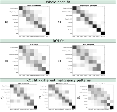 |
55 |
Comparison of diffusion MR models in lymph nodes at ultra high field
Andrada Ianus, Ines Santiago, Daniel Alexander, Celso Matos, Noam Shemesh
Diffusion MRI could play an important role for detection and characterisation of lymph nodes, however, the standard ADC metric lacks specificity and cannot accurately capture the signal decay. Multi-compartment models aim to represent the signal contribution of different water pools in order to better explain the acquired data. In this study, a rich dMRI dataset is acquired for ex-vivo lymph nodes and various diffusion models are fitted. The results show that including compartments which feature restricted diffusion improves the signal fit. Although such models have only a slight impact on lymph node differentiation, they could potentially provide more specific biomarkers.
|
|
5374.
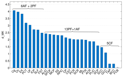 |
56 |
Heterochronous regional white matter development in pre-adolescent children: diffusion kurtosis imaging study
Farida Grinberg, Ezequiel Farrher, Kerstin Konrad, Irene Neuner, N. Jon Shah
Diffusion tensor imaging, and, more recently, diffusion kurtosis imaging enables the examination of white matter connectivity and of in vivo microstructural changes across the lifespan. However, only very few diffusion kurtosis imaging studies have addressed white matter maturation. Here we explore which specific brain regions continue to develop throughout late childhood into the adulthood as revealed by kurtosis tensor metrics. We show the potential of these metrics as maturation-sensitive biomakers capable of elucidating maturation heterochronicity of various white matter tract groups, such as association, projection, and commissural fibres.
|
|
5375.
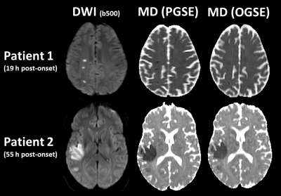 |
57 |
Oscillating Gradient Spin Echo Diffusion Tensor MRI of Acute Human Stroke
Christian Beaulieu, Leka Sivakumar, Ken Butcher, Sarah Treit, Derek Emery, Robert Stobbe
Different spatial scales of micro-structural alteration with disease can be interrogated by varying the time water has to interact with its surroundings (e.g. membranes, etc). Oscillating gradient spin echo (OGSE) diffusion MRI enables much shorter diffusion times (e.g. 6 ms) than typical pulsed gradient spin-echo (PGSE) (e.g. 40 ms). The large diffusion reduction typically observed with PGSE in ischemic lesions in human stroke was markedly less with OGSE, and the OGSE-PGSE difference was greatest for radial diffusivity in ischemic white matter. This result is consistent with the swelling and beading of the axons underlying diffusion contrast in cerebral ischemia.
|
|
5376.
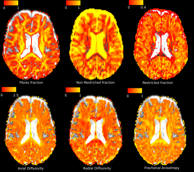 |
58 |
Non-invasive quantification of inflammation, axonal and myelin injury in MS: a 7T DBSI study
Simona Schiavi, Peng Sun, Sirio Cocozza, Maria Petracca, Mohamed Mounir El Mendili, Kornelius Podranski, Matilde Inglese, Sheng-Kwei Song
Diffusion basis spectrum imaging (DBSI) has successfully distinguished co-existing pathological processes in CNS diseases, such as multiple sclerosis (MS). The aim of our study was to determine the feasibility of DBSI in MS patients at 7T and to investigate the underlying pathological substrates of different lesion types and normal appearing white matter (NAWM). For the first time, we proved the feasibility of DBSI at 7T, validating the specificity of the different metrics to diverse pathological substrates. Additionally, we employed DBSI metrics to characterize focal and diffuse tissue damage in different MS phenotypes, confirming their utility as biomarkers of tissue-specific microstructural damage.
|
|
5377.
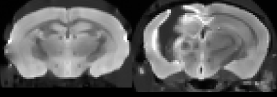 |
59 |
Diffusion Basis Spectrum Imaging and Diffusion Tensor Imaging in Mouse Model of Traumatic Brain Injury
Harri Merisaari, Tsen-Hsuan Lin, Gentian Toshkezi, Michele Kyle, Zhao Li-Ru, Sheng-Kwei Song
Traumatic brain injury (TBI) is a major cause of acquired brain injury in both children and adults. Traumatic axonal injury (TAI) is a major contributor to cognitive dysfunction followed by TBI. Diffusion Tensor Imaging (DTI) has shown to characterize TAI in non-invasive manner. However, DTI as a gross measure has limitations in its parameters in detection capability of underlying pathologies. Diffusion Basis Spectrum Imaging (DBSI) is an advanced imaging technique that has been applied to investigate nervous system pathology. Accurate characterization of brain pathologies in vivo, such as axonal injury, is extremely important in the study of changes that occur over time after TBI. To investigate these two methods in TBI data, we performed a comparison in mouse model between control and injured groups.
|
|
5378.
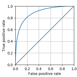 |
60 |
q-Space Novelty Detection in Short Diffusion MRI Scans of Multiple Sclerosis
Vladimir Golkov, Aleksei Vasilev, Francesco Pasa, Ilona Lipp, Wassim Boubaker, Eleonora Sgarlata, Franz Pfeiffer, Valentina Tomassini, Derek Jones, Daniel Cremers
Diffusion MRI can capture disease-related microstructural changes, but most methods use handcrafted data transformations that discard parts of the information and require quite long scan times. In contrast, q-space novelty detection (q-ND) circumvents these drawbacks, and does not require any knowledge whatsoever about the effect of disease on q-space measurements. Instead, q-ND highlights voxels that look unlike anything seen in a database of healthy scans. Here we show that novelty scores from q-ND largely coincide with multiple sclerosis lesions, and that q-ND also works at reduced scan times.
|
|
5379.
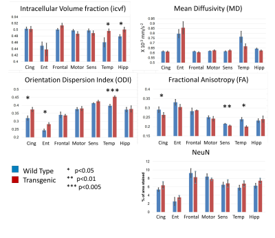 |
61 |
Diffusion-weighted imaging reveals microstructural alterations in a transgenic rat model of Alzheimer’s disease - validation against immunohistochemistry
Hamied Haroon, Ben Dickie, Matthias Vandesquille, Charlotte Auty, Herve Boutin, Geoffrey Parker, Laura Parkes
Diffusion-weighted imaging (DWI) holds the promise of detecting very subtle neuronal changes in Alzheimer’s disease (AD). We compare the sensitivity of different DWI metrics to microstructural change in a transgenic rat model of AD. We find genotype-related differences in regions known to be affected in AD, with NODDI metrics showing greater sensitivity than FA and MD. However, no genotype-related difference in neuronal density is detected using immunohistochemistry staining for NeuN suggesting that the DWI changes reflect alterations in the neuronal structure (dendritic density or myelination) rather than density. DWI metrics are however correlated with neuronal density on a regional basis.
|
|
5380.
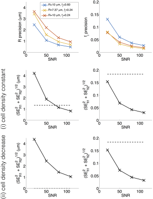 |
62 |
Towards a ‘resolution limit’ for DW-MRI tumour microstructural models: what microstructural changes can be distinguished?
Damien McHugh, Geoffrey Parker
The ability to detect specific changes in tumour tissue using diffusion-weighted (DW) MRI microstructural models depends on the accuracy and precision of parameter estimates, as well as the magnitude of specific biological changes. This work investigates the SNR requirements that data must meet in order to yield sufficiently precise estimates for detecting apoptotic cell shrinkage. Given the relative changes in cell size, R, and intracellular volume fraction, fi, as a result of apoptosis, simulations indicate that detecting changes in R require ~4-fold higher SNR than detecting changes in fi. Comparing the magnitude of biological changes with measurement accuracy and precision should form part of the validation process for potential biomarkers.
|
|
5381.
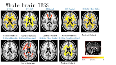 |
63 |
Demyelination or not: increased radial diffusivity in Parkinson's disease
Did Not Present
Ruimeng Yang, Pen Sun, Xiangling Zeng, Ajit George, Ajit George, xinhua Wei, xinqing jiang, Sheng-Kwei Song
White matter (WM) integrity alteration has been noted in Parkinson’s disease (PD). Whether the increased radial diffusivity (RD) revealed in previous study using diffusion tensor imaging (DTI) in PD patients is suggestive of demyelinating etiology remains to be established. To further investigate white matter (WM) microstructure changes in PD, diffusion tensor imaging (DTI) and diffusion basis spectrum imaging (DBSI) were analyzed using Tract Based Spatial Statistics (TBSS). Both whole brain voxel-based group analysis and reginal analysis on the corpus callosum were performed. DBSI results suggest that axonal injury is present in PD while no myelin injury was seen.
|
|
5382.
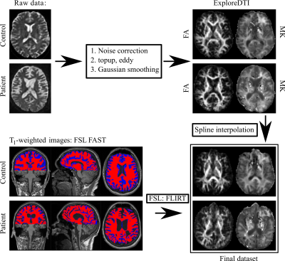 |
64 |
Diffusion kurtosis imaging in chronic disorders of consciousness: state estimation and treatment prognosis
Elena Kremneva, Lyudmila Legostaeva, Sofya Morozova, Elizaveta Mochalova, Dmitry Sinitsyn, Natalia Suponeva, Marina Krotenkova, Aleksandr Suslin, Mikhail Piradov, Ivan Maximov
Chronic disorders of consciousness (DOC) are severe neurological disorders due to lesions massively affecting the brain. The treatment and prognosis in the case of DOC is an actual problem demanding novel imaging approaches, in particular, allowing one to determine the extend of brain structural changes. Diffusion-weighted MRI is a powerful candidate for in vivo probing and visualization of brain damage severity in DOC patients. In order to investigate white matter microstructural changes associated with the chronic DOC we performed tract-based spatial statistics analysis and complementary brainstem diffusion metric comparisons between patients and healthy control groups. Our findings revealed large white matter changes in the DOC patients, in particular, using novel and sensitive biomarkers based on the kurtosis scalar metrics. We hope that our discoveries help us to improve a forthcoming treatment of DOC patients.
|
|
5383.
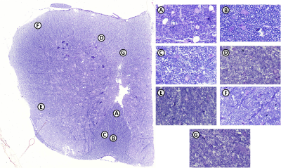 |
65 |
Spinal cord axonal diameter variations in the relapsing remitting EAE mouse model of multiple sclerosis
Ahmad Alghamdi, Ian Brereton, Nemat Khan, Maree Smith, Nyoman Kurniawan
This study aimed to determine the extent of axon diameter distribution changes in the lumbar spinal cord in C57BL/6 mice associated with experimental autoimmune encephalomyelitis (EAE). AxCaliber protocol was performed ex-vivo at 16.4T to analyze changes in mice with a range of EAE severity and the effect of treatment with alpha-lipoic acid (R-ALA), a nutraceutical compound that is recently known for suppressing microglial activation and CNS inflammation in EAE rodent models. AxCaliber showed axon diameter increases in mild relapsing remitting EAE group after R-ALA treatment. This might indicate a process of remyelination or other microstructural changes.
|
|
5384.
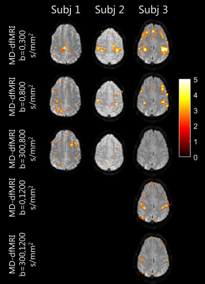 |
66 |
Brain activations during a motor cortex paradigm: a diffusion MRI signal perspective.
Alberto De Luca, Lara Schlaffke, Jeroen Siero, Martijn Froeling, Alexander Leemans
Brain activations during task are commonly investigated by gradient-echo functional MRI (GE-fMRI), which is sensitive to blood volume changes and blood deoxygenation. Recent studies have shown that diffusion MRI, which is regarded as static in the time-scale of minutes, might be sensitive to changes in activated areas. We investigated changes in signals acquired during a motor cortex paradigm using GE-fMRI and diffusion fMRI (dfMRI). We observed causality between the task and the dfMRI acquired at different diffusion weightings. These preliminary results suggest that the observed changes in the dfMRI signal might not be a pure result of blood volume alterations.
|
|
5385.
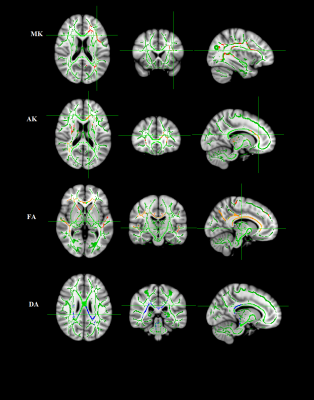 |
67 |
White matter micro-structural alterations in Hypothyroid: A Diffusion Kurtosis Imaging study
Did Not Present
Mukesh Kumar, Poonam Rana, Deepak Sharma, Ratnesh Kanwar, Tarun Sekhri, Maria D’souza, Subash Khushu
The aim of our study was to assess changes in brain tissue microstructures in hypothyroid patients using Diffusion Kurtosis Imaging (DKI). The water diffusion in living tissue is hindered by interactions with other molecules and cell membranes. Therefore, water movement in biological tissue is often non-Gaussian and this non-Gaussian behavior may contain useful information related to tissue structure and pathophysiology. Our findings demonstrate widespread reduced kurtosis indices MK, AK; diffusion index FA was decreased and AD was increased in major white matter pathways and such abnormal white matter structure may be linked to cognitive and behavioral impairment in hypothyroid patients.
|
|
5386.
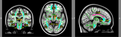 |
68 |
Alterations in white matter connectivity related to language comprehension in patients with Crohn’s disease in remission
Jiancheng Hou, Keith Dodd, Veena Nair, Poonam Beniwal-Patel, Vivek Prabhakaran, Sumona Saha
Reduced white matter integrity in patients with Crohn’s disease (CD) has been previously reported. However few studies have examined the behavioral implications of compromised WM integrity in these patients. Here we report results from an exploratory study investigating group differences in diffusivity measures in patients with Crohn’s disease in remission [compared to age and gender matched healthy control (HC) subjects] and examined their relationship to participants’ performance on a phonemic fluency task. CD patients demonstrate altered brain microstructural changes in regions associated with several cognitive functions including language processing.
|
|
5387.
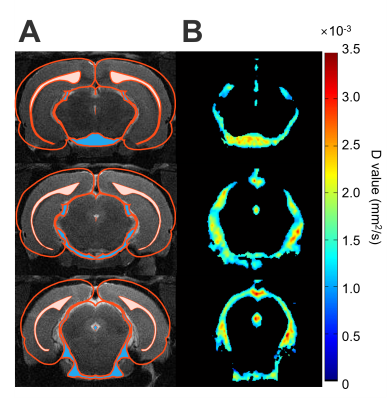 |
69 |
Alteration in Diffusion Characteristic of Cerebrospinal Fluid after Neurological Disease in Rats
Jun Tazoe, Yu-Chieh Kao, Chia-Feng Lu, Ping-Huei Tsai, Fei-Ting Hsu, Bao-Yu Hsieh, Cheng-Yu Chen
While the quantitative maps conducted by diffusion weighted or diffusion tensor imaging were applied to the brain parenchyma to describe the tissue type and fate, the diffusion coefficient in the cerebrospinal fluid (CSF) was not well explored. In this study, significant lower mean of diffusion constant (D-value) and change in the histogram of D-value were observed in stroke animals compared with intact rats and the mTBI animals, suggesting the brain lesion or damage may alter the diffusion characteristics in CSF.
|
|
5388.
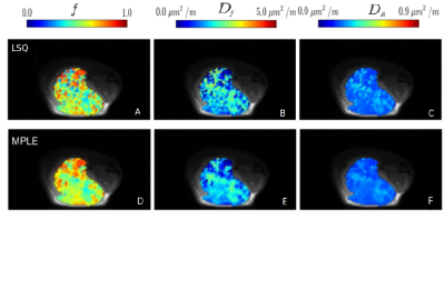 |
70 |
Spatially constrained mathematical models for diffusion weighted imaging of prostate cancer xenografts in mice: evaluation of therapy response
Video Permission Withheld
Parisa Movahedi, Harri Merisaari, Hanne Laakso , Ileana Montoya Perez, Heidi Liljenbäck, Hannu Aronen, Heikki Minn, Anne Roivainen, Timo Liimatainen, Ivan Jambor
Tumor growth in mice preclinical prostate cancer model (human prostate cancer cells, PC-3) was followed for 4 weeks by weekly DWI in control group (n=10) and treatment group (n=9) receiving Docetaxel. DWI data sets were acquired using 12 b-values the range of 0-2000 s/mm2. The DWI signal decays were fitted using monoexponential, biexponential, kurtosis and stretched exponential models/functions. Independent least squares fitting and spatially constrained Maximum Penalized Likelihood Estimation have been applied. The spatially constrained Maximum Penalized Likelihood Estimation revealed the effect of treatment in mice subjects, while conventional LSQ fitting failed to reveal significant difference between control and treatment.
|
|
5389.
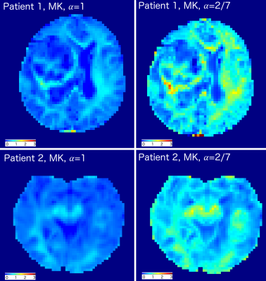 |
71 |
Generalised kurtosis imaging approach for glioblastoma evaluation
Eduard Pogosbekian, Artem Batalov, Alexander Turkin, Igor Pronin, Ivan Maximov
Primary brain gliomas are very a widespread type of intra-axial brain tumours. Brain gliomas have different cellular origins and can be differentiated by WHO 2016 classification. The most aggressive type of glioma is the glioblastoma (GB). Thus, a correct and reliable grading of gliomas, in particular, the GB, is critical for a patient treatment and prognosis. Diffusion kurtosis imaging (DKI) has been applied to glioma validation in order to perform non-invasive evaluation. DKI allowed one to obtain information about microstructure and inhomogeneity differentiation. In the present work we demonstrate advantages of generalised DKI approach for GB detection and evaluation.
|
|
5390.
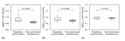 |
72 |
The Effect of Respiration on Apparent Diffusion Coefficient of the Brain
Video Permission Withheld
Naoki Ohno, Tosiaki Miyati, Yuki Hiramatsu, Ryoto Shimizu, Mitsuhito Mase, Satoshi Kobayashi, Toshifumi Gabata
ADC of the brain significantly changes during the cardiac cycle because of intracranial pressure changes and fluid fluctuations. We hypothesized that ADC of the brain changes during the respiratory cycle because intracranial pressure changes during the respiratory cycle. We determined the maximum and minimum ADC (ADCmax and ADCmin) and maximum change in ADC (ΔADC) in the respiratory cycle and non-synchronized periodical data. ADCmax and ΔADC in the white matter with respiratory-synchronization were significantly greater than those with non-synchronized periodical scan, whereas there was no significant difference in ADCmin between the scans. ADC of the brain changes during the respiratory cycle.
|
|
| Back |
| The International Society for Magnetic Resonance in Medicine is accredited by the Accreditation Council for Continuing Medical Education to provide continuing medical education for physicians. |
