|
Liver Imaging: What Can We Really Quantify?
Combined Educational & Scientific Session
ORGANIZERS: Kathryn Fowler
Monday, 18 June 2018
| S02 |
16:15 - 18:15 |
Moderators: Sudhakar Venkatesh, Ralph Sinkus |
Skill Level: Intermediate
Session Number: M-08
Overview
This session will focus on state-of-the-art quantitative liver imaging applications as they relate to assessing liver function, prediction and prognostication of tumors, and metabolism. The session will combine didactic talks with scientific abstracts.
Target Audience
Clinicians and physicists who participate in liver imaging.
Educational Objectives
As a result of attending this course, participants should be able to:
-Summarize current methods for quantitative imaging of the liver and their clinical applications;
-Describe cutting-edge and evolving practice applications of quantitative liver MRI; and
-List tumor imaging markers that are prognostic and predictive of outcomes.
16:15
|
|
 Measuring Liver Function-Technical Aspects Measuring Liver Function-Technical Aspects
Steven Sourbron
This talk will provide a broad introduction of MRI methods to measure the function of liver parenchyma, with more in-depth treatment of Dynamic Gadoxetate-Enhanced MRI for the quantification of hepatocellular transporter function. We will also cover some basic facts about the liver, review competing non-MRI techniques for assessing liver function, and present examples of applications in basic science, drug development and clinical practice.
|
16:45
|
0237.
 |
Clinical Assessment of Nonalcoholic Steatohepatitis (NASH) with Multi-parametric MRI
Jiahui Li, Alina Allen, Yi Sui, Dan Rettmann , Ann Shimakawa, Glenn Slavin, Kevin J. Glaser, Sudhakar K. Venkatesh, Taofic Mounajjed, Vijay Shah, Richard L. Ehman, Meng Yin
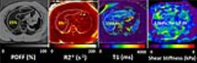
In 27 clinical patients, we performed multi-parametric hepatic MRI, including proton density fat fraction with R2* correction, MR Elastography (MRE), and T1 mapping to characterize nonalcoholic steatohepatitis (NASH). Fat fraction and multiple MRE-assessed mechanical parameters successfully diagnosed NASH (p<0.05 for all). Diagnostic abilities of all parameters were evaluated based on steatosis, inflammation and ballooning scores respectively. Spearman correlations were used to analyze the correlations between imaging parameters. We found that T1 relaxation time had a significantly positive correlation (ρ=0.72, p=0.0005) with fat fraction. In summary, multi-parametric MRI is a potential imaging surrogate for diagnosing NASH.
|
16:57
|
0238.
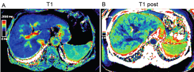 |
Implementation of T1 Mapping in Routine Clinical MR Liver Exam For The Detection of Hepatic Fibrosis and Portal Hypertension in Hepatitis C Patients
Ahmed Hamimi, Ronald Ouwerkerk, Theo Heller, Elliot Levy, Jatin Matta, Khaled Abd-elmoniem, Ahmed Gharib
Application of two short single breath hold T1 mapping shMOLLI technique (before and after Gadolinium injection) to a routine clinical MRI liver exam can detect both severe fibrosis and portal hypertension in Hepatitis C. The technique was prospectively validated in 29 patients with reference standard clinical methods including liver biopsy and direct portal venous pressure measurements. Utilizing this method would allow for a comprehensive anatomic and functional MRI study in a single session without substantial prolongation of scan time, thereby, allow non-invasive monitoring of therapy and/or progression of disease.
|
17:09
|
|
Prognosis & Prediction for Liver Tumors
Ihab Kamel
|
17:39
 |
0239.
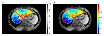 |
Preoperative Remnant Liver Function Evaluation using a Clinical-Available Gd-EOB-DTPA-Enhanced MR Imaging Protocol in HCC Patients
Yajie Wang, Lin Zhang, Jia Ning, Xinjing Zhang, Xuedong Wang, Shizhong Yang, Jiahong Dong, Huijun Chen
Accurate evaluating of remnant liver function preoperatively is important for surgery planning and reducing posthepatectomy liver failure (PHLF) rate in hepatocellular carcinoma (HCC) patients. In this study, an accurate remnant liver function evaluation method was proposed using a clinical-available MR imaging protocol. The remnant liver function measured by the proposed method showed significant difference between the patients with and without PHLF. More importantly, ROC analysis showed the proposed method has a larger AUC than remnant liver volume and ICG based parameters in predicting PHLF.
|
17:51
 |
0240.
 |
MRI texture features as predictors of histopathologic and genomic characteristics of hepatocellular carcinoma.
Stefanie Hectors, Sara Lewis, Cecilia Besa, Michael King, Juan Putra, Stephen Ward, Takaaki Higashi, Swan Thung, Yujin Hoshida, Bachir Taouli
The goal of this study was to assess the diagnostic value of texture features measured with MRI compared to qualitative imaging traits for the prediction of histopathologic and genomic characteristics of hepatocellular carcinoma (HCC) lesions. Texture features exhibited additional, complementary correlations with histopathology and genomics compared to qualitative imaging traits, including association with microvascular invasion and expression of immunotherapy target CTLA4. These promising results warrant further investigation of texture features as predictors of histopathologic and genomics measurements in HCC.
|
18:03
|
0241.
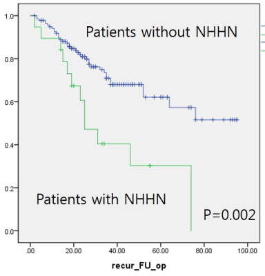 |
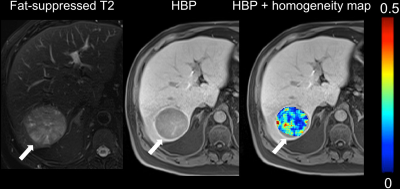
Presence of non-hypervacular hypointense nodule on hepatobiliary phase of gadoxetic acid enhanced MR: Risk of tumor recurrence after curative treatment for small single nodular HCC and guidance for selection of treatment option
Dong Ho Lee, Jeong Min Lee
Presence of non-hypervascular hypointense nodule on hepatobiliary phase of gadoxetic acid enhanced liver MR can stratify the risk of tumor recurrence after curative treatment for small single nodular HCC equal to or less than 3cm.
|
18:15
|
|
Adjournment & Meet the Teachers |
|
