Normal Pediatric Brain Development
Oral
Neuro
Tuesday, 19 June 2018
| W03/04 |
13:45 - 15:45 |
Moderators: Serena Counsell, Claire Kelly |
13:45
|
0523.
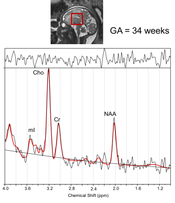 |
 Emerging Metabolic Trajectories in the Developing Fetal Brain Emerging Metabolic Trajectories in the Developing Fetal Brain
Subechhya Pradhan, Kushal Kapse, Reka Kovacs, Linda White, Catherine Limperopoulos
Knowledge of metabolite changes in the fetal brain will provide insight into the biochemical changes in the developing brain. Findings from measurement of metabolite concentration as a function of increasing GA are reported. Significant increases in NAA, Cr, Cho and scyllo-Inositol with increasing GA in the fetal brain was observed in this study. Changes in myo-Inositol with GA did not reach significance.
|
13:57
|
0524.
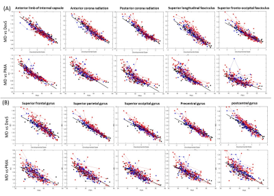 |
Developmental Score of the Neonatal Brains: Characterizing Diffusion MRI Changes in the Term- and Preterm-born Infants
Dan Wu, Linda Chang, Thomas Ernst, Jon Skranes, Kenichi Oishi
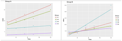
Postmenstrual age (PMA) is used as a time-scale to evaluate brain development, but it contains inaccuracy with regards to the estimated time of conception because the duration from the last menstrual period to conception varies. To address this variation, we designed a developmental score (DevS) that aligned individuals with similar development patterns together, and provides a linear trajectory of DTI-measurement as a function of an underlying brain developmental index. Compared to PMA, DevS showed an improved the regression with DTI-measurements, and it better separated the developmental differences between term- and preterm-born infants.
|
14:09
|
0525.
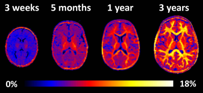 |
Quantitative assessment of pediatric brain myelination in a clinical setting using macromolecular proton fraction
Vasily Yarnykh, Nina Knipenberg , Olga Tereshchenkova
The fast macromolecular proton fraction (MPF) mapping method has been implemented as a part of a clinical pediatric MRI protocol for a 1.5T MRI scanner. 3D MPF maps were obtained from 31 pediatric patients aged from 2 weeks to 7 years without abnormal brain findings. MPF maps allowed robust quantitation of age-related changes in the myelin content. Temporal trajectories of regional brain myelination were analyzed using the general logistic model that enabled the formal description of distinctions in the onset and speed of myelination across a range of white and gray matter structures.
|
14:21
|
0526.
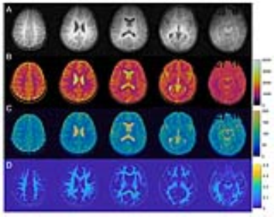 |
MR Fingerprinting enables quantitative measures of brain tissue relaxation times and myelin water fraction in early brain development
Yong Chen, Meng-Hsiang Chen, Weili Lin, UNC/UMN Baby Connectome Project Consortium
In this study, a high-resolution MR Fingerprinting technique was developed for rapid and simultaneous quantification of T1, T2 and myelin water fraction in pediatric neuroimaging. The method was applied to five subjects from 3 months to 52 months old and high quality quantitative maps were obtained from all subjects without evident motion artifacts. Our preliminary results also show a trend of decrease in T1/T2 and increase of myelin water fraction with age, which are consistent with finding in literature.
|
14:33
|
0527.
 |
Patterns of subcortical gray matter and white matter myelination from 4.5 months to 1 year-old: an observational study comparing the whole brain T1w/T2w ratio myelin mapping technique and an autopsy infant myelin-staining study
Joseph Yang, Jian Chen, Michelle Wu, Simone Mandelstam, Richard Leventer, Bonnie Alexander, Deanne Thompson, Marc Seal, Richard Beare
The whole brain T1w/T2w ratio technique enables in vivo quantitative myelin mapping using clinically acquired MRI sequences, which is potentially useful when investigating developmental myelination trajectories. Post-mortem infant myelin-staining studies provide useful references. This study characterizes myelination trajectories of 27 subcortical brain regions in typically developing 4.5 months to 1 year-old children using the T1w/T2w ratio technique and compared the patterns with those demonstrated by an autopsy infant myelin-staining study. Similar myelination trajectory patterns were observed in the structures that commenced myelination before birth. This highlights the T1w/T2w ratio as potential myelin imaging biomarker for these structures during this period.
|
14:45
|
0528.
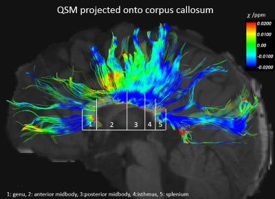 |
Profiling myelin maturation with quantitative susceptibility mapping (QSM) in pediatric brains
Lijia Zhang, Allen Song
Quantitative susceptibility mapping (QSM) has been increasingly used to access brain development, especially white matter myelination. In this study, diffusion tensor imaging (DTI) has been used to delineate the corpus callosum in the pediatric brains, followed by tract-based QSM analysis to accurately derive the baseline susceptibility across subjects. The baseline susceptibility is then used to evaluate white matter and myelin development.
|
14:57
|
0529.
 |
Neonate functional brain networks and distinctive intra-network connectivity
Qinmu Peng, Ouyang Minhui, Jiaojian Wang, Qinlin Yu, Chenying Zhao, Slinger Michelle, Hongming Li, Yong Fan, Bo Hong, Hao Huang
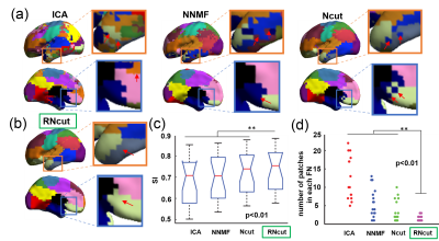
Little is known on the network organization in the neonatal brain. Here, we used a novel parcellation method, called regularized-Ncut (RNcut), to parcellate the neonate brain into functional networks with resting-state fMRI. RNcut effectively delineates the neonatal functional networks including the primary sensorimotor and higher-order networks. Based on RNcut parcellation of functional networks, intra-network connectivity was quantified. Distinctive intra-network connectivity was revealed for the first time. We found that the primary sensorimotor network has the highest intra-network connectivity while higher-order fronto-parietal network has lower intra-network connectivity. The distinctive intra-network connectivity pattern underlies heterogeneous emergence of early brain functional systems.
|
15:09
|
0530.
 |
Prenatal Maternal Cortisol Response Predicts One-month Infant White Matter Microstructure
Douglas Dean, Elizabeth Planalp, William Wooten, Nagesh Adluru, H Goldsmith, Andrew Alexander, Richard Davidson
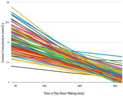
Exposure to differing concentrations of cortisol likely has a significant impact on brain development in childhood and adolescence; however, little is known about the time immediately following birth. Using multi-shell diffusion imaging data, we examined the associations between prenatal maternal diurnal cortisol patterns and infant white matter microstructure. Infant measures were associated with the slope of the maternal cortisol response across white matter, suggesting variations of cortisol within the intrauterine environment may have a significant influence on processes of early brain development.
|
15:21
|
0531.
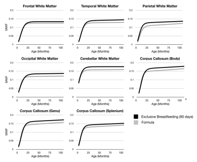 |
Influence of Infant Feeding on Longitudinal Brain and Cognitive Development
Sean Deoni, Douglas Dean, Viren D'Sa
Infancy and early childhood are sensitive and rapid periods of brain growth that coincide with the emergence of nearly all cognitive, behavioral, and social-emotional functions. Brain growth, including myelination, is modulated modulated by neural activity and are responsive to environmental, genetic, hormonal, and other influences, including nutrition. He we use longitudinal neuroimaging to examine the influence of early infant nutrition, specifically breastfeeding, on brain and cognitive development.
|
15:33
|
0532.
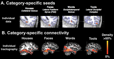 |
Early specific connections of the Visual Word Form Area during the first year of reading instruction: a longitudinal MRI study in children
Eric Moulton, Florence Bouhali, Karla Monzalvo, Cyril Poupon, Hui Zhang, Stanislas Dehaene, Ghislaine Dehaene-Lambertz, Jessica Dubois
Specialized in word recognition, the visual word form area (VWFA) is located in the posterior fusiform gyrus of the left hemisphere, regardless of one’s writing system. To test the hypothesis that this consistent location is determined by specific connections to linguistic regions, we studied 6-7year old children throughout the process of learning to read. Using diffusion and functional MRI, we analysed the white-matter connectivity of cortical regions processing visual categories (words, houses, faces, tools). We showed that the emerging VWFA has specific and stable connections to the inferior-dorsal parietal cortex, and these connections exhibit microstructural maturation related to reading improvement.
|
|
