Multiple Sclerosis: Lesions Everywhere
Oral
Neuro
Monday, 18 June 2018
| S06 |
08:15 - 10:15 |
Moderators: Irene Vavasour, Frederik Barkhof |
08:15
 |
0082.
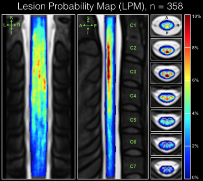 |
 Spatial Distribution of Multiple Sclerosis lesions in the Cervical Cord Spatial Distribution of Multiple Sclerosis lesions in the Cervical Cord
Dominique Eden, Charley Gros, Atef Badji, Sara Dupont, Josefina Maranzano, Ren Zhuoquiong, Yaou Liu, Jason Talbott, Elise Bannier, Anne Kerbrat, Gilles Edan, Pierre Labauge, Virginie Callot, Jean Pelletier, Bernard Audoin, Henitsoa Rasoanandrianina, Paola Valsasina, Massimo Filippi, Rohit Bakshi, Shahamat Tauhid, Ferran Prados, Marios Yiannakas, Hugh Kearney, Olga Ciccarelli, Constantina Treaba, Caterina Mainero, Russell Ouellette, Tobias Granberg, Sridar Narayanan, Julien Cohen-Adad
The study of the spatial distribution of multiple sclerosis (MS) lesions in the cervical spinal cord provides a means to further understand the disease pathophysiology. In this study we involve 358 patients across 7 sites, where cervical lesions were manually segmented. Using Spinal Cord Toolbox, lesion segmentations were registered to a common-space template and voxel-based lesion probability maps (LPMs) were computed across the patient population to assess lesion topography. Results revealed a predominance of lesions in the upper cord (C1-C3) and dorsal column, which confirms prior histopathology work and encourages further study of associations between cervical spine lesion volume and distribution with clinical status.
|
08:27
|
0083.
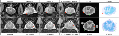 |
Spatiotemporal development of spinal cord lesions in a primate model of multiple sclerosis
Jennifer Lefeuvre, Pascal Sati, Cecil Yen, Seung Kwon Ha, Wen-Yang Chiang, Mathieu Santin, Steven Jacobson, Afonso Silva, Stéphane Lehéricy, Daniel Reich
The spatiotemporal development of spinal cord (SC) lesions in multiple sclerosis (MS) is poorly understood, despite the high prevalence of these lesions and their important contribution to patient disability. In this study, we report for the first time the serial imaging of SC lesions in a nonhuman primate model of MS. The results demonstrated substantial clinical and imaging features shared between this animal model and human MS. In particular, we observed focal and subpial demyelinating lesions that appeared at disease onset and proceeded to affect much of the entire cord over the course of several weeks to months.
|
08:39
|
0084.
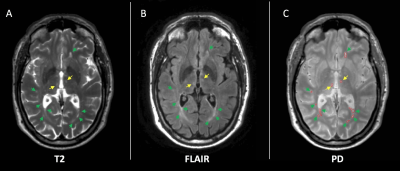 |
Thalamic lesions, thalamic volume and cognitive deficit in secondary progressive MS
Floriana De Angelis, Jonathan Stutters, Arman Eshaghi, Ferran Prados, Domenico Plantone, Anisha Doshi, Nevin John, David MacManus, Sebastien Ourselin, Sue Pavitt, Gavin Giovannoni, Richard Parker, Chris Weir, Nigel Stallard, Clive Hawkins, Basil Sharrack, Peter Connick, Siddharthan Chandran, Claudia Gandini Wheeler-Kingshott , Frederik Barkhof, Jeremy Chataway
We investigated the cross-sectional relationships between thalamic lesions (i.e. total thalamic lesion volume), thalamic volume, and cognitive deficit in 55 subjects with secondary progressive multiple sclerosis. We measured: total intracranial volume, T2 lesion volume (T2LV), thalamic volume, thalamic lesions, and symbol digit modalities test (SDMT). Thalamic lesions inversely correlated with thalamic volume and these two variables were independently associated with cognitive deficit as measured by SDMT. After adjusting for T2LV, thalamic volume was the strongest predictor of cognitive deficit. Thalamic lesions may have clinical relevance independent of thalamic volume and will be longitudinally investigated in a bigger sample size.
|
08:51
|
0085.
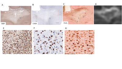 |
QSM identifies pro-inflammatory iron-positive MS lesions
Kelly Gillen, Mayyan Mubarak, Ishan Negi, Somiah Dahlawi, Thanh Nguyen, David Pitt, Yi Wang
Multiple sclerosis is an autoimmune disorder whose demyelinated plaques may be connected with elevated iron. We combined quantitative susceptibility mapping (QSM) with histopathological techniques to quantify iron, macrophages/microglia, and pro/anti-inflammatory markers to demonstrate that regions of high susceptibility on QSM correspond to pro-inflammatory iron-positive macrophages/microglia. QSM is therefore a valuable clinical tool to identify smoldering lesions not visible using conventional MRI techniques.
|
09:03
|
0086.
 |
A longitudinal study of lesion evolution in Multiple Sclerosis using multi-contrast 7T MRI
Kingkarn Aphiwatthanasumet, Olivier Mougin, Nicolas Geades, Nikos Evangelou, Molly Bright, Richard Bowtell, Penny Gowland
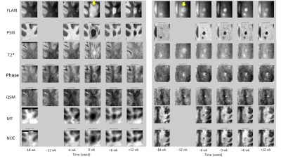
We use multi-contrast 7T MRI to evaluate longitudinal changes in white matter tissue properties prior to and after lesion appearance. Four MS patients were scanned 6 times at 6-week intervals, and 20 new lesions were identified on FLAIR images in that period. Of these, 35% showed a hypointense rim and 65% showed no rim in QSM data. Subtle changes in MT, NOE, and QSM relative to NAWM values could be detected 6 weeks prior to the first clinical appearance of new lesions. In future studies, these data will provide insight into specific tissue changes that precede lesion development in MS.
|
09:15
|
0087.
 |
A 3-year follow-up study of enhancing and non-enhancing multiple sclerosis (MS) lesions in MS patients with clinically isolated syndrome (CIS) using a multi-compartment T2 relaxometry (MCT2) model
Sudhanya Chatterjee, Olivier Commowick, Onur Afacan, Benoit Combès, Anne Kerbrat, Simon Warfield, Christian Barillot
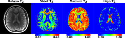
Obtaining information on condition of tissue microstructures (such as myelin, intra/extra cellular cells, free water) can provide important insights into MS lesions. However, MRI voxels are heterogeneous in terms of tissue microstructure due to the limited imaging resolution owing to existing physical limitations of MRI scanners. Here we evaluated a multi-compartment T2 relaxometry model and then used it to study the evolution of enhancing (USPIO and gadolinium positive) and non-enhancing lesions in 6 MS patients with CIS characteristics over a period 3 years with 7 follow-up scans after baseline.
|
09:27
|
0088.
 |
A multi-parametric study of MS lesions
Mara Cercignani, Camilla Vizzotto, Davide Esposito, Barbara Spano, Giovanni Giulietti, Marco Bozzali
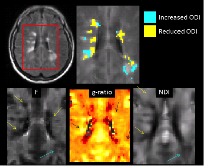
Counter-intuitively, reduced orientation dispersion has been reported in MS lesions, and confirmed by histology. Here we classify lesional tissue based on its orientation dispersion, and we compute a series of indices from from diffusion and magnetization transfer MRI to highlight potential differences in the pathological substrate of lesions with reduced vs increased orientation dispersion. We show that lesions with reduced dispersion are more likely to show extensive demyelination and axonal loss.
|
09:39
|
0089.
 |
The Age effect on Multi-parametric Magnetic Resonance Imaging changes in Multiple Sclerosis lesions
Elda Fischi-Gomez, Mário João Fartaria, Guillaume Bonnier, Cristina Granziera
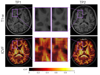
We assessed the effect of age on the longitudinal evolution of intralesional neurite density and orientation dispersion indices, magnetization transfer ratio and T1 relaxometry in a cohort of relapsing-remitting MS patients. While we observed a decrease of neurite dispersion in lesions and stable neurite density, MTR and qT1, age did not seem to influence those longitudinal changes in MS lesions.
|
09:51
|
0090.
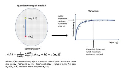 |
Linking macrostructural and microstructural damage in early MS: a geostatistical and diffusion MRI study
Carmen Tur, Robert Marschallinger, Ferran Prados, Sara Collorone, Daniel Altmann, Sébastien Ourselin, Claudia Gandini Wheeler-Kingshott, Olga Ciccarelli
Macroscopic white matter (WM) lesion volume has been extensively used to predict disability progression in multiple sclerosis (MS). However, currently used lesion-related metrics fail to capture the complexity of WM-lesion spatial distribution. Here we used geostatistics, an emerging approach to model spatial data projected onto a common coordinate space, to characterise the spatial distributional features of WM lesions of patients with their first MS attack, the clinical relevance of lesion distributional properties and their microstructural correlates, through diffusion MRI. We conclude that WM-lesion spatial distributional features reveal novel aspects of MS pathology, are clinically relevant and possess specific microstructural features.
|
10:03
|
0091.
 |
The relevance of cortical lesions in cortical thinning in multiple sclerosis by ultra-high field MRI
Video Permission Withheld
Constantina Treaba, Elena Herranz, Russell Ouellette IV, Tobias Granberg, Celine Louapre, Valeria Barletta, Ambica Mehndiratta, Jacob Sloane, Revere Kinkel, Caterina Mainero
Cortical lesions (CL) and cortical atrophy are frequent in multiple sclerosis (MS) and main determinants of disease progression. The relationship between them is still unknown, mostly due to the low sensitivity of clinical magnetic resonance imaging (MRI) to CL. Disconnection from white matter (WM) lesions has also been proposed as a pathogenic mechanism for cortical MS atrophy. Using 7 Tesla MRI that has shown increased sensitivity to CL than clinical MRI, we showed, in a large MS cohort that WM lesions are the main determinant of cortical thinning. Nevertheless, CL resulted as the main contributors of physical and cognitive disability.
|
|
