Parkinson's Disease
Oral
Neuro
Tuesday, 19 June 2018
| W05/06 |
08:15 - 10:15 |
Moderators: Shigeki Aoki, Kelly Tung |
08:15
 |
0418.
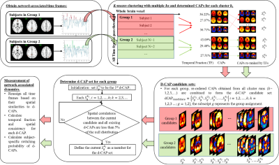 |
 Exploring Temporal Dynamics in Resting-State Networks using Co-Activation Pattern Analysis Exploring Temporal Dynamics in Resting-State Networks using Co-Activation Pattern Analysis
Xiaowei Zhuang, Ryan Walsh (co-first), Karthik Sreenivasan, Zhengshi Yang, Virendra Mishra, Dietmar Cordes
We propose a novel group CAP analysis method to investigate temporal dynamics of specific resting-state networks. Our data-driven method computes less spatially overlapping d-CAPs for each group. We compare network-dynamics between different populations using d-CAP based measurements. Using simulation, we demonstrate that the proposed method is able to determine spatially less overlapping d-CAPs for each group accurately. Using real fMRI data, we find reduced network-dynamics of most networks in PD subjects as hypothesized, which corroborates and expands upon previous electrophysiologic reports.
|
08:27
|
0419.
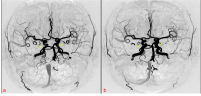 |
Findings from SNAP-MRA and PCASL in Patients with Parkinson’s Disease “OFF” and “ON” Levodopa
Yuhui Xiong, Zhangxuan Hu, Le He, Yu Ma, Wenjuan Cai, Zhensen Chen, Hua Guo
This study aimed to evaluate the effect of levodopa on cerebral arteries and gray matter cerebral blood flow (CBF) in patients with Parkinson’s disease (PD). We scanned 33 PD patients with “OFF” and “ON” levodopa using Simultaneous Non-Contrast Angiography and intraPlaque Hemorrhage (SNAP) MRA and Pseudo-Continuous Arterial Spin Labeling (PCASL), and used multiple statistical methods to analyze the data. Results from the statistical analysis show that after taking levodopa, patient’s treatment outcomes are highly correlated with his/her gray matter CBF. The results also suggest that levodopa can dilate cerebral arteries and improve CBF, but only in patients with mild symptoms.
|
08:39
|
0420.
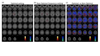 |
Functional MRI can distinguish between optimal and non-optimal frequencies in PD-DBS
Video Permission Withheld
Radhika Madhavan, Suresh Joel, Saikat Saha, Marisa DiMarziio, Eric Fiveland, Jeffrey Ashe, Michael Gillogly, Jennifer Durphy, Julia Prusik, Pilitsis Julie, Ileana Hancu
Deep Brain Stimulation is an effective treatment for Parkinson's disease symptoms. Despite its success, the underlying principle and the mechanisms are not yet fully understood. In this study, we recorded concurrent DBS-fMRI to 1) elucidate brain regions activated at the clinically optimal settings and, 2) determine the effect of changes in stimulation frequency on whole-brain activation. Optimal DBS frequencies showed activation in the thalamus and motor cortices. Further, there was a significant difference in activation in the sensorimotor cortices between the optimal and non-optimal frequencies, indicating potential use for fMRI as a tool for optimizing DBS parameters.
|
08:51
|
0421.
 |
Early Stage Parkinson’s Disease Shows Changes in Energy and Period Content of Resting-State Networks
Dietmar Cordes, Muhammad Kaleem, Xiaowei Zhuang, Karthik Sreenivasan, Zhengshi Yang, Sarah Banks, Brent Bluett, Zoltan Mari, Virendra Mishra
Low-frequency BOLD fluctuations of major resting-state networks in early Parkinson’s disease (PD) were studied and compared with matched normal controls. Empirical Mode Decomposition (EMD) was used to decompose the natural occurring frequency bands of major brain resting-state networks. The novelty of our approach lies in the data-adaptive decomposition of fMRI data using EMD, and identification of resting-state networks based on energy and period (inverse frequency) characteristics of intrinsic mode functions. For most networks studied that showed a large effect size, the frequency content of the associated network time series was found to be significantly reduced in PD.
|
09:03
|
0422.
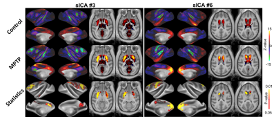 |
Resting state functional corticostriatal connectivity in Parkinsonian monkeys
Joonas Autio, Takayuki Ose, Kantaro Nishigori, Noboyoshi Tanki, Jun Takahashi, Matthew Glasser, Takuya Hayashi
Studies in animal models of Parkinson’s disease have established that excessive synchronization of neuronal activity in basal ganglia cortical loops is the hallmark of movement disorders in Parkinson’s disease. However, majority of the studies have focused on basal ganglia and motor areas in Parkinson’s disease models and it is still unclear how dopamine denervation influences other neocortical areas, which each exhibit distinct corticostriatal and thalamocortical connectivity profiles. To address this issue, we investigate the effects of dopaminergic neuronal loss on functional connectome using resting-state fMRI in anesthetized MPTP-treated monkeys.
|
09:15
 |
0423.
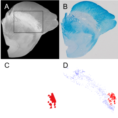 |
MR imaging of neuromelanin-iron cluster within human postmortem substantia nigra
Hansol Lee, Sun-Yong Baek, Se Young Chun, Jae-Hyeok Lee, HyungJoon Cho
The overall goal of this work was to provide a truth of neuromelanin-sensitive T1 weighted image with magnetization transfer effects and to segment the respective distribution of neuromelanin-iron complex and ferric iron within substantia nigra which are important to monitor Parkinson’s disease. Postmortem MR experiment at 7T and histological validation were performed for six normal midbrain samples. The correlation between T2*/T2 and neuromelanin pigments was highest compared to other MR parameters, especially compared to T2*, which shows specific distributions of paramagnetic molecules. Iron deposits were highly correlated with iron-sensitive T2 and T2* and the correlation was reduced for T2*/T2.
|
09:27
|
0424.
 |
Lateral-ventral tier nigral iron deposition and neuromelanin depletion in Parkinson's disease
Jason Langley, Daniel Huddleston, Stewart Factor, Bruce Crosson, Xiaoping Hu
The substantia nigra, a neuromelanin containing structure in the brainstem, contains a dense distribution of dopaminergic neurons which undergo degeneration in Parkinson’s disease. In this abstract, we examine spatial locations of neuromelanin depletion and iron deposition in the substantia nigra after onset of Parkinson’s disease. We found the lateral-ventral tier of substantia nigra to exhibit significant neuromelanin depletion whereas iron deposition was observed across the entire substantia nigra. These results accord with prior histological studies examining substantia nigra neuronal loss and may offer insight into the mechanisms behind this loss.
|
09:39
|
0425.
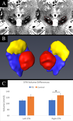 |
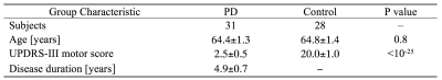
Fractional Anisotropy Within the Subthalamic Nucleus: An Imaging Biomarker for Early Parkinson’s Disease? A High-Resolution 7Tesla Magnetic Resonance Imaging Study
Remi Patriat, Jordan Kaplan, Jacob Jacob Niederer, Sommer Huffmaster, Matthew Petrucci, Noam Harel, Colum MacKinnon
In this study, we used high-resolution 7Tesla MRI to study whether subthalamic nucleus (STN) characteristics, such as volume and fractional anisotropy (FA), can be used as a biomarker for Parkinson’s Disease (PD). 7Tesla MRI data were acquired for twenty-nine PD patients and twenty-one controls. Right STN volume was significantly lower in PD patients and it was negatively correlated with the UPDRS motor score. The PD group also had significantly decreased FA values in bilateral STN. To our knowledge, this is the first study to report differences in volume and FA of the STN between people with PD and controls.
|
09:51
|
0426.
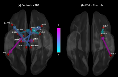 |
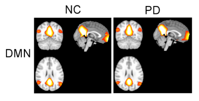
Disrupted Cortical and Subcortical Effective Connectivity in Early Parkinson’s Disease (PD): Insights from Parkinson’s Progressive Markers Initiative (PPMI) dataset
Karthik Sreenivasan, Ece Bayram, Virendra Mishra, Zhengshi Yang, Christopher Bird, Xiaowei Zhuang, Dietmar Cordes, Brent Bluett
This study utilized resting-state fMRI data to evaluate effective (directional) connectivity in newly diagnosed unmedicated patients with early PD to derive a comprehensive picture of the nature of cortico–striatal–thalamic loop connectivity in PD. We found decreased effective connectivity between the key areas in the cortico–striatal–thalamic loops in unmedicated early stage PD patients . These results were mostly lateralized to the hemisphere opposite the predominant side of parkinsonian symtoms. The findings of this study are mainly important, given the fact that, looking at changes in effective connectivity and its relation to different disease related factors may help us better understand the heterogeneity of PD and more accurately target therapeutic interventions (i.e. deep brain stimulation).
|
10:03
|
0427.
 |
Role of hippocampus in genesis of visual hallucinations in Parkinson’s disease: Insights from hippocampal sub-volumetry analysis
Abhishek Lenka, Apurva Shah, Jitender Saini, Madhura Ingalhalikar, Pramod Kumar Pal
The neurobiological underpinnings of psychosis, manifested through visual hallucinations (VH) in Parkinson’s disease (PD), are not fully elucidated, however, are linked to memory impairment and are associated with alterations in the hippocampus. To obtain profound understanding of the role of hippocampus in VH, a hippocampal subfield volumetric analysis on PD patients with (PD-P) and without psychosis (PD-NP) and healthy controls (HC) was performed. The results demonstrated a clear pattern with lowest volumes in PD-P, followed by PD-NP and the highest in HCs in several subfields. Moreover, the PD-P volumes in multiple sub-fields highly correlated with memory and attention scores.
|
|
