Neuro at Ultra-High Field
Oral
Neuro
Tuesday, 19 June 2018
| N02 |
16:15 - 18:15 |
Moderators: Priti Balchandani, Pratik Mukherjee |
16:15
 |
0578.
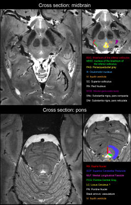 |
 High-resolution T2*-weighted imaging of subcortical brain enhanced by motion and field compensation High-resolution T2*-weighted imaging of subcortical brain enhanced by motion and field compensation
Alexander Aranovitch, Laetitia Vionnet, Simon Gross, Benjamin Dietrich, Lars Kasper, Bertram Wilm, Thomas Schmid, David Brunner, Klaas Pruessmann
We apply field control and prospective motion correction simultaneously to obtain a high-quality and high-resolution (0.3mm x 0.3mm x 1mm) T2* weighted depiction of subcortical brain structure which is a notoriously difficult region for T2* weighted scans at high field due to physiological field fluctuations. The combined approach of field control and motion correction achieved superior imaging results in the studied scenario.
|
16:27
|
0579.
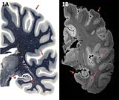 |
7T MRI allows detection of disturbed cortical layers in medial temporal lobe in patients with Alzheimer’s disease
Boyd Kenkhuis, Laura Jonkman, Marjolein Bulk, Mathijs Buijs, Jeroen J. Geurts, Wilma D.J. Berg, Louise Weerd
Using 7T T2*-w imaging, we scanned post-mortem hemispheres of aged controls and patients with Alzheimer’s disease (AD) to assess the potential of MRI for anatomical cortical parcellation of the medial temporal lobe based on myelo- and cytoarchitectural contrast. Segmentation was possible in both controls and AD scans and distortions in cortical lamination of AD patients could be observed in specific regions of the medial temporal lobe. Observed contrast correlated highly with myelination patterns on histology. 7T MRI may thereforedetect pathogenic cortical laminar distortions and provide new information on involved neuroanatomical structures and layers in AD.
|
16:39
|
0580.
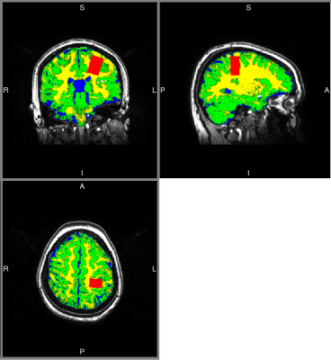 |
GABA concentration in sensorimotor area correlates with observed inhibitory response measured by magnetoencephalography.
Lucrezia Liuzzi, Bernard Lanz, Chen Chen, Gillian Roberts, Ryan Hill, Markus Bauer, Peter Morris, Matthew Brookes
Electrophysiological imaging suggests that task induced change in beta oscillations (13-30Hz) modulates with attention, and reflects synaptic inhibitory responses. GABA is known to mediate synaptic inhibition and hence may relate to these electrophysiological dynamics. Here, we determined GABA concentration in primary sensorimotor cortex, using MRS at 7T, and measured the electrophysiological response to a sensory attention task, using MEG, in the same region. We correlated the post-stimulus synchronisation in beta in the left sensory-motor cortex with GABA concentration detected using a STEAM sequence. Results show significant correlation (R = 0.48; p 0.010) across 28 participants.
|
16:51
|
0581.
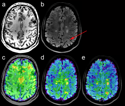 |
Ultra-high resolution MR spectroscopic imaging at 7 Tesla in Multiple Sclerosis: Initial results and comparison with clinical MR imaging
Wolfgang Bogner, Eva Heckova, Bernhard Strasser, Gilbert Hangel, Assunta Dal-Bianco, Elisabeth Springer, Michal Povazan, Petra Hnilicova, Paulus Rommer, Fritz Leutmetzer, Siegfried Trattnig, Stephan Gruber
Conventional T1/T2-weighted MRI has become indispensable for Multiple Sclerosis(MS) diagnosis and treatment monitoring, although it reflects only general macroscopic tissue damage. MRSI allows the additional mapping of pathological processes in MS on a biochemical level. In 39 relapsing-remitting MS patients and an age/sex-matched control group(n=10), we show that clinically feasible ultra-high resolution (100x100 matrix) FID-MRSI in ~6min reveals even well-delineated (sub-)cortical MS lesions down to ~3mm in regions inconspicuous on conventional MRI. Regions of mIns were often larger than on FLAIR and NAA maps, suggesting that mIns increase may be an earlier imaging biomarker for neuroinflammation/lesion development than conventional MRI.
|
17:03
|
0582.
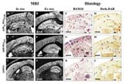 |
Contrast-enhanced MR microscopy of amyloid plaques in five mouse models of Alzheimer’s disease: comparison with amyloid plaques detection in human brains.
Clémence Dudeffant , Matthias Vandesquille, Kelly Herbert, Sandro Alves, Emmanuel Comoy, Fanny Petit, Marc Dhenain
Gadolinium(Gd)-stained MRI is based on Gd-contrast agent administration into the brain. This method significantly improves the detection of amyloid plaques, one of the lesions of Alzheimer's disease and a potential biomarker for its diagnosis. Here, we aimed to better understand the origin of contrast induced by amyloid plaques by determining critical parameters required for their detection using five mouse models of amyloidosis presenting with different plaque typologies. Then, we showed for the first time that Gd-stained MRI can detect amyloid plaques in postmortem human brain tissues and compared the detection achieved in mice with those obtained in human samples.
|
17:15
|
0583.
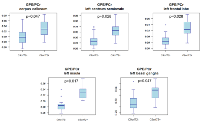 |
$$$^{31}$$$P MRSI of asymptomatic C9orf72 carriers and non-carriers at 7 Tesla
Graziella Donatelli, Henk-Jan Westeneng, Kevin van Veenhuijzen, Harold Tan, Peter Luijten, Dennis Klomp, Leonard van den Berg, Jannie Wijnen
Amyotrophic Lateral Sclerosis (ALS) is a progressive neurodegenerative disease with a largely unknown pathogenesis. The most common gene mutation in both familial and sporadic ALS is the C9orf72 repeat expansion. Investigating asymptomatic carriers of this mutation might give more insight into possible preclinical brain alterations. Using whole brain 31P MRSI at 7T, glycerophosphoethanolamine-to-phosphocreatine ratio (GPE/PCr) and uridine diphosphoglucose-to-phosphocreatine ratio (UDPG/PCr) were found to be higher in a number of brain regions in asymptomatic carriers compared with asymptomatic non-carriers. The increased GPE/PCr and UDPG/PCR might respectively indicate an increased catabolism of the cell membranes and an imbalance of energy metabolism.
|
17:27
|
0584.
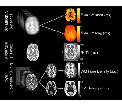 |
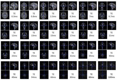
Interpreting fast and slow 23Na relaxation rates in human brain: comparisons with tissue microstructure at 7 Tesla.
Scott Kolbe, Yasmin Blunck, Rebecca Glarin, Syeda Taqdees, Bradford Moffat, Roger Ordidge, Leigh Johnston, Jon Cleary
23Na, a quadrupolar nucleus, exhibits bi-exponential relaxation in brain tissue. This study aimed to compare fast and slow sodium T2* relaxation times obtained using a 3D multi-echo radial sequence, to measures of tissue microstructure derived from advanced diffusion MRI and proton T1 mapping at 7T. We observed significant correlations between sodium T2* relaxation times and all measures of fibre and cellular density and T1. The results suggest that relative contribution of quadrupolar and dipolar relaxation pathways are related to the underlying tissue microstructure.
|
17:39
|
0585.
 |
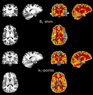
Whole Brain FLAIR Imaging at 7T Employing Universal Pulses
Eberhard Pracht, Vincent Gras, Nicolas Boulant, Tony Stöcker
In this work, we present a fluid suppressed Turbo-Spin-Echo sequence for ultra-high-field application. To obtain images comparable in contrast and homogeneity with 3T results, we replaced all RF pulses with universal parallel transmit kT-point pulses. During the imaging session, no pulse calculations or complex B1 shimming procedures are necessary, making this approach a promising tool for clinical application.
|
17:51
 |
0586.
 |
Parallel transmission kT-points and CAIPIRIHNA accelerated MP2RAGE for ultra-high field anatomical imaging
Valentin Kemper, Federico De Martino, Elia Formisano, Tobias Kober, Benedikt Poser
The MP2RAGE imaging sequence is widely used for T1 weighted anatomical imaging, especially at 7T, where B1 inhomogeneity may degrade the image quality. This study demonstrates an improved MP2RAGE sequence featuring kT-points parallel transmission excitation and CAIPIRINHA 2D acceleration for improved T1 weighted anatomical images of the living human brain at 7 T and 9.4 T. High resolution (0.6 mm and 0.45 mm isotropic) data with high contrast-to-noise ratio allow for improved cortical segmentation, e.g. for cortical laminar investigations. We further demonstrate a 6-fold accelerated whole brain CAIPIRINHA acquisition at 0.6 mm resolution in only 4:10 min.
|
18:03
|
0587.
 |
Compartmental brain sodium concentrations in human brain tissues in white matter and grey matter: a multi-echo ultra-high field 23Na-MRI study
Ben Ridley, Armin Nagel, Mark Bydder, Adil Maarouf, Jan-Patrick Stellmann, Soraya Gherib, Jeremy Verneuil, Patrick Viout, Maxime Guye, Jean-Philippe Ranjeva, Wafaa Zaaraoui
Using multi-echo 23Na-MRI at 7T acquired in 13 healthy subjects and modelling the relationship between signal and reference concentration and applying it to in vivo 23Na-MRI signal, we quantify both T2* decay times and concentrations associated with short and long components for the first time. Relaxation times and concentrations differed between grey and white matter and subregions of differing tissues, suggesting sensitivity of 23Na toward features of tissue composition. As such, these results raise the prospect of multi-echo 23Na-MRI as an adjunct source of information on biochemical mechanisms in both physiological and pathophysiological states.
|
|
