Traumatic Brain Injury
Oral
Neuro
Tuesday, 19 June 2018
| S05 |
13:45 - 15:45 |
Moderators: Andre Obenaus, Guangming Lu |
13:45
 |
0503.
 |
 Multi-parametric MRI data fusion reveals acute and persistent brain changes in concussed female varsity rugby players Multi-parametric MRI data fusion reveals acute and persistent brain changes in concussed female varsity rugby players
Kathryn Manning, Alberto Llera, Robert Bartha, Gregory Dekaban, Christy Barreira, Arthur Brown , Lisa Fischer, Tatiana Jevreomvic, Kevin Blackney, Timothy Doherty, Douglas Fraser, Jeff Holmes, Christian Beckmann, Ravi Menon
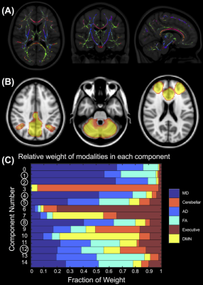
Multi-parametric 3T MRI data was acquired in female varsity rugby players during the in- and off-season and compared to concussed teammates acutely (24-72 hours) and longitudinally (3- and 6-months) after a mild traumatic brain injury (mTBI). Using linked independent component analysis, we found acute and prolonged resting state functional connectivity and diffusion white matter microstructure changes that persisted beyond symptomatic recovery and clearance to return to play. These fused components also significantly correlated with aspects of concussion history and were robust and consistent across model orders and within individual concussed athletes longitudinally.
|
13:57
|
0504.
 |
Chronic Neurovascular Dysfunction in a Mouse Model of Repeated Mild Traumatic Brain Injury
Conner Adams, Paolo Bazzigaluppi, Tina Beckett, Jossana Bishay, Joe Steinman, Lydiane Hirschler, Jan Warnking, Emmanuel Barbier, JoAnne McLaurin, John Sled, Bojana Stefanovic
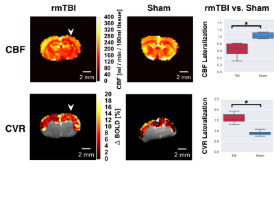
In-situ assessments of brain function following repeated mild traumatic brain injury (rmTBI) have yet to be seen. Herein, we report the first functional imaging study in the chronic phase of a mouse model of rmTBI. Pseudo-continuous arterial spin labelling MRI revealed a reduction in basal cerebral blood flow and cerebral reactivity, while electrophysiological recordings of evoked responses were greatly reduced in injured brain. Finally, immunohistochemical analysis of vascular endothelium, astrogliosis, and neurons was performed to investigate cellular populations.
|
14:09
|
0505.
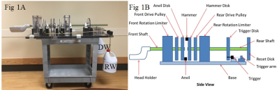 |
A Weighted Head Accelerator Mechanism (WHAM) for imaging brain tissue deformation during mild head rotation using a tagged MRI sequence
Ronald Pratt, Greg Lee, Aaron McAllister, Greg Myer, Thomas Klein, Christopher Ireland, Wolfgang Loew, Matt Lanier, Charles Dumoulin
A new research tool, the Weighted Head Accelerator Mechanism (WHAM), is described. The WHAM, used together with a custom MRI sequence, permits in depth investigations into brain biomechanics during mild head rotations and the evaluation of mild traumatic brain injury mitigating strategies by directly evaluating their effects on brain biomechanics in vivo.
|
14:21
|
0506.
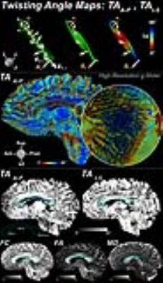 |
Ribbon Tractography Reveals Reorientation of White Matter in the Corpus Callosum Following Severe Traumatic Brain Injury
Choukri Mekkaoui, Brian Edlow, William Kostis, Marcel Jackowski, Kawin Setsompop, Thomas Witzel, Qiuyun Fan, Ned Ohringer, Javier Cabrera, Timothy Reese, Ona Wu, Susie Huang
Traditional streamline tractography does not capture the changes in fiber twisting resulting from traumatic brain injury. We used ribbon tractography to quantify the acute directional changes in white matter tracts in patients with traumatic brain injury. We identified significant changes in the corpus callosum of these patients both as compared to healthy controls and between the acute and follow-up scans of a subset of patients. These findings reveal alterations in the architecture of the corpus callosum within and beyond the area of injury and offer a potential marker for structural changes that are not detected by standard techniques.
|
14:33
|
0507.
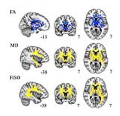 |
The Evolution of White Matter Microstructural Changes after Mild Traumatic Brain Injury: A Longitudinal DTI and NODDI Study
Eva Palacios, Julia Owen, Esther Yuh, Mary Vassar, Geoffrey Manley, Pratik Mukherjee
Problem: Mild traumatic brain injury (mTBI) can result in long-term sequelae. Lack of sensitive biomarkers makes diagnosis challenging. Methods: Cross-sectional and longitudinal study of 40 mTBI patients at 2 weeks and 6 months after injury. Diffusion tensor imaging and multishell neurite orientation dispersion and density imaging (NODDI) parameters were assessed. Results: Cross-sectional analysis between patients at 2-weeks and controls revealed a decrease of fractional anisotropy and increase of mean diffusivity in the patient group together with elevated free water values. Longitudinally, after mTBI, a decline in neurite density was observed. Conclusions: NODDI measurements are sensitive imaging biomarkers for the subtle underlying white matter pathology after mTBI.
|
14:45
|
0508.
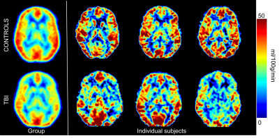 |
5-Year Arterial Spin Labeling MR Perfusion sequelae of concussive blast traumatic brain injury
Swati Rane, Jalal Andre, Jason Barber, Nancy Temkin, Christine MacDonald
Concussive blast traumatic brain injury (cbTBI) may benefit from evaluation with advanced MR imaging techniques. Here we report the results of a 5-year follow-up prospective, observational, longitudinal human cohort study evaluating cbTBI using arterial spin labeling, correlated with durable measures of long term outcome taken from an extensive battery of neurobehavioral, neuropsychological, and psychiatric evaluations. Specifically, service members who sustained combat-related cbTBI exhibited significant, regional hypoperfusion 5 years post-injury compared to combat-deployed controls, associated with measures of long term outcome.
|
14:57
|
0509.
 |
Redundancy and resiliency of arousal mechanisms in traumatic coma patients
Marta Bianciardi, Saef Izzy, Bruce Rosen, Lawrence Wald, Brian Edlow
Traumatic brain injury to brainstem/thalamic/hypothalamic/basal-forebrain arousal nuclei is implicated in the pathogenesis of coma. However, due to difficulty in localizing these nuclei with conventional MRI, it is unknown which lesioned nuclei cause coma, and which are compatible with recovery of arousal/consciousness. We mapped our recently developed brainstem arousal-nuclei atlases and current thalamic/hypothalamic/basal-forebrain atlases to 3Tesla susceptibility-weighted-images in twelve acute traumatic-coma patients, who recovered full arousal/consciousness within six-months. We identified multiple combinations of injured brainstem/thalamic arousal-nuclei that cause acute coma, yet are compatible with a full recovery of arousal/consciousness. Thus, there is redundancy and resiliency of arousal mechanisms in traumatic coma.
|
15:09
|
0510.
 |
Assessing Mild Traumatic Brain Injury (TBI) Induced Optic Neuropathy in Novel Closed-head Injury Mouse Model Using Diffusion Basis Spectrum Imaging (DBSI)
Tsen-Hsuan (Abby) Lin, Andrew Sauerbeck, Terrance Kummer, Sheng-Kwei Song
Traumatic brain injury (TBI) is a major cause of death and disability worldwide, and mild TBI (mTBI) is the most common type injury. TBI-induced optic neuropathy (ON) has recently raised attention due to subjective complaints of visual impairment. Currently, non-invasive and longitudinal direct assessment in TBI-induced ON is still limited. In the current study, we applied diffusion basis spectrum imaging (DBSI) to a novel surgery-free and closed-head impact model of TBI called modCHIMERA at 3 days post-injury to study TBI-induced ON with histological validation on our imaging metrics.
|
15:21
|
0511.
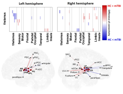 |
Association Between Concussion-induced Dizziness and Damage of Thalamo-cortical Connectivity after mild Traumatic Brain Injury
Chia-Feng Lu, Yu-Chieh Kao, Li-Chun Hsieh, Sho-Jen Cheng, Ho-Fang Huang, Wen-Jin Hsieh, Fei-Ting Hsu, Ping-Huei Tsai, Cheng-Yu Chen
Dizziness is a frequent symptom of concussion, however neuroimaging evidence that supports clinical symptoms after mTBI was less explored. This study demonstrated the post-concussion damages of thalamo-cortical networks revealed by the axonal injuries of specific fiber tracts and the altered functional connectivity. The estimates of Dizziness Handicap Inventory significantly correlated to the functional connectivity of thalamic nuclei.
|
15:33
|
0512.
 |
Metabolic concentrations in normal appearing brain tissue: 3 month after severe pediatric TBI.
Petr Menshchikov, Natalia Semenova, Andrei Manzhurtsev, Maxim Ublinskii, Ilya Melnikov, Tolib Akhadov
In this study for the first time cerebral NAA, Asp and Glu concentrations of major inhibitory were simultaneously estimated in patients severe TBI . This work revealed significant reduction NAA and Asp concentrations in frontal lobe (on 65 and 61% respectively) in patients with severe TBI as compare to control group. At the same time, there was no significant change in Glu concentration. Our findings indicate that the main reason for the [NAA] reduction in the chronic severe TBI is caused by a decrease in [Asp]. Precursor of synthesis [NAA]. [Asp] reduction along with an unchanged Glu level may be caused by the dysfunction of one of the most important metabolic regulation systems - the malate-aspartate shuttle (MAS).
|
|
