Application of Neurovascular Methods
Oral
Neuro
Tuesday, 19 June 2018
| N02 |
08:15 - 10:15 |
Moderators: Luis Hernandez-Garcia, Seong-Gi Kim |
08:15
 |
0368.
 |
 Unraveling Cardiac and Respiratory Contributions to Brain Tissue Motion using Single Shot 2D DENSE at 7T MRI. Unraveling Cardiac and Respiratory Contributions to Brain Tissue Motion using Single Shot 2D DENSE at 7T MRI.
Jacob Jan Sloots, Ayodeji Adams, Peter Luijten, Geert Jan Biessels, Jaco Zwanenburg
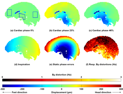
The cardiac cycle and respiration both influence CSF dynamics and therefore the displacement of brain tissue. In this work we unravel their contribution to brain tissue displacement using a single shot 2D cine displacement-encoded imaging method employing stimulated echoes (DENSE) for brain motion measurements. Displacement-encoded data sets in the Feet-to-Head direction of seven volunteers were fitted to a linear model. Consistent trends in displacements were observed. The developed DENSE sequence results showed similar sized contributions to brain tissue displacement. Relating these displacements to contributions to the clearance system remains future work.
|
08:27
 |
0369.
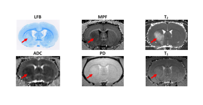 |
Macromolecular proton fraction closely correlates with myelin loss in the rat ischemic stroke model
Alena Kisel, Marina Khodanovich, Dmitriy Atochin, Andrey Akulov, Lilia Mustafina, Anna Naumova, Vasily Yarnykh
Non-invasive quantitative assessment of myelin damage in ischemic stroke is currently unavailable. The goal of this study was to evaluate a recently proposed myelin imaging technique, macromolecular proton fraction (MPF) mapping in the rat stroke model and compare it with histological myelin quantitation. MPF decrease in the brain infarct closely correlated (R = 0.81, p<0.001) with luxol fast blue staining on the 1st, 3rd, and 10th day after stroke. Further improvement in accuracy of myelin quantitation (R = 0.98) was achieved with the use of bivariate linear regression model including T2 to correct errors related to edema.
|
08:39
|
0370.
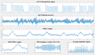 |
The effect of breath-hold on cardiovascular pulse in the brain - a multimodal MREG study.
Lauri Raitamaa, Viola Borchardt, Niko Huotari, Heta Helakari, Janne Kananen, Vesa Korhonen, Vesa Kiviniemi
MREG enables critical sampling of human brain physiology. This study reveals novel pulsation changes during repeated breatholds of 32 seconds. Cardiorespiratory control centers in the brain stem show increased pulsation amplitude during the end of the breathold. Also the overall cardiac pulse propagation in the brain alters markedly by breathold, especially when repeated. The findigs show that the MREG data enables spatially accurate estimation of the status of the cardiovascular pulsation which is the primary driver of glymphatic brain clearance.
|
08:51
|
0371.
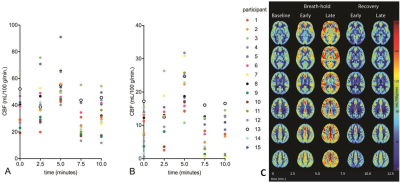 |
ASL imaging and 31P/1H-MRS during prolonged breath-hold among experienced freedivers: Insights into cerebrovascular and metabolic responses
Video Permission Withheld
Vera Keil, Henri Mutsaerts, Lars Eichhorn, Frank Träber, Wolfgang Block, Burkhard Mädler, Kim van de Ven, Jeroen Siero, Bradley MacIntosh, Jan Petr, Hans Schild, Elke Hattingen
This pCASL and 31P/1H-MRS study explored the cerebrovascular reactivity (CVR) and its efficacy on brain metabolic stability during a five-minute breath-hold in fifteen experienced freedivers. Cerebral blood flow (CBF) increase occurred later than the decrease of the recently discovered arterial transit time correlate, spatial CoV. The latter may thus be an early CVR biomarker. CBF varied between vessel territories, gray and white matter and usually lowered with more experience. MRS showed near stable physiological cerebral pCr, ATP and pH concentrations despite peripheral lactate acidosis. In conclusion, this trial revealed that CVR sufficiently compensates the metabolic challenge of a five minute breath-hold. In conclusion, this trial revealed that cerebral perfusion increase sufficiently compensates the metabolic challenge of a five-minute breath-hold.
|
09:03
|
0372.
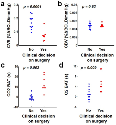 |
Sensitivity and specificity of cerebrovascular reactivity in predicting surgical decisions in Moyamoya patients
Peiying Liu, Babu Welch, Binu Thomas, Yang Li, Marco Pinho, Judy Huang, Hanzhang Lu
Moyamoya disease (MMD) is characterized by chronic occlusion of the distal intracranial internal carotid arteries and can be treated by revascularization surgery. At present, surgical decisions are primarily based on symptomatology and imaging studies such as DSA and SPECT. However, current procedures are costly, invasive, and qualitative. Here we applied a novel iVas-MRI technique that provides quantitative assessment of multiple hemodynamic parameters in a single scan of 9 minutes, and examined its sensitivity and specificity in predicting surgical decisions. Our results showed that iVas-MRI has an overall accuracy of 0.93 in predicting surgical decisions in MMD.
|
09:15
|
0373.
 |
Baseline Risk Factors Suggestive of Advanced Vascular Disease Predict Differences in Brain CBF Changes After Carotid Revascularization Surgery
Salil Soman, Kyuwon Lee, Weiying Dai, Elizabeth Hitchner, Michael Moseley, Greg Zaharchuk, Allyson Rosen, Wei Zhou
Carotid stenosis is a risk factor for stroke. A number of risk factors are associated with stroke. Our work shows specific risk factors that predict differences in brain perfusion using ASL MRI technique after carotid revascularization surgery. Specifically elevated systolic blood pressure, chronic renal insufficiency, and history of prior stroke have mpacts on initial baseline to immediately post surgery prefusion increase, and baseline elevated cholesterol and body mass index show differences in 1 day to 6 months post operation. This is in contrast to hypertension, smoking and diabetes, which did not show significant predict relationships. Our work suggests that these risk factors may be more closely related to cerebrovascular dysfunction.
|
09:27
|
0374.
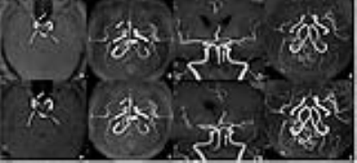 |
Clinical Feasibility Study of an Accelerated 3D Intracranial Magnetic Resonance Angiography Using Compressed-Sensing Algorithm
Zhiyong Lin, Xiaodong Zhang, Ke Wang, Yuan Jiang, Xiaoyu Hu, Yong Huang, Shuai Ma, Yi Liu, Lina Zhu, Zhizheng Zhuo, Jing Liu, Xiaoying Wang
Accelerated 3D intracranial magnetic resonance angiography(MRA) using Compressed-Sensing algorithm could be clinically valuable not only for improving the image quality and having almost the same diagnostic performance compared to conventional intracranial MRA, but also for reducing the scanning time which could improve the overall workflow of MRA imaging. It is a feasible protocol in intracranial MRA imaging.
|
09:39
|
0375.
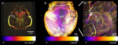 |
Multiscale multimodal imaging of the mouse cerebral vascular architecture.
Rukun Hinz, Jan Detrez, Lore Peeters, Caroline Berghmans, Marleen Verhoye, Annemie Van der Linden, Winnok De Vos, Georgios Keliris
We present a multiscale multimodal atlas of the mouse cerebral vasculature co-registered to the Allen Brain atlas space. To this end, we combined MRI anatomical imaging with the macro vasculature detected with TOF-MRA as well as the micro vasculature acquired from 3D microscopic imaging of a cleared mouse brain. To achieve a better homogeneity, we used a double labeling of the vasculature using isolectin to stain the vascular wall combined with an albumin staining of the blood vessel lumen. The atlas provides a reference space across multiple imaging modalities and a link to other functional, anatomical and genetic information.
|
09:51
|
0376.
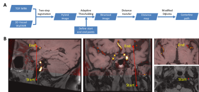 |
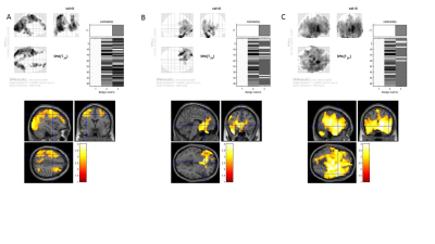
Hybrid Vessel Centerline Tracking: Towards Automated Lesion Analysis on 3D Intracranial Vessel Wall MR
Feng Shi, Wenhao Xu, Haili Wu, Zixiao Tian, Qi Yang, Debiao Li, Zhaoyang Fan
We proposed a hybrid vessel centerline tracking method on 3D intracranial vessel wall MR. TOF MRA was used as a complementary modality that provides additional information for potential vessel path extraction. Results show high agreement of results from the proposed method and manual rater.
|
10:03
|
0377.
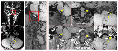 |
Three Dimensional Intra- and Extra-cranial Arterial Vessel Wall Joint Imaging in Patients with ischemic stroke
Na Zhang, Lin Jia, Jinhao Lyu, Lei Zhang, Wenxiao Jia, Hairong Zheng, Xin Liu
Intracranial and carotid atherosclerotic plaques are responsible for about 75% of ischemic stroke, the leading cause of mortality and morbidity worldwide. A 3D intra- and extra-cranial arterial vessel wall joint imaging was developed for simultaneously evaluating intracranial and carotid arterial plaques. The aim of this study was to assess the clinical potential of the 3D joint imaging technique in a large-scale patients with recent cerebrovascular symptoms. In general, this joint imaging method allows satisfactory image quality and comprehensive evaluation of atherosclerotic disease, and it has great potential to be used for optimizing treatment.
|
|
