Brain Tumours: Pre-Treatment
Oral
Neuro
Wednesday, 20 June 2018
| W03/04 |
16:15 - 18:15 |
Moderators: Meiyun Wang, Sammy Badr |
16:15
|
0953.
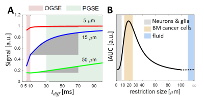 |
 Selective Size Imaging using Filters via diffusion Times (SSIFT): A new contrast-free highly-specific MR cancer imaging method Selective Size Imaging using Filters via diffusion Times (SSIFT): A new contrast-free highly-specific MR cancer imaging method
Junzhong Xu, Albert Attia, Lori Arlinghaus, Austin Kirschner, Evan Osmundson, Hakmook Kang, Guozhen Luo
We propose a novel, exogenous-agent-free, highly-specific, and high-resolution cancer imaging technique termed SSIFT. Based on different diffusion time dependence on length scales, SSIFT creates filters via appropriately chosen diffusion times to selectively enhance detection sensitivity to cancer cells with simultaneous suppression of sensitivity to normal brain cells, vasogenic edema, and cystic fluid. In the first applications in metastatic brain cancer patients, SSIFT is capable of significantly enhancing tumor conspicuity and delineation, and more importantly capable of differentiating tumor recurrence from radionecrosis, which is not reliably achievable by current MRI methods.
|
16:27
|
0954.
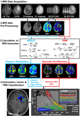 |
Tumor microenvironment (TME) mapping: MRI of intratumoral heterogeneity of oxygen metabolism and neovascularization uncovers two survival relevant subgroups of IDH1 wild-type glioblastoma
Andreas Stadlbauer, Max Zimmermann, Arnd Dörfler, Roland Coras, Stefan Oberndorfer, Michael Buchfelder, Karl Rössler
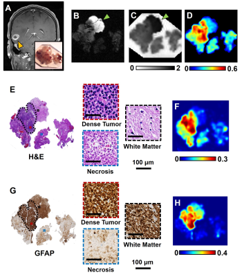
The dismal prognosis of glioblastoma is largely attributed to hypoxic and perivascular niches in the tumor microenvironment (TME) which are essential for elucidation of pathophysiological mechanisms behind therapy resistance and recurrence. Here, we combined MRI biomarkers for oxygen metabolism and neovascularization with an automatic classification strategy for localization of hypoxic and vascular niches within the heterogeneously structured TME. Correlation with the metabolic pathway for energy production uncovered two different phenotypes for glioblastoma IDH1wt: A glycolytic phenotype with stable functional neovasculature, and a necrotic/hypoxic phenotype with defective neovasculature and a more aggressive tumor behavior. The glycolytic phenotype showed longer progression-free survival.
|
16:39
|
0955.
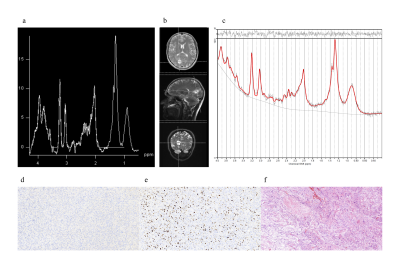 |
Magnetic Resonance Spectroscopic Differences of Diffuse Glioma Groups Classified by IDH and TERT Promoter Mutations at 3T
Esin Ozturk-Isik, Sevim Cengiz, Alpay Ozcan, Cengiz Yakicier, M. Necmettin Pamir, Koray Ozduman, Alp Dincer
Overall survival of gliomas has been reported to be highly associated with the presence of isocitrate dehydrogenase (IDH) and telomerase reverse transcriptase (TERT) promoter mutations. The aim of this study is to define MR spectroscopic (MRS) differences of diffuse glioma subgroups classified by IDH and TERT promoter mutations at 3T. TERT-only mutated and TERT wild type IDH wild type (TW-IDHW) gliomas had similar metabolic profiles. Besides well-reported 2HG, our study indicated the importance of glycine, glutathione, glutamate, and glutamine in identification of IDH-mutant gliomas. TERT-only gliomas had the highest glutamate and glutathione, which might be indicators of poor overall survival.
|
16:51
|
0956.
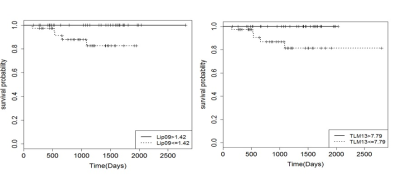 |
Magnetic Resonance Spectroscopy markers of survival in paediatric brain tumours: A 3T Multi-Centre investigation
Ben Babourina-Brooks, Lesley MacPherson, Laurence Abernethy, Theodoros Arvanitis, Simon Bailey, Nigel Davies, Daniel Gutierrez , Tim Jaspan , Dipayan Mitra, Paul Morgan, Barry Pizer , Richard Grundy , Dorothee Auer, Andrew Peet
Brain tumours have a high mortality rate and are the most common solid tumour of childhood. The non-invasive imaging technique, MRS, measures tumour metabolites which can provide additional prognostic information to aid in clinical management. MRS metabolites Glycine, Scyllo-Inositol, NAA and Lipids have been associated with prognosis for pediatric brain tumour patients in a single centre 1.5T study. This study aimed to validate these MRS survival markers in a 3T multicentre setting. In this preliminary study Lipids were validated as a survival marker across childhood brain tumours.
|
17:03
 |
0957.
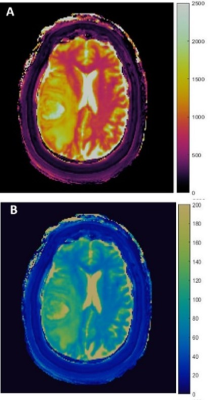 |
RADIOMIC ANALYSIS OF 3D MR FINGERPRINTING IN ADULT BRAIN TUMORS
Louisa Onyewadume, Ozden Kilinc, Satyam Ghodasara, Debra McGivney, Samuel Frankel, Dan Ma, Sara Dastmalchian, Jeffrey Sunshine, Marta Couce, Mark Griswold, Vikas Gulani, Jill Barnholtz-Sloan, Andrew Sloan, Chaitra Badve
Though conventional and advanced MR imaging studies such as perfusion and MR spectroscopy are useful for evaluating brain tumors, there remains a need for a rapid, quantitative, and non-invasive method. Magnetic Resonance Fingerprinting (MRF) utilizes pseudo-randomized acquisition parameters to simultaneously quantify multiple tissue properties including T1 and T2 relaxation times. A previous 2D MRF study quantitatively differentiated between solid tumor and peri-tumoral white matter regions of various brain tumors. In this ongoing study we demonstrate the capability of volumetric 3D MRF to improve lesion characterization between adult intra-axial brain neoplasms using first- and second- order radiomic analysis.
|
17:15
|
0958.
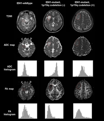 |
Whole-Tumor Histogram and Texture Analyses of Diffusion Tensor Imaging for Evaluation of IDH1 Mutation and 1p/19q Codeletion Status in WHO grade II gliomas
Yae Won Park, Kyunghwa Han, Sung Soo Ahn, Sohi Bae, Yoon Seong Choi, Jong Hee Chang, Se Hoon Kim, Seok-Gu Kang, Eui Hyun Kim, Seung-Koo Lee
We analyzed the histogram and texture features of apparent diffusion coefficient (ADC) and fractional anisotropy (FA) based on entire tumor to determine isocitrate dehydrogenase 1 (IDH1) mutation and 1p/19q codeletion status in WHO grade II gliomas. Regions of interest were drawn on ADC and FA maps of 93 grade II gliomas. Histogram and texture analyses were performed. The areas under the curve (AUC) for IDH1-wildtype prediction was 0.853, and AUC for 1p/19q codeletion prediction was 0.807. The whole-tumor histogram and texture features of ADC and FA maps are useful in predicting the molecular status in WHO grade II gliomas.
|
17:27
 |
0959.
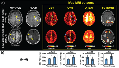 |
Integrated vascular (iVas) MRI in brain tumors
Yang Li, Peiying Liu, Shruti Agarwal, Xirui Hou, Sherry Shen, Jay Pillai, Hanzhang Lu
Perfusion and functional MRI, yielding markers such as cerebral blood volume, cerebrovascular reactivity, and functional connectivity, play an important role in the diagnosis and treatment of brain tumors. However, a major limitation is that collection of all this information requires separate scans and, in some cases, separate visits. Here we applied a novel iVas-MRI technique that provides quantitative assessment of multiple hemodynamic parameters and functional connectivity in a single scan of 9 minutes. It was found that the multi-parametric maps can reliably differentiate tumor from normal tissue and can further predict tumor grade.
|
17:39
|
0960.
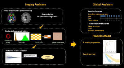 |
Radiomics utilizing Fractional Anisotropy in Peritumoral Nonenhancing Region Predicts Local Progression and Overall Survival in Patients with Glioblastoma
Eun-Jung Choi, Min Jae Yoon, Ho Sung Kim, Jongho Lee, Ji Eun Park
We explored the radiomic features of peritumoral nonenhancing lesion in newly diagnosed glioblastoma patients to predict local progression and overall survival using fractional anisotropy (FA) at 3 Tesla. Among 1618 extracted radiomic features, 8 FA features were significantly associated with 6-month progression and overall survival (OS). The cross-validated area under the ROC curve (AUC) for 6-month progression was 0.71 and C-index for OS was 0.75. FA radiomics in nonenhancing lesion has the potential for predicting local progression and overall survival in glioblastoma.
|
17:51
|
0961.
 |
Simultaneous pH- and oxygen-weighted metabolic MRI of human gliomas at 3T using multi-echo amine proton chemical exchange saturation transfer spin-and-gradient-echo echoplanar imaging (CEST-SAGE-EPI)
Benjamin Ellingson, Jingwen Yao, Ararat Chakhoyan, Phioanh Nghiemphu, Albert Lai, Whitney Pope, Linda Liau, Timothy Cloughesy
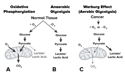
Glycolysis is enhanced in cancers, even in the presence of abundant oxygen, leading to accumulation of lactic acid. We present a technique for fast pH- and oxygen-weighted MR imaging using multi-echo amine proton chemical exchange saturation transfer echo spin-and-gradient echoplanar imaging (CEST-SAGE-EPI) on a clinical 3T MRI system. In phantom and human experiments, we investigate the ability to simultaneously measure MTRasym at 3ppm, a measure dependent on pH, and R2’, which is sensitive to oxygen extraction. Results suggest T2 hyperintense tumor is acidic, but not hypoxic; whereas contrast enhancing tumor is acidic and hypoxic, consistent with known cancer biology.
|
18:03
|
0962.
 |
Grading Brain Tumor Using ADC as a Marker of Cellularity: Fact or Fiction
Zezhong Ye, Joshua Lin, Richard Price, Jeff Viox, Michael Wallendorf, Sonika Dahiya, Albert Kim, Sheng-Kwei Song
Here we demonstrate that ADC is not a reliable imaging biomarker of tumor cellularity in high grade glioma since ADC does not consistently correlate with histology determined tumor cellularity. In contrast, diffusion MRI Histology (D-Histo) derived restricted isotropic diffusion fraction demonstrated a significantly positive correlation with histology determined cellularity in high grade glioma.
|
|
