Brain Tumours: Post-Treatment
Oral
Neuro
Thursday, 21 June 2018
| S04 |
15:30 - 17:30 |
Moderators: Michaël Belloy, Sabrina Ronen |
15:30
|
1231.
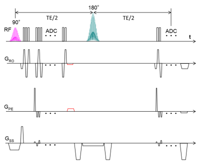 |
 A High Resolution Gradient-Echo/Spin-Echo EPI Sequence for Vessel Architecture Imaging A High Resolution Gradient-Echo/Spin-Echo EPI Sequence for Vessel Architecture Imaging
Ke Zhang, Seong Dae Yun, Simon Triphan, Volker Sturm, Lukas Buschle, Artur Hahn, Sabine Heiland, Martin Bendszus, Heinz-Peter Schlemmer, N. Jon Shah, Christian Ziener, Felix Kurz
To obtain vessel architectural imaging (VAI), a dual gradient-echo/spin-echo EPI sequence is needed to simultaneously track the dynamic signal changes in both gradient echo and spin echo contrasts. However, brain coverage and in-plane matrix size in previous brain studies were limited. In this study, the multiband excitation and blipped-CAIPI techniques were applied to improve the slice coverage. To enhance the in-plane resolution, two rephasing gradients were inserted after the GE readout, to return the data acquisition to the k-space center before the SE readout and enable parallel imaging techniques.
|
15:42
|
1232.
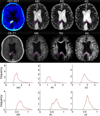 |
Combined 18-FET-PET and diffusion kurtosis imaging study in treated glioblastoma patients: differentiation between metabolically active tumours and treatment-induced tissue abnormalities
Farida Grinberg, Francesco D’Amore, Jörg Mauler, Norbert Galldiks, Ezequiel Farrher, Ganna Blazhenets, Gabriele Stoffels, N. Jon Shah, Karl-Josef Langen
MRI and diffusion MRI techniques provide important diagnostic information regarding anatomic structure and microstructural features in brain tumours with high spatial resolution. 18F-FET-PET enables identification of the metabolically active regions in spatially heterogeneous lesions, information lacking in any MRI techniques. In this work we report a novel approach combining metabolic information gained from PET and microstructural information obtained from diffusion kurtosis histogram analysis. This approach was shown to provide sensitive biomarkers allowing for differentiation between progressing tumours and treatment-induced tissue abnormalities in glioblastoma patients. *The first two authors contributed equally to the work.
|
15:54
|
1233.
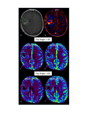 |
Moving Towards A DSC-MRI Consensus: A new single dose option for standardized rCBV.
Kathleen Schmainda, Melissa Prah, Robert Wujek, Jennifer Connelly
DSC-MRI is the most common approach used for brain tumor perfusion imaging. Consensus is being reached regarding a recommended method for data acquisition, one that requires a contrast agent preload and the application of leakage correction. However, recent simulations suggest that if a low flip angle is used for DSC-MRI data collection then a contrast agent preload might not be necessary. In this study DSC-MRI data collected in 19 patients with brain tumors supports this hypothesis and further suggests that the application of leakage correction as well as a standardization algorithm further improves the results.
|
16:06
 |
1234.
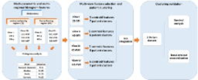 |
Multi-Parametric and Multi-Regional Histogram Analysis of MRI: Revealing Imaging Phenotypes of Glioblastoma Correlated with Patient Survival
Chao Li, Shuo Wang, Angela Serra, Turid Torheim, Florian Markowetz, Stephen Price
Glioblastoma is characterized by its remarkable heterogeneity and dismal prognosis. Histogram features of MRI modality show potential in measuring the intratumoral heterogeneity. We integrate multi-parametric and multi-regional MRI histogram features to divide patients into groups and assess the relevance to treatment outcome. The results demonstrated that integrating multi-parametric and multi-regional MRI histogram features may help to stratify patients. The feature selected in this process also displayed prognostic values in the multivariate survival analysis. The histogram features selected from the proposed approach may be used as potential imaging markers in personalized treatment strategy and response determination.
|
16:18
|
1235.
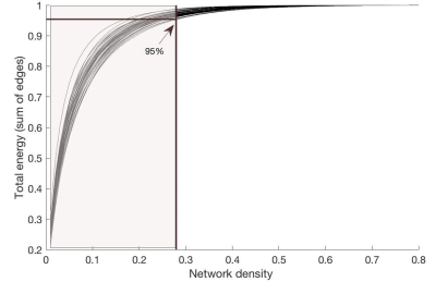 |
Supratentorial reorganization after treatment for childhood infratentorial tumors from a graph theoretical perspective
Charlotte Sleurs, Dafnis Batalle, Jurgen Lemiere, Daan Christiaens, Jacques-Donald Tournier, Stefan Sunaert, Anne Uyttebroeck, Sandra Jacobs, Serena Counsell, Sabine Deprez
In this study, structural brain topology was investigated in young adults who were treated for infratentorial tumors during childhood, using graph theory metrics based on whole-brain tractography. Comparisons with healthy controls yielded significant differences in global efficiency measures, independently from network density threshold. This cost-corrected measure was significantly related with IQ scores. By contrast, local efficiency and average nodal strength appeared different for core networks only. This finding suggests that connectivity of highly connected supratentorial hubs are predominantly affected following treatment for infratentorial tumours.
|
16:30
|
1236.
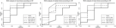 |
Effect of Vascular Input Function Selection on Quantitative Dynamic Contrast-Enhanced MRI for Differentiating Pseudo-progression from True Progression in Glioblastomas
Kevin Wang, Melissa Chen, Ho-Ling Liu
Vascular input function (VIF) is a major source of error in the pharmacokinetic modeling of dynamic contrast-enhanced (DCE) magnetic resonance imaging (MRI) data. We investigated the influence of VIF variability on forward volumetric transfer constant, Ktrans, one of the kinetic parameters of DCE MRI. The diagnostic power of DCE MRI to discriminate between pseudo-progression and true progression of glioblastoma following chemoradiation therapy was determined by deriving Ktrans from five different VIF sources (population-based, middle cerebral artery, superior sagittal sinus—the latter two calculated by applying either an assumed or a measured T1) and using receiver operating curve analysis.
|
16:42
|
1237.
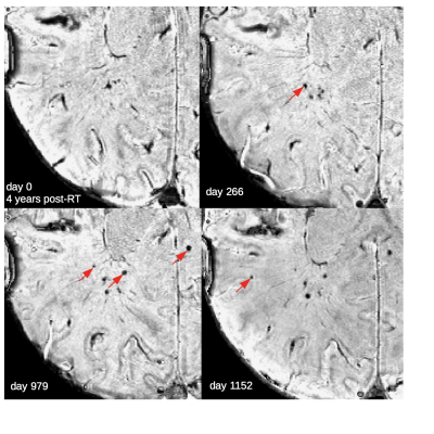 |
Clinical predictors of serial changes in radiotherapy-induced microbleed burden and their relationship to white matter damage in 133 patients with glioma
Melanie Morrison, Angela Jakary, Devika Nair, Christopher Hess, Jennifer Clarke , Nicholas Butowski, Susan Chang, Annette Molinaro, Janine Lupo
In the treatment of adult brain tumors, radiation therapy is associated with long-term effects including vascular injury in the form of cerebral microbleeds (CMBs), and white-matter changes. Ultra high-field MRI was used to detect CMBs, and predictors of CMB development were identified. Changes in individual and total CMB burden were characterized from serial imaging data with white-matter changes. Time since RT, multiple surgeries, tumor type and location were all predictors of development. The total number and volume of CMBs increased over time, while individual CMB size decreased and the surrounding white-matter showed signs of degradation.
|
16:54
|
1238.
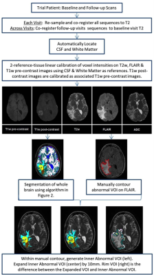 |
MRI Predictors of Response to Pembrolizumab, Bevacizumab and Hypofractionated Stereotactic Irradiation in Patients with Recurrent High Grade Gliomas
Samuel Hawkins, Olya Stringfield, Nicolas Rognin, John Arrington, Michael Yu, Heiko Enderling, Solmaz Sahebjam, Natarajan Raghunand
Standard MRI scans of patients with recurrent high grade gliomas treated with pembrolizumab were analyzed to build a model to predict time-to-progression. Images from five standard MRI sequences were co-registered across multiple scan dates per patient and automatically segmented into normal and pathologic tissue types based on calibrated pixel intensities. 308 radiomic features describing size, shape, and texture were extracted per image type. The four most predictive features were used in a linear regression model that could predict time-to-progression to within an average of three months of actual progression in test patients.
|
17:06
|
1239.
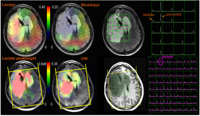 |
Evaluating patients with Glioma using Multi-modal Hyperpolarized C-13 and H-1 Metabolic Imaging
Yan Li, Adam Autry, Jeremy Gordon, Ilwoo Park, Duan Xu, Susan Chang, Daniel Vigneron, Sarah Nelson
Gliomas are heterogeneous and infiltrative brain tumors. Noninvasive evaluation of the extent and activity of tumor is important for making decisions about how to manage patients with glioma. In this study, we are examining the relationship between dynamic hyperpolarized C-13 data and H-1 MRSI parameters. Eleven patients with glioma were studied with both hyperpolarized [1-13C]pyruvate imaging and 3D H-1 lactate-edited MRSI. The H-1 data were affinely registered to the C-13 data, which allows voxel-by-voxel comparison. While these initial results indicate that there may be a trend towards an inverse relationship between C-13 lactate/pyruvate and steady state normalized lactate within the choline-to-NAA (CNI) lesion, further analysis is needed in a larger population of patients.
|
17:18
|
1240.
 |
Longitudinal Evaluation of Glioblastoma (GBM) Response to Chemo-radiation with Quantitative Magnetization Transfer (qMT)
Hatef Mehrabian, Sten Myrehaug, Hany Soliman, Arjun Sahgal, Greg Stanisz
Assessing response of glioblastoma (GBM) to standard 6-week chemo-radiation at early stages of the treatment allows for changing or adjusting therapy in patients with progressive tumors. Quantitative magnetization transfer (qMT) which probes the properties of the macromolecular content of the tumor and their interactions with free water protons is more sensitive to treatment-induced effects and is capable of differentiating progressors from non-progressors as early as two weeks into the treatment. Moreover, certain qMT parameters (i.e. amount of MT and direct effect of free water pool) were capable of characterizing GBM aggressiveness even before the start of the treatment.
|
|
