Myelin Imaging: Applications
Oral
Neuro
Wednesday, 20 June 2018
| S04 |
16:15 - 18:15 |
Moderators: Valentin Prevost, Jeffrey Stanley |
16:15
|
0923.
 |
 Correlations between quantiative myelin imaging using macromolecular proton fraction, neurogenesis, and oligodendrogenesis in the murine model of cuprizone-induced demyelination Correlations between quantiative myelin imaging using macromolecular proton fraction, neurogenesis, and oligodendrogenesis in the murine model of cuprizone-induced demyelination
Marina Khodanovich, Anna Pishchelko, Valentina Glazacheva, Edgar Pan, Andrey Akulov, Vasily Yarnykh
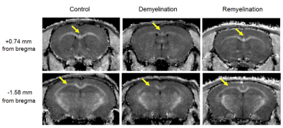
This study aimed to identify associations between neurogenesis in the neurogenic niches, oligodendrogenesis, and myelination in the murine cuprizone demyelination/remyelination model. Myelination was quantified by the recently proposed macromolecular proton fraction (MPF) mapping method. Neurogenesis and oligodendrogenesis were assessed by immunohistology. Negative correlations were found between oligodendrogenesis and neurogenesis in both the subventricular zone and dentate gyrus. Correlation between MPF and oligodendrogenesis was also negative, whereas correlation between MPF and neurogenesis was positive. Associations between MPF and neurogenesis/oligodendrogenesis reveal the feasibility of using MPF as a surrogate marker of reparative processes in demyelinating diseases.
|
16:27
|
0924.
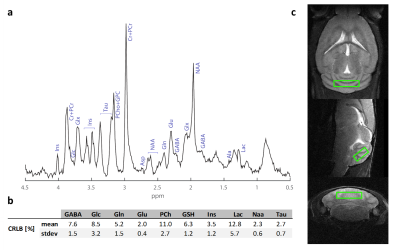 |
Neurochemical signature of the metabolic mechanisms underlying de- & re-myelination in the mouse’s cerebellum
Georges Hankov, Aline Seuwen, Giovanna Ielacqua, Anna Mechling, Eva Mracsko, Andreas Bruns, Basil Künnecke, Markus von Kienlin, Markus Rudin, Thomas Mueggler
Because of an increasing therapeutic need to understand the underlying mechanism of myelin damage and repair in pathologies such as multiple sclerosis (MS), we used 1H-MRS to longitudinally characterize the metabolic changes in the cerebellum associated with de- and re-myelination in the cuprizone mouse model. Our results, in line with similar findings in the corpus callosum, suggest that a group of metabolites provide a unique neurochemical signature of cuprizone induced de- and re-myelination. Additionally, we observe a reversible and robust increase of GABA levels upon cuprizone feeding that goes in contradiction with current trends in clinical studies of MS patients.
|
16:39
|
0925.
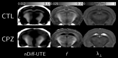 |
A comparison of magnetization transfer, diffusion tensor imaging and ultrashort TE measurements in a murine model of demyelination
Lucas Soustelle, Cristina Antal, Julien Lamy, François Rousseau, Jean-Paul Armspach, Paulo Loureiro de Sousa
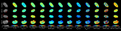
Exploration of myelin content in the brain and spinal cord is essential for monitoring pathologies such as multiple sclerosis. Quantitative MRI methods such as diffusion tensor imaging (DTI) and quantitative magnetization transfer imaging (qMT) already demonstrated efficiency in characterizing demyelination. Ultrashort echo time sequences were previously investigated for myelin characterization within long-T2 suppression condition. In this work, we propose to compare parameters from DTI, qMT, and Diff-UTE in ex-vivo brains, using a murine model of demyelination.
|
16:51
|
0926.
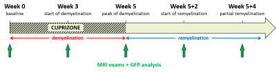 |
Inhomogeneous Magnetization Transfer (ihMT) sensitivity to myelin impairments in cuprizone mouse model
Valentin Prevost, Myriam Cayre, Victor Carvalho, Samira Mchinda, Gopal Varma, Jean Philippe Ranjeva, Jean Pelletier, David Alsop, Pascale Durbec, Olivier Girard, Guillaume Duhamel
Inhomogeneous magnetization transfer (ihMT) is a new MR imaging modality weighted by the dipolar relaxation time (T1D), which has demonstrated a strong linear correlation with myelin-specific fluorescence signal measured in genetically-modified mice, hence validating the technique as a myelin biomarker. In the current study, the ihMT sensitivity has been evaluated on a cuprizone mouse model, a widely used model of de- and remyelination. A longitudinal analysis has been performed in vivo and quantitative signal was compared to myelin amount measured by GFP quantification at remarkable time points in order to evaluate the ihMT sensitivity for monitoring in vivo myelin state.
|
17:03
 |
0927.
 |
Are we seeing any better? A comprehensive comparison of myelin biomarkers in the healthy and multiple sclerosis post mortem spinal cord
Marco Battiston, Torben Schneider, Francesco Grussu, Geert Schenk, Stig Wergeland, Mohamed Tachrount, Marios Yiannakas, Carmen Tur, Jeroen Geurts, Claudia Wheeler-Kingshott, Rebecca Samson
Conventional MRI of the multiple sclerosis (MS) spinal cord offers low specificity to underlying pathological processes taking place. Quantitative MRI metrics able to characterize damage at the microstructural level are required. Of particular interest are those known to be sensitive to myelin content, which can be generated via various different contrast mechanisms. It is important to assess the specificity of such prospective myelin biomarkers to MS spinal cord pathology and validation studies comparing MRI with histological findings are essential for this purpose. Here a comparison of myelin-sensitive quantitative MRI metrics measured in MS and healthy ex vivo cord are presented.
|
17:15
|
0928.
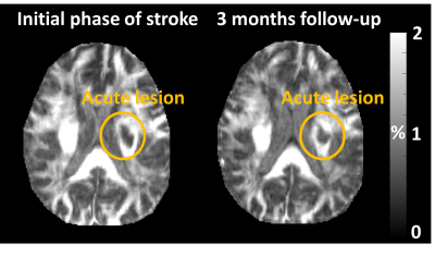 |
Evaluation of recovery in myelin water signal after acute ischemic stroke
Joon Yul Choi, Do Yeon Kim, Jongho Lee, Seung-Hoon Lee
This study investigated the recovery in myelin water signals after acute ischemic stroke. The results showed an increase of myelin water signals in acute lesions after 3 months of treatment. Furthermore, mild stroke patients recovered more than severe stroke patients when comparing measures of myelin water signals in acute lesions.
|
17:27
|
0929.
 |
Neuroplastic Changes of Myelin Microstructure With Video Game Play
Douglas Dean, Austin Patrick, Thomas Gorman, C Green, Andrew Alexander
Mounting evidence suggests that changes in the brain can occur within hours, however, the mechanisms underlying these changes remain unknown. In this work, we utilized multicomponent relaxometry (mcDESPOT) to examine the effects of both short and long term video game playing on myelinated white matter. Short and long term changes in quantitative longitudinal relaxation times as well as long term changes of myelin water fraction were observed. These results add to the growing literature that neuroplastic effects can take place over the short term while also suggesting long term changes may involve mechanisms of myelination.
|
17:39
|
0930.
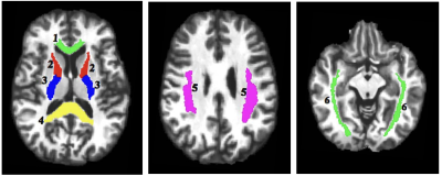 |
Evidence of altered myelination in adolescents with Attention-Deficit/Hyperactivity Disorder: A multi-echo T2 imaging study
Jennifer Losiowski, Phil Easter, David Rosenberg, Jeffrey Stanley
Attention-deficit/hyperactivity disorder (ADHD) is a highly prevalent childhood disorder with considerable evidence of a developmental basis for the etiology of ADHD. Structural neuroimaging studies, though limited in its interpretation, have implicated cortical and subcortical white matter in ADHD. Here we examine myelin content and axonal size/packing density in adolescents with ADHD in six white matter tracts using multi-echo T2 (ME-T2) imaging. Results show reduced myelin content as well as smaller axonal size (increased axonal packing density), which is suggestive of a lack of progressive myelination in commissural and projection tracts in ADHD.
|
17:51
|
0931.
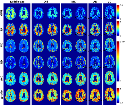 |
Myelin water fraction, diffusion tensor imaging, and g-ratio measurements characterize myelin changes in normative aging, mild cognitive impairment, and dementia
Video Permission Withheld
Mustapha Bouhrara, Abinand Rejimon, Diana Lee, Richard Spencer
In previous work, we showed evidence of myelin loss with mild cognitive impairment (MCI) using a direct and specific MRI measure of myelin water fraction, a surrogate for myelin content. Here, we extend this by investigating changes in myelin content in normative aging, MCI, and dementia using myelin content, diffusion tensor imaging, and g-ratio measurements. Our results showed decrease in myelin content in several brain regions of the subjects diagnosed with MCI or dementia in comparison with the old healthy subjects. Higher myelin content in middle-aged subjects was also observed, in agreement with the literature.
|
18:03
|
0932.
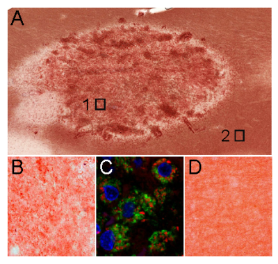 |
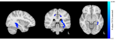
Myelin digestion during multiple sclerosis lesion formation contributes to increase on QSM
Kofi Deh, Gerald Ponath, Zaki Molvi, Gian-Carlo Parel, Kelly Gillen , Shun Zhang, Thanh Nguyen , Pascal Spincemaille, Yinghua Ma , Ajay Gupta , Susan Gauthier , David Pitt, Yi Wang
During the initial processes in the formation of MS lesions, myelin is immediately digested in macrophages after phacocytosis. Chemical bond breakdown in myelin basic proteins (MBP) and among lipid bilayers (LB) can increase magnetic susceptibility. Here we measure susceptibility increase in phantom experiments of MBP breakdown and myelin LB breakdown. We investigate myelin degradation in the first few weeks of MS lesion formation by performing histology on MS brain samples and by in vivo imaging of enhancing lesions in MS patients.
|
|
