Neurofluids & Glymphatic System
Oral
Neuro
Thursday, 21 June 2018
| S02 |
13:15 - 15:15 |
Moderators: Nivedita Agarwal, Shigeki Aoki |
13:15
|
1137.
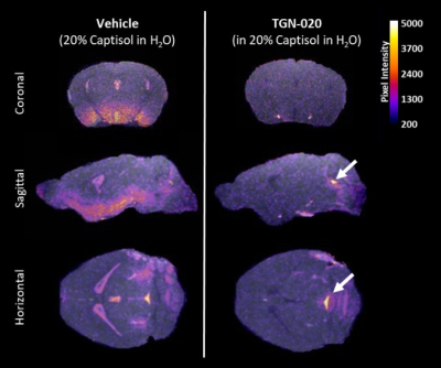 |
 Imaging Brain Clearance in the Mouse Brain: Aquaporin-4 Modulates Glymphatic Inflow Imaging Brain Clearance in the Mouse Brain: Aquaporin-4 Modulates Glymphatic Inflow
Ian Harrison, Ozama Ismail, Yolanda Ohene, Payam Nahavandi, Jack Wells, Mark Lythgoe
The ‘glymphatic’ system, which has recently been identified using MRI, is a brain-wide pathway for removal of waste solutes. This system is implicated in Alzheimer’s disease (AD), due to discovery that both amyloid-β and tau, accumulations that define AD, are cleared from the brain via this pathway. Using contrast-enhanced MRI, we demonstrate that glymphatic function is dependent upon aquaporin-4, a water channel located on astrocytic endfeet surrounding blood vessels in the brain. Herein, using a novel pharmacological approach, we show that aquaporin-4 represents a suitable drug target for manipulation of glymphatic function in the brain, and in treatment of AD.
|
13:27
|
1138.
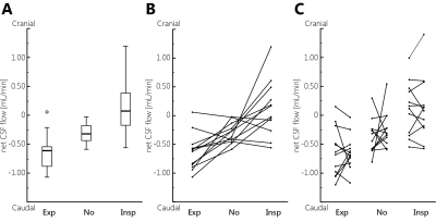 |
PC-MRI measurements of net CSF flow through the cerebral aqueduct strongly depend on respiration
Jolanda Spijkerman, Lennart Geurts, Jeroen Siero, Jeroen Hendrikse, Peter Luijten, Jaco Zwanenburg
In this work the influence of respiration on net CSF flow measurements was investigated. In 12 volunteers net CSF flow was measured in the cerebral aqueduct using PC-MRI at 7T, with respiratory gating on expiration and on inspiration, and without respiratory gating. Repeated measurements were performed. Net flow over the cardiac cycle was determined.
Net CSF flow (mean±sd) was -0.64±0.32 mL/min (caudal) during expiration, +0.12±0.49 mL/min (cranial) during inspiration, and -0.31±0.18 mL/min without respiratory gating. Two outliers, with reversed (cranial) net CSF flow, were observed when no respiratory gating was performed. Repeatability was best for gating on inspiration.
|
13:39
|
1139.
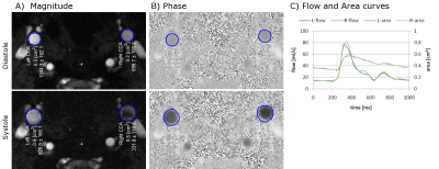 |
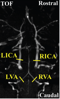
Cine MRI assessment of carotid artery pulse wave velocity and reflected wave amplitude in children with sickle cell disease
Jackie Leung, Datta Goolaub, Christopher Macgowan, Lindsay Cahill, John Sled, Andrea Kassner
The mechanical and morphological properties of the cerebral vasculature can be studied by measuring blood flow dynamics of the feeding arteries. In this study, we used high resolution phase contrast MRI to measure the blood flow and vessel area of the common carotid arteries of children with and without sickle cell disease (SCD). Preliminary results show that pulse wave velocity and wave reflection is significantly higher in patients with SCD, suggesting increased vessel stiffness and downstream abnormalities in the vasculature. This method is clinically feasible and may provide useful insight into the vascular properties of children with cerebrovascular disease.
|
13:51
|
1140.
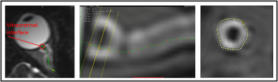 |
Optic Nerve Sheath Volume Changes Acutely in Response to Intracranial Pressure Change after Lumbar Puncture and CSF Drainage
John Oshinski, Muhammad Islam, Chansu Kim, Amit Saindane
This study used 3D, T2W imaging of the brain to evaluate the optic nerve sheath (ONS) in 10 patients with idiopathic intracranial hypertension (IIH). Patients were imaged pre- and immediately post-lumbar puncture/CSF drainage. Images were reformatted to be perpendicular to the optic nerve and the volume of the ONS pre- and post-lumbar puncture was calculated. The study found that ONS volume decreased significantly within an 82 minute window in response to a reduction in CSF pressure, and this change can be quantitatively evaluated using MRI.
|
14:03
|
1141.
 |
Association between Gray Matter alterations and CSF biomarkers in amnestic MCI patients
Domenico Zacà, Giuliano Giari, Moira Marizzoni, Clarissa Ferrari, Samantha Galluzzi, Diego Albani, Claudio Babiloni, Mira Didic, Josè Luis Molinuevo, Flavio Mariano Nobili, Lucilla Parnetti, Pierre Payoux, Paolo Maria Rossini, Peter Schönknecht, Andrea Soricelli, Magda Tsolaki, Pieter Jelle Visser, Jens Wiltfang, Regis Bordet, Libera Cavaliere, Jill Richardson, Olivier Blin, Giovanni B. Frisoni, Jorge Jovicich
In this study the association between CSF and structural imaging based early biomarker of Alzheimer Diasease (AD) was investigated in amnestic Mild Cognitive Impairment (aMCI) patients. Voxel based morphometry and partial least square correlation were used to analyze differences in local and whole brain GM profiles (covariance) between two groups of aMCI patients divided in two groups based on CSF amyloid (Aβ42) levels. Both voxel-wise and structural covariance GM differentiated Aβ positive (prodromal AD) from Aβ negative patients. These results indicate that GM measures may provide a sensitive metric for tracking AD progression.
|
14:15
|
1142.
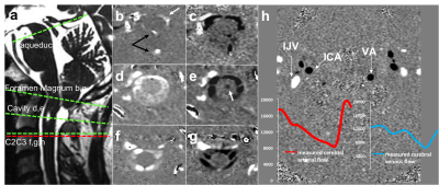 |
2D phase contrast MRI to quantify CSF dynamics change after surgery of Chiari “malformations"
olivier baledent , pauline padovani, anthony fichten, patrick toussaint, catherine gondry-jouet, serge metanbou, johann peltier, cyrille capel
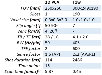
Diagnosis of Chiari malformation (CM) is mainly based on morphological. The objective of this study was to quantify the evolution of CSF and blood flows dynamics before and after surgery treatment of CM. 21 patients who underwent surgery for CM, and presented good clinical outcome at 12 months were retrospectively included. We have shown that in CM, CSF flow is mainly reduced at the foramen magnum whereas it is normal at the cervical level. CM reduces jugular outflow. Complementary to the morphological investigation 2D PCMRI should be take in account to improve the evaluation of the severity of the Chiari Malformation.
|
14:27
|
1143.
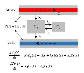 |
Quantitative Modeling of Glymphatic Pathways of the Brain Using MRI
Esmaeil Davoodi-bojd, Guangliang Ding, Li Zhang, Qingjiang Li, Lian Li, Michael Chopp, Quan Jiang
The dynamic exchange of CSF with ISF is identified as the glymphatic system. The impairment of the glymphatic clearance is involved in the development of neurodegenerative conditions. Despite many recent studies that investigated the glymphatic system, few studies have tried to use a mathematical model to describe this system, quantitatively. In this study, we aim to model the glymphatic system from the kinetics of GD-DTPA tracer in order to 1) map the glymphatic system pathways, 2) derive kinetic parameters of the glymphatic system, and 3) provide quantitative maps of the structure and function of this system.
|
14:39
|
1144.
 |
Phase-contrast MRI for estimating global cerebral blood flow of mice at 11.7T: optimization and age-dependence
Zhiliang Wei, Lin Chen, Zixuan Lin, Jiadi Xu, Peter van Zijl, Hanzhang Lu
Cerebral blood flow (CBF) is a crucial physiological parameter reflective of brain functioning and metabolism. In this study, our goal was to systematically optimize a phase-contrast (PC) quantitative flow technique for CBF assessment in mice. We also demonstrated the sensitivity of this technique to longitudinal CBF changes in aging. This optimized PC protocol may serve as a helpful non-invasive tool for CBF measurement in preclinical studies of brain physiology and pathophysiology with mouse disease models.
|
14:51
|
1145.
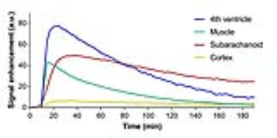 |
Impact of the glymphatic system on the kinetic and distribution of gadodiamide in the rat brain: Observation by dynamic MRI and effect of circadian rhythm on tissue gadolinium concentrations.
Toshiaki Taoka, Gregor Jost, Shinji Naganawa, Hubertus Pietsch
We tried to elucidate the role of the glymphatic system on the distribution and kinetics of a linear gadolinium based contrast agent in the rat brain. Dynamic MRI signal changes of the brain and the cerebrospinal fluid (CSF) indicated that intravenously administrated gadodiamide enter from blood into the CSF, indicating the involvement of the glymphatic system. The long term presence of gadolinium in the brain after repeated administration of gadodiamide quantified by ICP-MS indicates the involvement of the glymphatic system on the clearance of gadolinium from brain tissue in terms of the influence of circadian rhythm and anesthesia during administration.
|
15:03
|
1146.
 |
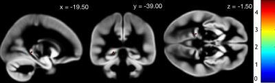
Increased pulsatility in cerebral perforating arteries in patients with lacunar infarction or deep intracerebral hemorrhage, an explorative 7T MRI study
Lennart Geurts, Geert Jan Biessels, Peter Luijten, Karin Klijn, Jaco Zwanenburg
Cerebral small vessel disease is a major cause of dementia and stroke with unknown pathophysiology. Increased blood flow pulsation is hypothesized to be an underlying pathology of small vessel disease, but has not been investigated in cerebral small vessels. Using 2D phase contrast MRI at 7T, we measured the pulsatility index in cerebral perforating arteries of patients with lacunar infarction or deep intracerebral haemorrhage and a group of healthy controls. We showed that both patient groups have a higher pulsatility index in small vessels than controls. This warrants further research into the possible role of pulsatility in small vessel disease.
|
|
