Brain Connectivity
Oral
Neuro
Thursday, 21 June 2018
| N03 |
15:30 - 17:30 |
Moderators: Fernando Calamante, Baete Steven |
15:30
 |
1211.
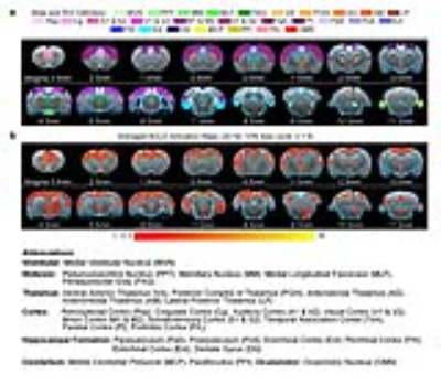 |
 Optogenetic fMRI dissection of brain-wide vestibular pathways Optogenetic fMRI dissection of brain-wide vestibular pathways
Alex T. L. Leong, Xunda Wang, Russell W. Chan, Xiong Cao, Ed X. Wu
The vestibular system is essential to our sense of balance and spatial orientation. fMRI mapping of the vestibular system has been challenging due to the physical constraints limiting a subject’s ability to perform motion, balance and orientation related tasks within an MRI scanner. At present, our knowledge of the brain-wide cortical and subcortical regions that participate in processing the vestibular sense is scarce. Here, we combine fMRI and optogenetic stimulation of vestibular excitatory neurons and, for the first time, successfully map the multiple brain-wide vestibular pathways.
|
15:42
|
1212.
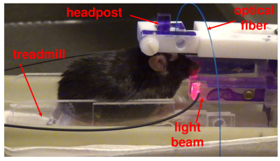 |
A novel approach for fMRI of rodents actively performing optogenetic self-stimulation
Hanbing Lu, Christopher Cover, Andrew Kesner, Elliot Stein, Satoshi Satoshi Ikemoto , Yihong Yang
Linking the dynamics of neural circuits to behavior is arguably a central theme in neuroscience. fMRI in rodent animals offers the opportunity to combine systems-level brain readout with modern in-vivo cell biology tools, such as DREADDS and optogenetics, to dissect circuit dynamics. Despite efforts to image awake rodents to avoid confounds from anesthesia, there has been no report on fMRI of rodent animals being actively engaged in a goal-directed behavior. We report a method that permits fMRI of brain dynamics while a mouse is actively self-administering optogenetic stimulation, opening the door to real-time imaging of animal behavior using MRI.
|
15:54
 |
1213.
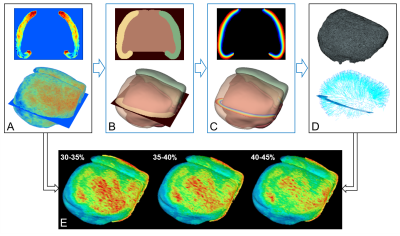 |
MouseStream: A Software Suite for Mapping and Analyzing Mouse Cortical Functional Architecture In Vivo Using Magnetic Resonance Microscopy
Jia Guo, Xinyang Feng, Hannah Sigmon, Frank Provenzano, Scott Small
The functional architecture of the cortex has never been mapped in vivo with the fidelity necessary to distinguish “functional unit”. Mapping the functional architecture of the cortex, reflecting the observed regional and layer differences in synaptic density and its correlate energy metabolism, has been very challenging. Here we set out to address this issue by in vivo high-resolution cerebral blood volume (CBV) mapping of the mouse cortex. Tailored software, MouseStream, was developed to reconstruct the cortical CBV data through the normalized curved cortical coordinate (NCCC). NCCC allows projection of functional architectures mapped using CBV onto the cortical surface across the whole cortex and from different cortical depths.
|
16:06
|
1214.
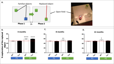 |
Longitudinal alterations of resting-state functional connectivity in Alzheimer’s disease in a tauopathy mouse model
Laetitia Degiorgis, Meltem Karatas, Marion Sourty, Thomas Bienert, Marco Reisert, Chantal Mathis, Anne-Laurence Boutillier, Frédéric Blanc, Jean-Paul Armspach, Laura-Adela Harsan
Alzheimer’s disease is the most widespread cause of dementia and constitutes one of the biggest challenges for society. Among dominant mechanisms of the disease is the abnormal accumulation of the protein tau leading to tauopathy. In this study we explored in vivo the longitudinal evolution of the brain functional connectome, in the Thy-Tau22 mouse, a model of tauopathy. We used resting-state functional MRI in correlation with behavioral analysis to show the remodeling functional circuitry over-time including default mode network and memory networks in transgenic mice.
|
16:18
|
1215.
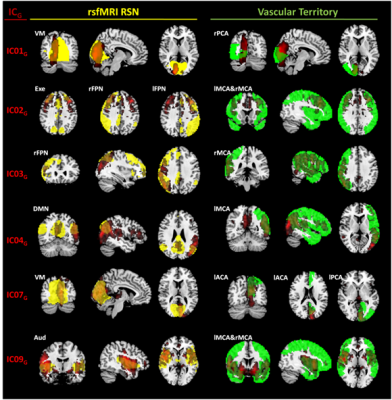 |
Brain perfusion patterns and their relationships with fMRI resting state networks and arterial vascular territories
Maria Marcella Lagana, Laura Pelizzari, Alice Pirastru, Niels Bergsland, Mario Clerici, Pietro Cecconi, Francesca Baglio
In this study, we aimed to assess the consistency of the perfusion patterns that can be extracted using independent component analysis (ICA) on cerebral blood flow (CBF) maps derived from arterial spin labeling (ASL) data. Furthermore, we aimed to evaluate the similarity between the CBF-derived components and the well-known spatial patterns of functional MRI resting state networks (RSN) and cerebral vascular territories (VT). Our results showed that good spatial constancy of perfusion patterns can be extracted from CBF maps. Almost all the derived components overlapped with RSN or specific VT.
|
16:30
|
1216.
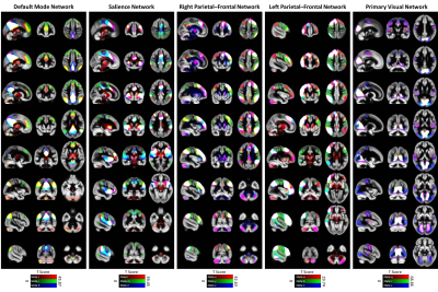 |
An approach to capture time-varying spatial connectivity in resting fMRI networks
Armin Iraji, Maziar Yaesoubi, Anees Abrol, Zening Fu, Yuhui Du, Srinivas Rachakonda, Vince Calhoun
The analysis of time-varying connectivity has become an important part of neuroscience discussions. The majority of such studies have focused primarily on the temporal variations of functional connectivity among fixed regions of interest (ROIs) or brain networks. However, the brain reorganizes itself on both spatial and temporal scales. Approaches that capture spatial and temporal coupling variations are needed. Here, we describe a novel approach capable of identifying the base states of brain networks and capturing their spatial variations over time. It also provides a unique opportunity to characterize the temporal variations of the brain at both network and global scales.
|
16:42
|
1217.
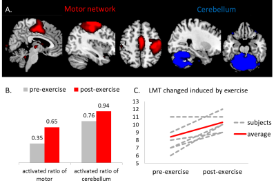 |
Physical Exercise Increased Involvement of Motor Networks as compensatory mechanism under challenging cognitive task
Lanxin Ji, Godfrey Pearlson, Keith Hawkins, David Steffens, Lihong Wang
Neuroimaging studies show reorganization of neural resources in older adults may compensate for cognitive decline. To effectively evaluate neural compensation, we proposed a data-driven independent component analysis method, and tested the measure through a longitudinal study. Twenty-six healthy older adults participated in a 6-week physical exercise program. Gait speed, cognitive function, and fMRI during a challenging memory task were measured before and after the program. Results showed a positive correlation between the compensatory ability measure and gait speed at baseline. Physical exercise improved gait speed, cognition, and compensatory ability through increased involvement of motor-related networks in conducting the cognitive task.
|
16:54
|
1218.
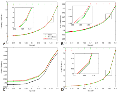 |
Are prediabetes and type 2 diabetes associated with white matter connectivity alterations? The Maastricht Study
Laura Vergoossen, Walter Backes, Miranda Schram, Jacobus Jansen, on behalf of The Maastricht Study
Type 2 diabetes (T2DM) is associated with cognitive decline, while prediabetes may already show comparable cognitive decrements. We investigated whether white matter network integrity is associated with prediabetes and/or T2DM in a large population-based cohort study. For calculation of white matter volumes and graph measures, 3T structural and diffusion MRI (dMRI) were performed. Prediabetes had lower clustering coefficient and local efficiency compared to NGM. Communicability was significantly higher in T2DM, but not in prediabetes, which suggests that alternative white matter connections are used to compensate for structural disturbances and white matter decline, which may not be present yet in prediabetes.
|
17:06
|
1219.
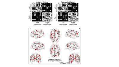 |
Exploring diffusion derived connectivity patterns between cognitively impaired and nonimpaired active professional fighters
Virendra Mishra, Karthik Sreenivasan, Zhengshi Yang, Xiaowei Zhuang, Sarah Banks, Dietmar Cordes, Charles Bernick
In this study, we utilized the diffusion MRI (dMRI) data of cognitively impaired and nonimpaired active professional fighters from the Professional Fighters Brain Health Study and investigated the structural connectivity patterns between the two groups. Our study showed a disrupted connectivity pattern between cognitively impaired and nonimpaired active professional fighters, mainly due to short-range fibres. Further, a complex heterogeneous pattern was speculated due to fighting. These findings may help the clinicians better understand the effect of repeated head trauma on structural connectivity pattern and its association with later mental defects due to fighting.
|
17:18
|
1220.
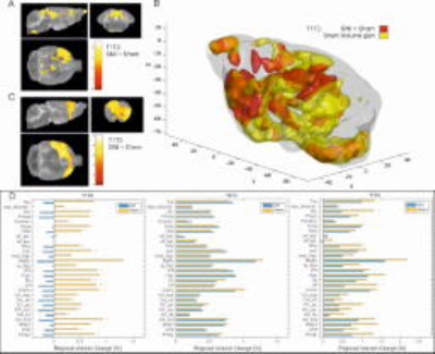 |
Longitudinal structural and functional brain network alterations in a mouse model of neuropathic pain
Claudia Falfan-Melgoza, Ainhoa Bilbao, Robert Becker, Sarah Leixner, Markus Sack, Gabriele Ende, Alexander Sartorius, Rainer Spanagel, Wolfgang Weber-Fahr
We combined multimodal (rsfMRI,M MRS, VBM) neuroimaging to longitudinally monitor changes in brain metabolism, structure and connectivity using the spared nerve injury (SNI) mouse model of chronic neuropathic pain. Voxel-based morphometry demonstrated volume decrease in all brain sites assessed. Global and local network changes after SNI disappeared over time, except the nucleus accumbens, prefrontal cortex and hippocampus. Connectivity changes were accompanied by enhanced glutamate levels in the hippocampus. We suggest that hippocampal hyperexcitability may alter synaptic plasticity within the nucleus accumbens, circadian motor activity and emotionality during pain chronification.
|
|
