Quantitative Neuroimaging
Oral
Neuro
Wednesday, 20 June 2018
| S02 |
13:45 - 15:45 |
Moderators: Hongyu An |
13:45
 |
0793.
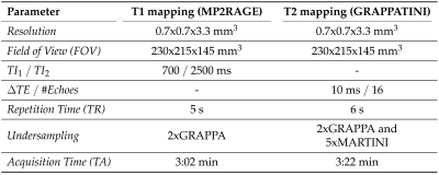 |
 Quantitative T1 and T2 Brain Atlases for the Detection of Abnormal Relaxation Times Quantitative T1 and T2 Brain Atlases for the Detection of Abnormal Relaxation Times
Gian Franco Piredda, Tom Hilbert, Cristina Granziera, Guillaume Bonnier, Reto Meuli, Filippo Molinari, Jean-Philippe Thiran, Tobias Kober
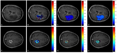
This work aims at the detection of abnormal relaxation times in the human brain. To that end, a pipeline for creating normative atlases was established. High-resolution atlases of normative T1 and T2 values were created based on mapping data from 52 healthy subjects. A regression model including gender and age was introduced and z-score maps calculated to compare T1 and T2 maps of a single-subject to the derived norms. Initial results based on multiple sclerosis patient data detect not only lesional tissue but also presumably altered normal-appearing white matter regions.
|
13:57
|
0794.
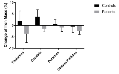 |
Assessment of the total iron mass using Quantitative Susceptibility Mapping (QSM): Deep gray matter iron depletion in multiple sclerosis?
Ferdinand Schweser, Jesper Hagemeier, Michael Dwyer, Niels Bergsland, Akshay Dhamankar, Bianca Weinstock-Guttman, Robert Zivadinov
It is often overlooked that iron concentrations, as determined, e.g., via Quantitative Susceptibility Mapping (QSM), reflect the mass of iron per unit volume. Consequently, structural atrophy alone (i.e. decreased volume) increases the tissue iron concentration if the total mass of iron remains constant. In this work, we present a technique to assess the mass of regional tissue iron in milligrams (mg). We retrospectively applied the technique to data from a recently published 2-year longitudinal study, in which we had investigated iron concentration changes in Multiple Sclerosis (MS).
|
14:09
|
0795.
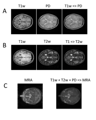 |
MRI2MRI: A deep convolutional network that accurately transforms between brain MRI contrasts
Sa Xiao, Yue Wu, Aaron Lee, Ariel Rokem
Different brain MRI contrasts represent different tissue properties and are sensitive to different artifacts. The relationship between different contrasts is therefore complex and nonlinear. We developed a deep convolutional network that learns the mapping between different MRI contrasts. Using a publicly available dataset, we demonstrate that this algorithm accurately transforms between T1- and T2-weighted images, proton density images, time-of-flight angiograms, and diffusion MRI images. We demonstrate that these transformed images can be used to improve spatial registration between MR images of different contrasts.
|
14:21
|
0796.
 |
Absolute Cerebral Blood Flow Derived from Low GBCA Dose DCE-MRI in Patients with Type 2 Neurofibromatosis
Ka-Loh Li, Daniel Lewis, Xiaoping Zhu, Alan Jackson
A newly developed low dose T1W-DCE-MRI method, ACcomb, was used to estimate the absolute CBF of vestibular schwannomas (VS) and normal appearing brain tissue in a group of 12 consecutive type 2 neurofibromatosis (NF2) patients, undergoing anti-angiogenic bevacizumab treatment. This new method consistently displayed excellent gray-white matter flow contrast and produced mean GM and WM CBF estimates consistent with previous literature values. Use of this new method showed that at 90 days post bevacizumab treatment, there was increased positive correlation between CBF and estimated plasma volume within the VS, and a significant increase in CBF within normal appearing white matter.
|
14:33
 |
0797.
 |
Comparison of the Tofts and the Shutter Speed Model for DCE-MRI in patients with Brain Glioma
Marianna Inglese, Lesley Honeyfield, Eric Aboagye, Adam D Waldman, Matthew Grech-Sollars
DCE-MRI allows interrogation of patho-physiological insular micro-environments through the passage of contrast agents and model-based pharmacokinetic analysis. In this study, we analysed data from 14 patients with suspected primary glioma who underwent DCE-MRI. Using both the Tofts model and the shutter speed model (SSM), we evaluated the performance and variability of each extracted parameter. We then analysed the ability of the two models to discriminate between tumour and healthy tissue to test the differences between the two methods. Results showed higher performance for the SSM, with a high robustness for the mean capillary water molecule lifetime τi.
|
14:45
|
0798.
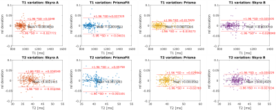 |
Multicenter and multiscanner reproducibility of Magnetic Resonance Fingerprinting relaxometry in the brain
Gregor Körzdörfer, Rainer Kirsch, Kecheng Liu, Josef Pfeuffer, Bernhard Hensel, Yun Jiang, Dan Ma, Mark Griswold, Vikas Gulani, Mathias Nittka
Magnetic Resonance Fingerprinting (MRF) is a technique for generating tissue property maps by matching pseudo randomly varying MR signal time courses in each voxel with a precalculated dictionary of possible evolutions. In this study, the repeatability of MRF results on the same scanner as well as the reproducibility on different scanners is evaluated for the human brain.
|
14:57
|
0799.
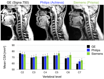 |
Consensus acquisition protocol for quantitative MRI of the cervical spinal cord at 3T
Stephanie Alley, Guillaume Gilbert, Claudia Gandini Wheeler-Kingshott, Rebecca Samson, Francesco Grussu, Allan Martin, Elise Bannier, Virginie Callot, Seth Smith, Junqian Xu, Blake Dewey, Kenneth Weber II, Todd Parrish, Donald McLaren, Gareth Barker, Nico Papinutto, Maryam Seif, Patrick Freund, Robert Barry, Samantha By, Sridar Narayanan, Julien Cohen-Adad
We present a consensus recommendation for best practices in high quality data acquisition of quantitative MRI (qMRI) of the cervical spinal cord at 3T. We propose protocols for computing cross-sectional area (CSA), magnetization transfer ratio (MTR) and diffusion tensor imaging (DTI) from three main vendors. We demonstrate these protocols by repeated scans of a single subject, from which the data and analysis scripts are made available. We hope harmonized and publicly-available spinal cord imaging protocols will promote reproducibility and thus accelerate the progress for spinal cord measurements to be more widely accepted as MRI biomarkers in multicenter studies.
|
15:09
|
0800.
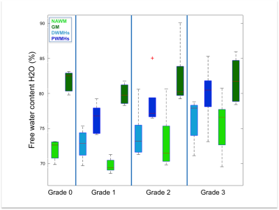 |
Quantitative Magnetic Resonance Imaging of the Human Brain with White Matter Hyperintensities: a New Approach towards Understanding the Underlying Pathology
Elene Iordanishvili, Melissa Schall, Ricardo Loucao, Svenja Caspers, Ketevan Kotetishvili, Jon Shah, Ana-Maria Oros-Peusquens
The interest in white matter hyperintensities (WMHs) has increased recently as they have been associated with various clinical disorders such as stroke and dementia. Although postmortem histopathological studies reported various underlying pathophysiological mechanisms, in vivo studies have not been specific enough to conform those results. We attempted to fill this gap with quantitative MRI of water content, T1, T2*, and MT parameters. These parameters were analyzed for white and grey matter globally and for manually segmented WMHs. Based on the changes identified for WMHs and their trend with WMH load, we were able to complement histopathological findings.
|
15:21
|
0801.
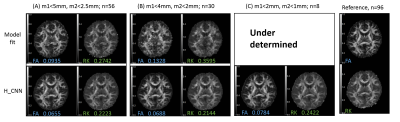 |
Robust diffusion parametric mapping of motion-corrupted data using a deep-learning-based method
Ting Gong, Hongjian He, Zhiwei Li, Qiqi Tong, Zhichao Lin, Yi Sun, Feng Yu, Jianhui Zhong
Motion occurring during the acquisition of diffusion-weighted image volumes is inevitable. Deficient accuracy of volumetric realignment and within-volume movements cause the quality of diffusion model reconstruction to deteriorate, particularly for uncooperative subjects. Taking advantage of the strong inference ability of neural networks, we reconstructed diffusion parametric maps with remaining volumes after the motion-corrupted data removed. Compared to conventional model fitting, our method is minimally sensitive to motion effects and generates results comparable to the gold standard, with as few as eight volumes retained from the motion-contaminated data. This method shows great potential in exploiting some valuable but motion-corrupted DWI data.
|
15:33
|
0802.
 |
Crosstalk of atrophic trigeminal nerve to abnormal brain structure in idiopathic trigeminal neuralgia
Yuan Wang, Bethany Remeniuk, David Seminowicz, Ming Zhang
Idiopathic trigeminal neuralgia (ITN) is characterized by intermittent, lancinating attacks in the branches of the trigeminal nerve (TGN). 40 ITN patients and 40 matched controls underwent MRI sessions and clinical pain assessment. TGN volume of cisternal segment and whole brain grey matter volume (GMV) were evaluated using voxel-based morphometry, ect. Reduced GMV was found in the insula, dorsal anterior cingulate cortex, precuneus, and several areas of the temporal lobe in ITN subjects. Correlation analysis revealed that decreased GMV of the left insula and decreased TGN volume were associated with increased pain ratings, providing new insight into pathophysiology of the disease.
|
|
