CMR Innovations
Oral
Cardiovascular
Thursday, 21 June 2018
| S03 |
08:00 - 10:00 |
Moderators: Markus Henningsson, Dana Peters |
08:00
|
1047.
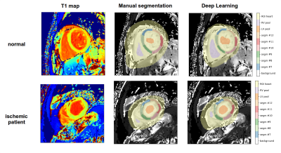 |
 Automatic AHA model segmentation of cardiac T1 maps with deep learning Automatic AHA model segmentation of cardiac T1 maps with deep learning
Nicola Martini, Daniele Della Latta, Gianmarco Santini, Gabriele Valvano, Andrea Barison, Francesco Avogliero, Daniele De Marchi, Luigi Landini, Dante Chiappino
We proposed a fully automated approach for the segmental analysis of T1 mapping using a fully convolutional neural network architecture. T1 maps acquired using the MOLLI sequence from 394 subjects were considered. Excellent segmentation results are demonstrated by high Jaccard (0.969±0.023) and a Dice (0.984±0.012) indexes. No significant difference in the obtained segmental T1 values compared to manual measurements was found, with a mismatch percentage ranging from 0.95% to 3.14% across segments.
|
08:12
 |
1048.
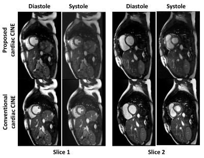 |
Single Breath-held, ECG-Free Cardiac CINE MRI using Parallel Imaging and Deep Learning Combined Image Reconstruction
Fei Han, Ziwu Zhou, Vahid Kouzehkonan, Yu Gao, Yingli Yang, Peng Hu
Cardiac CINE MRI is widely used for evaluating ventricular wall motion and cardiac function. Conventional cardiac CINE consists of ECG-triggered k-space segmented 2D acquisitions, each performed within a breath-hold. In this study, we propose an ECG-free, cardiac CINE protocol that covers the entire LV within a single breath-hold. Our solution is based on a highly accelerated real-time imaging that is enabled by our recently proposed parallel imaging and deep learning combined (PI-DL) image reconstruction. In this study, we evaluated the proposed solution in healthy volunteers and compare its performance with cardiac CINE images acquired using conventional protocol.
|
08:24
|
1049.
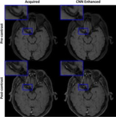 |
Deep Convolutional Neural Network Enhanced 3D High Resolution Turbo Spin Echo Intracranial Vessel Wall Imaging
Zechen Zhou, Shuo Chen, Jiayi Wu, Xihai Zhao, Peter Börnert, Chun Yuan
Turbo spin echo (TSE) imaging with variable flip angle (VFA) is commonly used for three-dimensional (3D) high resolution intracranial vessel wall imaging. However, different tissues may experience various blurring effects particularly for longer TSE factor. In this study, a deep convolutional neural network is trained to provide a solution for this special deblurring problem. Combined with a signal-to-noise ratio (SNR)-priority VFA design scheme, the developed technique can provide a better tradeoff across scan efficiency, point spread function and SNR for 3D TSE acquisitions. Preliminary results have demonstrated its improvement for sharper delineation of intracranial vessel wall and plaque boundaries at isotropic 0.5mm resolution.
|
08:36
|
1050.
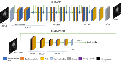 |
Super Resolution MRI Using 3D Generative Adversarial Network: Towards Single Breath-Hold Coronary MR Angiography
Video Permission Withheld
Yibin Xie, Ruiyuan Lin, Yuhua Chen, Yubo Zhang, Feng Shi, Yanan Fei, Zixin Deng, Derenik Haghverdian, Madhvi Kannan, Hyuk-Jae Chang, C.-C. Jay Kuo, Debiao Li
Coronary MRA is an attractive imaging tool to offer noninvasive, radiation-free evaluation of coronary artery disease. However, long scan time and sensitivity to motion limit its current clinical applications. In this paper, we propose a super resolution reconstruction framework based on 3D generative adversarial network (GAN) to allow substantial acceleration (10x plus) and potentially whole-heart coronary MRA within a breath-hold. Preliminary results demonstrated significantly improved vessel sharpness and image quality metrics in highly under-sampled coronary MRA dataset.
|
08:48
|
1051.
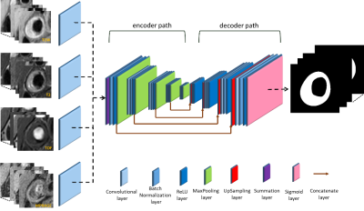 |
Automatic Segmentation of Carotid Vessel Wall in Multi-Contrast Blackblood Images using Deep Learning
Jifan Li, Shuo Chen, Xihai Zhao, Chun Yuan, Rui Li
In this work, we proposed an automatic approach for segmentation of carotid vessel wall in multi-contrast blackblood images, using a fine-tuning U-net neural network. The U-net network consists of an encoder path that captures context and reduces data dimension and a symmetric decoder path that enables precise localization and high resolution. The fine-tuning was utilized to accommodate multi-contrast images input. The pixel-level sensitivity, specificity and IoU of our model achieved 0.869, 0.987 and 0.751 on the test data set, respectively.
|
09:00
|
1052.
 |
Fetal whole-heart 3D cine reconstruction using motion-corrected multi-slice dynamic imaging
Joshua van Amerom, David Lloyd, Maria Kuklisova Murgasova, Anthony Price, Shaihan Malik, Milou Van Poppel, Kuberan Pushparajah, Mary Rutherford, Reza Razavi, Joseph Hajnal
Cine fetal cardiac imaging with whole heart coverage presents numerous challenges due to maternal and fetal motion as well as high heart rate and lack of reliable independent synchronisation signal. To overcome these challenges, highly accelerated multi-slice 2D dynamic images were retrospectively reordered in time and aligned in space to produce a 3D cine using scattered interpolation methods with outlier rejection. The proposed methods were tested on ten human fetal subjects and validated using a numerical phantom.
|
09:12
|
1053.
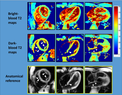 |
Dark-blood T2 mapping for improved assessment of the left ventricular subendocardium, right ventricle and left atrium
Chenxi Hu, Dana Peters
The accuracy of cardiac T2 mapping in small structures, such as the subendocardium, right ventricle, and left atrium, is impaired by the partial-voluming effect between myocardium and blood. In this work, we propose a dark-blood T2 mapping method that nulls the blood prior to the T2 quantification, so that partial-voluming is reduced and T2 estimation accuracy in these small structures is improved. Both conventional and the proposed dark-blood T2 mapping methods were performed in healthy subjects. The results demonstrated the potential of dark-blood T2 mapping to improve the clinical assessment of the subendocardium, right ventricle, and left atrium.
|
09:24
|
1054.
 |
Myocardial Infarct Border Zone Metabolism Measured by Hyperpolarized 13C-Pyruvate MRI
Did Not Present
James Pilla, Gabor Mizsei, Jerry Zsido II, Norman Butler, Yoshiaki Saito, Gabrielle Pilla, Akito Imai, Keitaro Okamoto,, Christopher Gade, Mehrdad Pourfathi, Kai Ruppert, Stephen Kadlecek, Sarmad Siddiqui, Rahim Rizi, Joseph Gorman III, Robert Gorman, Terence Gade
Adverse remodeling after a myocardial infarct has been linked to the elevated wall stress in the myocardium adjacent to the infarct (i.e. border zone). Perturbed metabolism in this region could drive the transition from compensated to adverse remodeling. To evaluate regional metabolism this study compared the uptake and intracellular conversion of [1-13C]pyruvate using hyperpolarized 13C. An infarct model of ventricular remodeling was used to investigate region metabolism. Pharmacologic stress produced an increase in remote metabolite flux compared to border zone region which may provide a metabolic mechanism for the established association of mechanical stress and adverse cardiac remodeling following infarct.
|
09:36
|
1055.
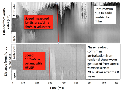 |
Transient intrinsic torsional shear wave propagation demonstrates a difference in Left Ventricular Myocardial stiffness between volunteers and patients with HFpEF
Jessica Webb, Jurgen Runge, Omar Darwish, Alessandro Polcaro, Torben Schneider, Gerald Carr-White, Jordi Martorell, David Nordsletten, Reza Razavi, Ralph Sinkus
Heart Failure with preserved Ejection Fraction is common, associated with high morbidity and mortality, and is challenging to diagnose. We have developed a novel patient friendly non-invasive technique to quantify myocardial stiffness using transient MR Elastography. Aortic valve closure results in the propagation of a transient shear wave through the myocardium. Torsional wave propagation can be visualised using a 1D pencil beam navigator positioned longitudinally along the myocardial septum, using four breath holds each 15 seconds. Providing a temporal resolution of 0.3ms, we observe increased myocardial stiffness in HFpEF patients compared to healthy volunteers (torsional speed 5.5 ± 1.1 m/s in volunteers and 10.0 ± 0.7 m/s in Patients; stiffness 36 ± 12.0 kPa and 108 ± 15.2 kPa, respectively) .
|
09:48
|
1056.
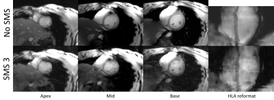 |
Simultaneous Multi-Slice (SMS) Cardiac CINE using embedded Hadamard-encoded reference data at 7 Tesla
stanislas Rapacchi, Thomas Troalen, Maxime Guye, Monique Bernard, Alexis Jacquier, Frank Kober
To perform high-resolution cardiac function MRI, thin-slice Cine MRI at 7T is accelerated using Simultaneous Multi-Slice (SMS) technique. Additionally, Hadamard encoding strategy along the temporal dimension is superimposed on the CAIPIRINHA phase shift. The Hadamard-decoded data serve as embedded reference for SMS image reconstruction. Additional in-plane L1-SPIRiT reconstruction allows for limited noise amplification. Results show excellent slice separation, satisfactory SNR and CNR for assessment of cardiac function. However, SAR restrictions impose a lower flip angle for SMS acquisitions that result in poorer blood-to-myocardium contrast.
|
|
