Velocity & Flow
Oral
Cardiovascular
Wednesday, 20 June 2018
| N02 |
08:15 - 10:15 |
Moderators: Emilie Bollache, Alejandro Roldan-Alzate |
08:15
|
0683.
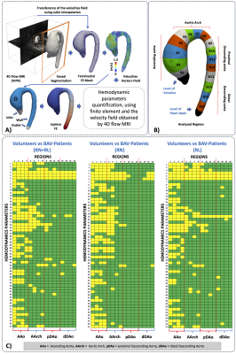 |
 Comprenhensive analysis of Hemodynamics parameters in patients with bicuspid aortic valve using 4D flow data and a finite element method. Comprenhensive analysis of Hemodynamics parameters in patients with bicuspid aortic valve using 4D flow data and a finite element method.
Julio Sotelo, Lydia Dux-Santoy, Andrea Guala, Aroa Ruiz-Muñoz, Arturo Evangelista, Joaquín Mura, Daniel Hurtado, José Rodríguez-Palomares, Sergio Uribe
Bicuspid aortic valve (BAV) is the most common congenital cardiac defect. Current criteria to support surgical decision in BAV patients has been recently questioned, which motivates the development of better biomarkers to assess the disease stage of development. In this work, we extensively analyze several hemodynamics parameters in volunteers and BAV patients from 4D flow MRI using a finite-element-based quantification framework. We found that the backward velocity fraction, the circumferential WSS, and turbulence parameters, can be used together with the diameters of the vessel as a new risk marker, as it can strongly discriminate subjects from BAV patients.
|
08:27
 |
0684.
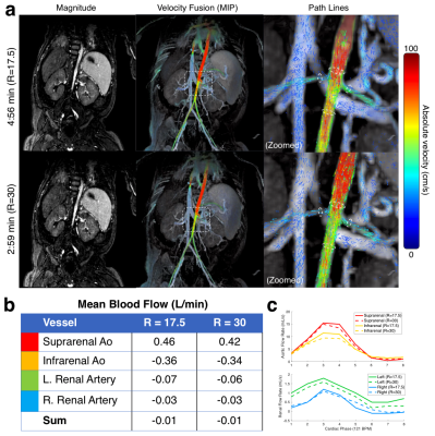 |
Accelerated abdominal 4D flow MRI using 3D golden-angle cones trajectory
Christopher Sandino, Joseph Cheng, Marcus Alley, Michael Carl, Shreyas Vasanawala
4D flow MRI enables comprehensive abdominal evaluation, but long acquisition times and motion corruption limit its clinical applicability. To address these limitations, we present a 4D flow sequence with a 3D golden-angle reordered cones sampling trajectory. Cones has high sampling efficiency to allow for vastly accelerated scan times, and excellent aliasing properties that diffuse respiratory and bowel motion artifacts. To further improve motion-robustness, respiratory signals are estimated from each cone readout, and then used to suppress motion during reconstruction. We show that these techniques can be combined to achieve high quality abdominal 4D flow renderings in under 5 minutes.
|
08:39
 |
0685.
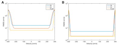 |
PC-MRI with Phase Recovery from Multiple Wrapped Measurements (PRoM)
Shen Zhao, Lee Potter, Ning Jin, Yingmin Liu, Orlando Simonetti, Rizwan Ahmad
In traditional phase-contrast MRI (PC-MRI), the strength of velocity encoding gradient (VENC) offers a tradeoff between the velocity-to-noise ratio (VNR) and the extent of phase wrapping. In contrast, dual-VENC (DV) acquisition achieves the VNR associated with lower of the two VENCs, with higher VENC measurement solely used to perform phase unwrapping1. Here, we demonstrate that the phase unwrapping can be more effective from two low-VENC measurements, where both VENC values are below the peak velocity. The proposed method, called Phase Recovery from Multiple wrapped measurements (PRoM), enables computationally simple yet near-optimal estimation of unwrapped phase (velocity) from multiple wrapped measurements.
|
08:51
|
0686.
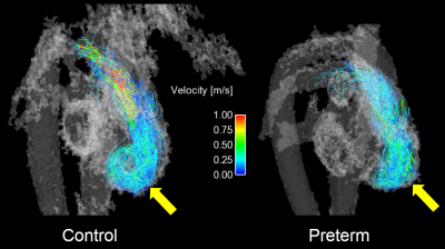 |
Inefficient Right Heart Function in Preterm Adults as shown with 4D Flow MRI
Jacob Macdonald, Greg Barton, Arij Beshish, Kara Goss, Marlowe Eldridge, Christopher Francois, Oliver Wieben
Infants born preterm often have impaired pulmonary function, but little is known regarding long-term implications on right-heart function as these patients reach adulthood. We performed 4D flow MRI during rest and exercise in young adults born preterm and age-matched controls to compare their right heart function and efficiency. While flow and velocity in the pulmonary artery appeared similar in both groups, preterm subjects demonstrated increased kinetic energy in the right ventricle (RV) per unit of ejected blood. Pathline visualizations in the right ventricle suggested less structured filling during diastole. These data suggests decreased RV efficiency in preterm adults.
|
09:03
|
0687.
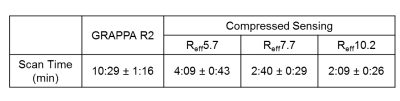 |
Highly accelerated 4D flow in the aorta with compressed sensing, respiratory controlled adaptive k-space reordering and inline reconstruction
Ning Jin, Liliana Ma, Kelvin Chow, Christoph Forman, Andreas Greiser, Susanne Schnell, Alex Barker, Michael Markl
The clinical application of 4D flow is limited by its long acquisition time. Recently, sparse sampling and compressed sensing (CS) reconstruction have been combined with 4D flow to speed up the image acquisition. However, most of CS reconstructions are implemented offline with long reconstruction times, making them infeasible for use in clinical settings. We developed a highly accelerated 4D flow with CS and Respiratory Controlled Adaptive k-space Reordering (ReCAR) acquisition to enable a 3-minute aortic protocol with isotropic 2.5mm voxels and fast 5-minute inline reconstruction. Initial volunteer tests show a good match of flow quantification results with the conventional technique.
|
09:15
|
0688.
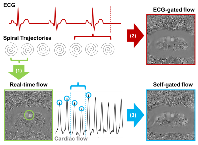 |
Real-time and self-gated flow using a golden-angle spiral trajectory
Rajiv Ramasawmy, Adrienne Campbell-Washburn, Robert Lederman, Daniel Herzka
Beat-to-beat measurements of cardiac output have been demonstrated as a valuable diagnostic tool during interventional cardiovascular MR procedures with physiological provocations such as exercise. This preliminary study investigates the potential of golden-angle spiral imaging for combined real-time and retrospectively-binned flow measurements at rest and during exercise. In flow phantoms and human subjects, good agreement was observed between reference Cartesian measurements and self-gated spiral acquisitions at rest. During exercise, real-time and self-gated spiral imaging measured an increase in cardiac output, demonstrating this technique’s potential application to interventional MRI.
|
09:27
|
0689.
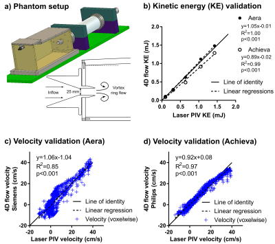 |
Multi-vendor validation and reproducibility of 4D flow MRI using 4-fold parallel imaging acceleration with and without respiratory gating
Johannes Töger, Jelena Bock, Sebastian Bidhult, Karin Markenroth Bloch, Mikael Kanski, Håkan Arheden, Frederik Testud, Andreas Greiser, Einar Heiberg, Marcus Carlsson
4D-flow sequences on two 1.5T scanners from different vendors (Philips Achieva dStream and Siemens MAGNETOM Aera) were validated head-to-head. 4D-flow in a pulsatile flow phantom showed high accuracy and precision compared to reference laser particle image velocimetry for both scanners. 2D-flows in ascending and descending aorta and pulmonary trunk were compared to 57 4D-flow scans in 10 subjects. Lower bias for flow volumes was found on Aera (-4.7±12.5%) compared to Achieva (-18.0±20.1%). Kinetic energy showed lower bias for repeated examinations at the same scanner compared to different scanners the same day. 4D-flow without respiratory gating on Aera showed acceptable quality.
|
09:39
|
0690.
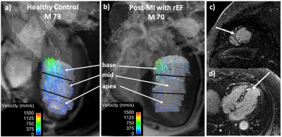 |
Hemodynamic Assessment of the Post-Myocardial Infarction Left Ventricle with 4D Flow MRI
Philip Corrado, Niti Aggarwal, Jacob Macdonald, Jonathan Weinsaft, Christopher Francois, Oliver Wieben
This work employed high resolution time resolved (4D) flow cardiac MRI (CMR) to characterize altered left ventricular (LV) flow physiology after anterior myocardial infarction (MI). 4D Flow CMR was used to quantify LV velocity (parallel to the LV long axis) at pre-specified landmarks in the basal, mid, and apical LV. Post-MI patients with impaired global LV function had reduced peak systolic velocity in all regions compared to age-matched controls (p<0.05). A difference in flow patterns between the LV base and apex was also discerned in post-MI patients, characterized by a marked reduction and prolongation of forward flow in the apex.
|
09:51
|
0691.
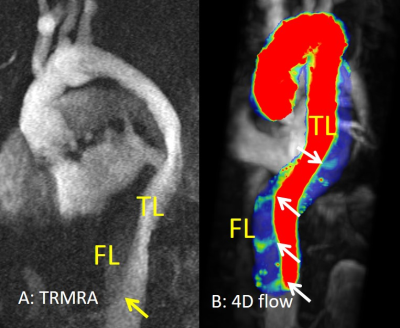 |
4D flow MRI Improves Dissection Flap Fenestration Detection in Type B Aorta Dissection
Bradley Allen , Amir Rahsepar, Alex Barker, James Carr, Jeremy Collins, Michael Markl
Type B aortic dissections involve the aortic arch/descending aorta with false lumen patency/thrombosis, and size of entry tears identified as predictors of adverse events. These parameters suggest a complex hemodynamic environment that is poorly assessed by current diagnostic tools. The presence of additional fenestrations in the dissection flap likely also alters false lumen hemodynamics. 4D flow MRI offers a comprehensive assessment of 3D aortic hemodynamics in type B dissection. Our results demonstrate that 4D flow can detect nearly two-times the number of dissection flap fenestrations relative to time-resolved MRA, suggesting the potential for improved hemodynamic characterization of these patients.
|
10:03
|
0692.
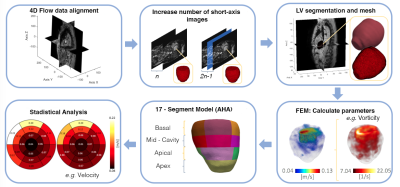 |
Comprehensive 3D flow characterization in patients with Dilated Cardiomyopathy from 4D flow MRI data using a finite element method and 17-Segment Bullseyes
Pamela Franco, Julio Sotelo, Bram Ruijsink3, David Nordsletten, Eric Kerfoot, Joaquín Mura, Daniel Hurtado, Sergio Uribe
Dilated Cardiomyopathy (DCM) is a disease of the heart muscle characterized by the enlargement of the left ventricle and systolic/ diastolic dysfunction. Cardiac Magnetic resonance (CMR) is the gold standard to measure cardiac function using b-SSFP CINE images1. However, hemodynamic parameters within the ventricles have received less attention. In this work, we present a method that allows a comprehensive assessment of different flow parameters in the left ventricle of DCM patients using a finite element framework over 4D flow data sets.
|
|
