Atherosclerosis & Vessel Wall Imaging
Oral
Cardiovascular
Tuesday, 19 June 2018
| S03 |
13:45 - 15:45 |
Moderators: Claudia Calcagno, Kevin DeMarco |
13:45
|
0483.
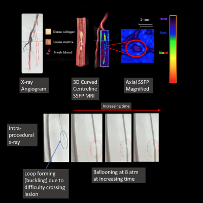 |
 Relationship between MR characteristics of peripheral artery lesions and difficulty of peripheral endovascular procedures Relationship between MR characteristics of peripheral artery lesions and difficulty of peripheral endovascular procedures
James Zhou, Trisha Roy, Hou-Jou Chen, Andrew Dueck, Graham Wright
The most common mode of failure for percutaneous vascular interventions (PVI) is the inability to cross hard lesions with a guidewire. This study uses magnetic resonance (MR) lesion characterization to predict the difficulty of PVI. Steady state free precession (SSFP) MR angiography and ultrashort echo time imaging were used to categorize lesions as “hard” (e.g. calcium, collagen), or “soft” (thrombus, lipids). 17 patients were imaged prior to PVI. MRI-defined hard lesions required significantly longer time to cross (14.81 min vs 1.61 min) and required stenting more often.
|
13:57
|
0484.
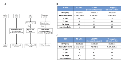 |
Antibiotic treatment affects vascular elastin remodeling both focally and distally to the site of aortic injury in a murine model
Begoña Lavin Plaza, Alkystis Phinikaridou, Marcelo Andia, René Botnar
Atherosclerosis is a systemic, inflammatory disease of the large and medium-sized arteries. Although vascular interventions aim at treating focal stenosis, they may trigger systemic responses that accelerate lesions elsewhere. Elastin remodeling plays a crucial role in vessel wall thickening with monocytes and vascular smooth muscle cells (VSMC) being the primary sources of elastin synthesis. In this study, we used an elastin-binding MR contrast agent to assess whether (1) vascular injury in the abdominal aorta accelerates atherosclerosis in the brachiocephalic artery located distally to the site of injury (2), whether antibiotic treatment alters vascular elastin remodeling and (3) whether antibiotic treatment alters VSMC migration and proliferation and monocyte recruitment and polarization.
|
14:09
|
0485.
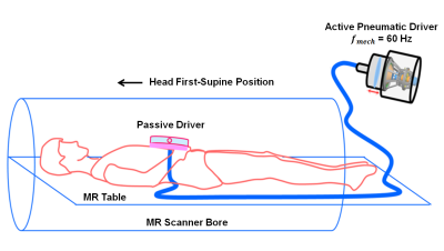 |
In Vivo Quantification of Aortic Stiffness in Abdominal Aortic Aneurysm Patients using MR Elastography: A Longitudinal Study
Huiming Dong, Brian Raterman, Xiaokui Mo, Prateek Kalra, Richard White, Arunark Kolipaka
Abdominal aortic aneurysm (AAA) rupture causes death in 90% of AAA patients. Clinically, the rupture potential is evaluated using AAA diameter. Aortic stiffness can potentially provide more accurate rupture risk assessment. Therefore, the aim of this study is to use non-invasive MR elastography (MRE) to estimate aortic stiffness in AAA patients, and study the relationship between stiffness and AAA diameter. Results showed that aortic stiffness varied during the serial follow-up, demonstrating (1) no correlation between AAA stiffness and diameter, and (2) AAA diameter may not be an accurate indicator for rupture potential.
|
14:21
|
0486.
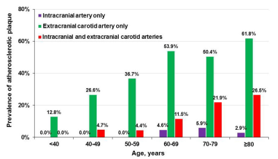 |
Increases in the Prevalence of Subclinical Cerebrovascular Atherosclerosis with Age: A 3.0 T Magnetic Resonance Imaging Study of Community-based Adults
Xihai Zhao, Gaifen Liu, Runhua Zhang, Xiaoyi Chen, Dongye Li, Yong Jiang, Yilong Wang, Yongjun Wang, Chun Yuan
It is well established that atherosclerotic diseases occurring in intracranial and extracranial carotid arteries are associated with ischemic cerebrovascular events. It is important to find a surrogate risk factor for subclinical cerebrovascular atherosclerosis in asymptomatic subjects for stroke prevention. This study sought to investigate the association between age and subclinical cerebrovascular atherosclerosis in community-based adults using MR vessel wall imaging. We found that the prevalence of cerebrovascular atherosclerotic plaques increased with age. The association between age and cerebrovascular atherosclerosis suggests that age was an independent indicator for subclinical cerebrovascular atherosclerosis.
|
14:33
 |
0487.
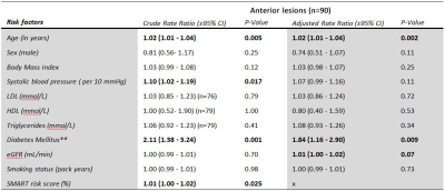 |
Vascular risk factors and intracranial atherosclerosis at 7T vessel wall MRI in Caucasian ischemic stroke and TIA patients
Arjen Lindenholz, Anja van der Kolk, Irene van der Schaaf, Bart van der Worp, Anita Harteveld, Nikki Dieleman, Michiel Bots, Jeroen Hendrikse
There is a lack of knowledge on the association between cardiovascular risk factors and intracranial atherosclerotic lesion burden in the Caucasian population. In this study 105 Caucasian stroke and TIA patients underwent 7T intracranial vessel wall imaging within 3 months after symptom onset. Poisson regression analysis showed that older age, higher systolic blood pressure, diabetes mellitus and a higher SMART risk score (wherein multiple cardiovascular risk factors are combined) were associated with a higher number of vessel wall lesions. Controversially, dyslipidemia and cigarette smoking were not associated with the severity of intracranial vessel wall lesions.
|
14:45
|
0488.
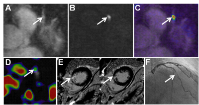 |
Preliminary study of PET/MR 18F-NaF imaging combined with CATCH sequence of high-risk plaque
Xiao Bi, Wei Dong, Yibin Xie, Jiajin Liu, Xiaojun Zhang, Zhiwei Guan, Damini Dey, Jing An , Piotr Slomka, Daniel Berman, Debiao Li, Baixuan Xu
This study aimed to investigate the value of PET/MR 18F-NaF combined with CATCH technology in the diagnosis of high-risk plaque preliminarily. We compared if it had the statistical difference of PMR and TBRmax between the culprit vascular plaque and the control,and evaluated if it had correlation between PMR and TBRmax. There were higher PMR and TBRmax in culprit plaque. PMR and TBRmax had a weak correlation. PET/MR 18F-NaF combined with the CATCH technique can noninvasively determine the location of high-risk plaque in coronary artery, and is valuable for the diagnosis of high-risk plaque.
|
14:57
|
0489.
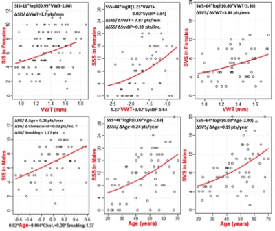 |
Gender-Related Difference in Atherosclerosis: Magnetic Resonance Coronary Artery Wall Thickness is a Predictor of Coronary Plaque Burden in Asymptomatic Young Women
Khaled Abd-Elmoniem, Ahmed Ghanem, Ahmed Hamimi, Jatin Matta, Reham Elgarf, Ranganath Muniyappa, Michael McConnell, Colleen Hadigan, Ahmed Gharib
This study evaluates the potential association of coronary plaque burden with traditional atherosclerosis risk factors and coronary wall thickness (VWT) measured with time-resolved phase-sensitive dual-inversion black-blood TRAPD-MRI. The study demonstrates that there is substantial coronary plaque burden in asymptomatic subjects at low and intermediate Framingham score. In addition, there was evidence of a gender-dimorphic association between the predictors and plaque burden. In women, coronary wall thickness was the strongest and common significant predictor for all plaque burden scores and the presence of calcification. Meanwhile in men, age was the strongest and common predictor of all plaque burden scores and the presence of calcification. This suggests that atherogenesis in women may differ from men and that VWT could supplement traditional risk scores for CAD risk stratification in women. This is in line with the previous studies that demonstrated the impaired utility of these CAD risk score models for women compared to men.
|
15:09
|
0490.
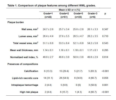 |
Association between Vulnerability of Carotid Artery Atherosclerotic Plaques and White Matter Lesions in Asymptomatic Elderly Population: A MR Vessel Wall Imaging Study
Ying Cai, Yang Liu, Qiang Zhang, Weizhong Tian, Chun Yuan, Cheng Li, Wei Wang, Xihai Zhao
This study investigated the relationship between morphological and compositional characteristics of carotid artery atherosclerotic plaques and WML in asymptomatic elderly population using 3D multicontrast MR vessel wall imaging. We found that carotid artery plaque compositional characteristics, especially calcification, intraplaque hemorrhage, and high risk plaque, were significantly associated with the severity of WML, suggesting that vulnerable atherosclerotic plaque in carotid artery might be an independent indicator for cerebral ischemic lesions in asymptomatic elderly adults.
|
15:21
|
0491.
 |
Intraplaque Hemorrhage and Calcification Detection with Quantitative Susceptibility Mapping
Chaoyue Wang, Saifeng Liu, Yongsheng Chen, Sagar Buch, Zhaoyang Fan, E. Mark Haacke, Qi Yang
In carotid atherosclerosis, the vulnerable feature that has drawn the most attention is intraplaque hemorrhage (IPH). Clinically, MPRAGE has been used to detect IPH. However, IPH has several drawbacks. The susceptibility difference between arterial blood, vessel wall and different types of plaque makes QSM a potential tool for carotid plaque imaging. The purpose of this work was to develop an optimized protocol for both intraplaque hemorrhage and calcification imaging using Quantitative Susceptibility Mapping (QSM) and demonstrate initial clinical validation of QSM as a tool for characterizing components of carotid plaque.
|
15:33
|
0492.
 |
Identification of high-risk plaque features in intracranial atherosclerosis: initial experience using high-resolution MRI with a quantitative radiomics approach
Zhang Shi, Chengcheng Zhu, Jianping Lu, Tao Jiang, David Saloner, Qi Liu
This study aims to evaluate a radiomic approach including texture analysis based on HR-MRI to differentiate acute symptomatic plaque from asymptomatic plaque. 94 patients with basilar artery stenosis underwent HR-MRI. The stenosis value, plaque area/burden, lumen area, intraplaque hemorrhage (IPH), contrast enhancement ratio and 94 quantitative radiomic features were extracted. Multivariate logistic analysis and a random forest model were performed. Results: IPH and enhancement ratio were independently associated with acute symptoms. Radiomic features in T1 and CE-T1 images were associated with acute symptoms. The combined T1 and CE-T1 radiomic approach had a significantly higher AUC of 0.940. Conclusion: Radiomic analysis accurately distinguished between acutely symptomatic plaques and asymptomatic plaques.
|
|
