Thoracic MRI: Lung & Mediastinum
Oral
Body: Breast, Chest, Abdomen, Pelvis
Thursday, 21 June 2018
| W05/06 |
08:00 - 10:00 |
Moderators: Simon Veldhoen, Yoshiharu Ohno |
08:00
 |
1077.
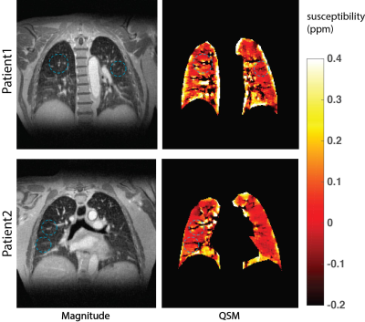 |
 Motion-resolved UTE based Pulmonary Quantitative Susceptibility Mapping Motion-resolved UTE based Pulmonary Quantitative Susceptibility Mapping
Xucheng Zhu, Hongjiang Wei, Kevin Johnson, Scott Nagle, Wenwen Jiang, Joseph Cheng, Shreyas Vasanawala, Michael Lustig, Chunlei Liu, Peder Larson
Pulmonary MRI is challenging due to many factors, such as short T2* relaxation time and respiratory motion corruption. However, the large susceptibility differences in the lungs from blood oxygenation and O2 content might provide more information related to pulmonary function. In this work, we combined ultra-short TE(UTE) acquisition, quantitative susceptibility mapping(QSM), and motion-resolved reconstruction techniques together to look at the susceptibility contrast in the lung and changes in different motion states. According to the results, this technique provides extra contrast information compared to traditional intensity images, and shows susceptibility changing of lung in different respiration states.
|
08:12
|
1078.
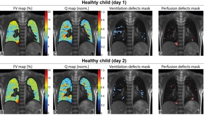 |
Repeatability of ventilation and perfusion impairment assessed with matrix pencil decomposition MRI and lung function in children with cystic fibrosis
Grzegorz Bauman, Sylvia Nyilas, Orso Pusterla, Enno Stranzinger, Kathryn Ramsey, Florian Singer, Sophie Yammine, Carmen Casaulta, Philipp Latzin, Oliver Bieri
This study examines the repeatability of functional lung MRI using matrix pencil decomposition and lung function tests with multiple breath washout technique in children with cystic fibrosis and age-matched healthy controls. We found a good degree of the agreement between outcomes measured on two consecutive days in both groups. Furthermore, a strong correlation between functional MRI and global ventilation inhomogeneity index from multiple breath washout was observed. The results of this study highlight the potential of matrix pencil decomposition MRI for the assessment of disease progression in cystic fibrosis
|
08:24
|
1079.
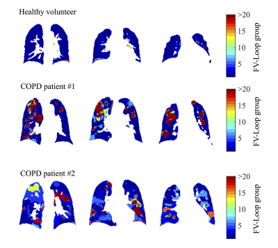 |
Imaging-Based Spirometry in Chronic Obstructive Pulmonary Disease (COPD) Patients using Phase Resolved Functional Lung Imaging (PREFUL)
Andreas Voskrebenzev, Filip Klimeš, Marcel Gutberlet, Till Kaireit, Christian Schönfeld, Julius Renne, Heike Biller, Jens Hohlfeld, Frank Wacker, Jens Vogel-Claussen
Recently, an alternative proton lung MRI Fourier Decomposition (FD) method for phase resolved functional lung imaging (PREFUL) was proposed. Using a sine model, respiratory and cardiac cycles with increased temporal resolution are obtained, enabling the assessment of dynamic parameters. Similar to flow-volume loops in spirometry, the regional ventilation can be quantified in terms of fractional ventilation (FV) loops. In this study the FV loops of six healthy volunteers and 16 chronic obstructive pulmonary disease patients are evaluated. As a metric the cross-correlation to a healthy reference is used. The results suggest potential benefits for early detection or treatment monitoring.
|
08:36
|
1080.
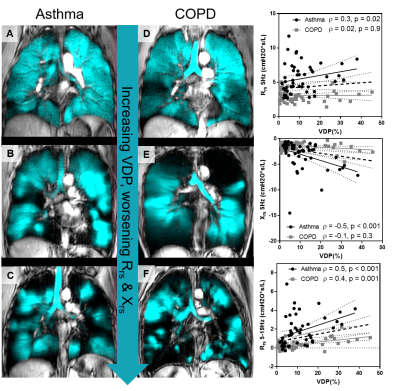 |
Pulmonary MRI Measurements of Ventilation Heterogeneity in Obstructive Lung Disease: Relationship to Oscillometry, Quality of Life and Disease Control
Heather Young, Fumin Guo, Rachel Eddy, Geoffrey Maksym, Grace Parraga
We measured ventilation heterogeneity in 100 patients (50 asthma, 50 COPD) with hyperpolarized 3He MRI, oscillometry and quality-of-life questionnaires. We showed that MRI-measured ventilation heterogeneity is significantly related to FOT-measured small airways resistance and worsened quality-of-life for asthma and COPD patients. We also showed that MRI-measured ventilation heterogeneity is significantly increased in asthmatic patients with poor disease control. This study directly demonstrates the relationships between 3He MRI ventilation heterogeneity with small airway dysfunction and patient quality-of-life.
|
08:48
|
1081.
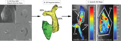 |
Altered Right-Heart 3D Blood Flow and Kinetic Energy in Chronic Obstructive Pulmonary Disease. The MESA COPD Study
Haben Berhane, Ozair Rahman, Pallavi Balte, Kenichiro Suwa, Stephen Dashnaw, David Bluemke, Martin Prince, Bharath Venkatesh, Joao Lima, James Carr, Antoinette Gomes, Karol Watson, Cynthia Rigsby, R. Graham Barr, Michael Markl
Chronic Obstructive Pulmonary Disease (COPD) affects over 65 million people worldwide and is the third leading cause of death in the US. The aim of this feasibility study was to employ 4D-flow MRI for the comprehensive assessment of the effects of COPD on right-sided hemodynamics. Measures of blood kinetic energy (KE) were used to quantify hemodynamic changes in COPD patients compared to controls. Findings showed a significant increase in KE in the right atrium across increasing categories of COPD severity. Right-sided 4D flow MRI maybe a promising tool for the detection of hemodynamic disruptions in COPD and other vasoconstrictive diseases.
|
09:00
|
1082.
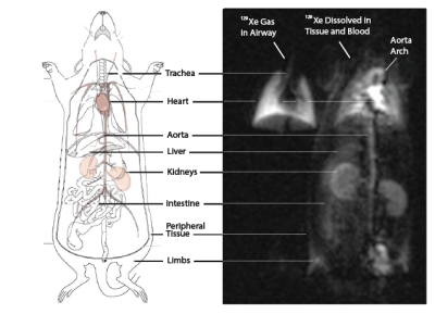 |
A Technique for Quantitatively Measuring Gas Uptake in the Lung and its Distribution to the Kidneys Using Hyperpolarized Xenon-129 MR Imaging
Hooman Hamedani, Stephen Kadlecek, Kai Ruppert, Yi Xin, Mehrdad Pourfathi, Sarmad Siddiqui, Faraz Amzajerdian, Luis Loza, Harrilla Profka, Ryan Baron, Tahmina Achekzai, Shampa Chatterjee, Maurizio Cereda, Rahim Rizi
Hyperpolarized 129Xe was used before to image lung function and structure. It was also shown to be a very good marker of oxygen diffusion and uptake from the lung, and a surrogate for alveolar wall thickness. Here, we demonstrated a technique for quantitatively measuring gas uptake in the lung and its distribution to the kidney and calculate the arrival time of the gas to the kidneys. Appropriate acquisition parameters will enable the same technique to be used to measure the gas’ arrival at the heart, specific vasculature, or other organs/tissue as appropriate to specific disease states.
|
09:12
|
1083.
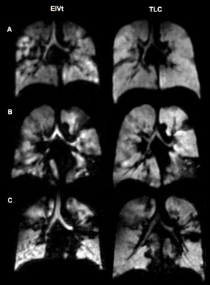 |
Hyperpolarised gas MRI shows a decrease in lung ventilation defects at increased inspiratory lung volumes in Cystic Fibrosis
Laurie Smith, Paul Hughes, Helen Marshall, Guilhem Collier, Noreen West, Alex Horsley, Jim Wild
The effect of lung inflation level on ventilation defects, using hyperpolarised (HP) gas ventilation MRI, has not been assessed. The most commonly adopted method of inhaling a volume of 1L from functional residual capacity (FRC) will result in a lung volume closer to total lung capacity (TLC) for smaller people, compared with taller people. We assessed HP-MRI in 21 people with cystic fibrosis at both end inspiratory tidal volume (EIVt) and at TLC. Ventilation defects decreased in all subjects at TLC when compared to EIVt and therefore the inspiratory volume should be carefully considered when interpreting ventilation-imaging results.
|
09:24
|
1084.
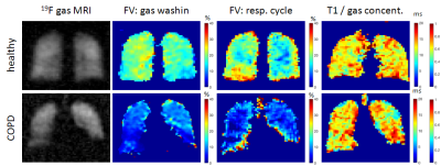 |
Quantification of gas concentration and fractional ventilation using high temporal resolution MRI of pulmonary fluorinated (19F) gas washin dynamics in free breathing
Marcel Gutberlet, Arnd Obert, Andreas Voskrebenzev, Filip Klimes, Frank Wacker, Jens Vogel-Claussen
19F gas washin/ washout MRI allows quantification of regional lung ventilation in free breathing even in obstructed lungs. By increasing the temporal and spatial resolution of dynamic 19F gas MRI at adequate image quality lung ventilation imaging was improved. In addition to measuring regional gas washin/ washout times, gas density variations during breathing were determined, giving further information of gas dynamics and therefore lung function. Additionally, the concentration of the fluorinated gas tracer and therefore its distribution in the lung was quantified. The new approaches were tested in a healthy volunteer and a COPD patient.
|
09:36
|
1085.
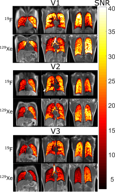 |
Comparing 19F C3F8 Lung Ventilation Imaging with Hyperpolarized 129Xe: Similarities and Limitations
Adam Maunder, Paul J.C. Hughes, Ho-Fung Chan, Graham Norquay, Guilhem Collier, Oliver Rodgers, Fraser Robb, Madhwesha Rao, Jim Wild
Fluorinated gases are a cheaper alternative to hyperpolarized gas for lung ventilation imaging. However, lower resolution images are necessitated by the inherently smaller MR signal from 19F. Within this study we determine the feasibility of using C3F8 for lung ventilation imaging by comparing quantitative metrics of lung function obtained from ventilation images of 19F with those obtained from ventilation images of hyperpolarized 129Xe. A reproducibly lowered coefficient of variation distribution was observed due to lower resolution of 19F imaging. However, ventilation images acquired with 19F were of comparable SNR to those using 129Xe.
|
09:48
|
1086.
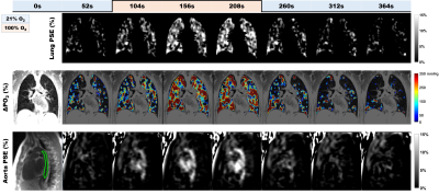 |
Dynamic 3D Isotropic Resolution Imaging of Human Lungs Using Oxygen-enhanced Radial UTE MRI
Wei Zha, Robert Cadman, Andrew Hahn, Kevin Johnson, Sean Fain
Oxygen-enhanced 3D radial UTE MRI (UTE OE-MRI) has the potential for dynamic imaging of oxygen wash-in and wash-out in the lungs with isotropic resolution and full chest coverage. Four normal subjects underwent DESPOT1 and ~13-min dynamic OE-MRI using 3D radial UTE MRI. Temporal variations in median percent signal enhancement (MPSE) strongly correlated (p<0.0001) in the lungs and aorta. Wash-in (0.61±0.13 min) and wash-out (0.30±0.18 min) time constants for partial pressure of oxygen (ΔPO2) time curves are comparable to values in the literature. This study shows feasibility for dynamic imaging with whole lung coverage and isotropic spatial resolution for clinical research.
|
|
