Hepatobiliary: Neoplasm
Oral
Body: Breast, Chest, Abdomen, Pelvis
Monday, 18 June 2018
| S04 |
08:15 - 10:15 |
Moderators: Satoshi Goshima, Ihab Kamel |
08:15
|
0072.
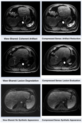 |
 View-Sharing Artifact Reduction with Retrospective Compressed Sensing Reconstruction in the Context of Contrast-Enhanced Liver MRI for Hepatocellular Carcinoma (HCC) Screening View-Sharing Artifact Reduction with Retrospective Compressed Sensing Reconstruction in the Context of Contrast-Enhanced Liver MRI for Hepatocellular Carcinoma (HCC) Screening
Jamil Shaikh, Paul Stoddard, Evan Levine, Stephanie Chang, Albert Roh, Brian Hargreaves, Shreyas Vasanawala, Andreas Loening
View-sharing (VS) increases spatiotemporal resolution in dynamic contrast-enhanced (DCE) MRI by temporally sharing high frequency k-space data across contrast phases. However, this temporal sharing results in respiratory motion occurring in any single phase to propagate artifacts across all phases. Compressed sensing (CS) can eliminate need for VS by recovering missing k-space data from pseudorandom under-sampling, reducing temporal blurring while maintaining spatial resolution. We tested CS versus VS in the setting of DCE MRI for HCC. CS reduced respiratory artifacts, produced images with a more synthetic appearance, and did not result in a difference in lesion detection.
|
08:27
|
0073.
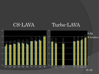 |
Dynamic Gd-EOB-DTPA enhanced MR imaging of the liver: Value of High Temporal-resolution Images with Parallel imaging and Compressed Sensing
Takayuki Masui, Motoyuki Katayama, Mitsuteru Tsuchiya, Masako Sasaki, Kenshi Kawamura, Yuki Hayashi, Takahiro Yamada, Naoyuki Takei, Yuji Iwadate, Kang Wang, Dan Rettmann
With ARC and CS, breath-hold dynamic Gd-EOB-DTPA enhanced MR imaging for the liver can be successfully performed with acceptable image quality and lesion recognitions on a clinical 3T magnet. High temporal resolution images with CS-LAVA for dynamic contrast MR study may give us benefits in comparison of Turbo-LAVA with lower temporal resolutions.
|
08:39
|
0074.
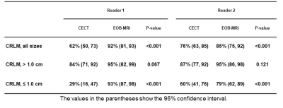 |
Prospective Comparison of Gadoxetic Acid-Enhanced Liver MRI And Contrast-Enhanced CT With Histopathological Correlation For Preoperative Detection Of Colorectal Liver Metastases Following Chemotherapy And Potential Impact On Surgical Plan
Kartik Jhaveri, Sandra Fischer, Hooman Hosseini Nik, Ravi Menezes, Steven Gallinger, Carol-Ann Moulton
Complete resection of colorectal cancer liver metastases increases survival and is a recommended therapeutic option. Thus accurate detection of liver metastases is crucial. Many patients receive preoperative chemotherapy which often causes hepatic steatosis and decreases sensitivity of CT in detecting liver metastases. This prospective study with histopathological correlation compared the diagnostic performance of gadoxetic acid Liver MRI in the preoperative detection of liver metastases following chemotherapy including the influence of hepatic steatosis and lesion size. We also evaluated the potential change in the hepatic resection plan due to inclusion of gadoxetic acid MRI compared to CT.
|
08:51
|
0075.
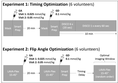 |
Combined Gadoxetic Acid and Gadobenate Dimeglumine Enhanced Liver MRI for Liver Metastasis Detection: A Parameter Optimization Study
Gesine Knobloch, Timothy Colgan, Xiaoke Wang, Tilman Schubert, Diego Hernando, Scott Reeder
The detection of small perivascular metastatic lesions can be challenging with gadoxetic acid-enhanced liver MRI because both, blood vessels and metastases appear hypointense during the hepatobiliary phase. We sought to demonstrate the feasibility of combined gadoxetic acid (GA)/gadobenate dimeglumine (GD) liver MRI for improved lesion detection and optimize the imaging protocol regarding GA-dosing, imaging time after GD-injection and flip angle. Preliminary results show a homogenously enhanced liver and vasculature (“plain-white-liver”) 1-3min after GD-bolus detection with optimal contrast using flip angles of 25-35°. The combined GA/GD protocol has potential to improve the diagnostic performance of hepatobiliary phase liver MRI.
|
09:03
|
0076.
 |
Gadoxetate-enhanced abbreviated MRI is reliable and effective for HCC surveillance in high-risk patients.
Ryan Brunsing, Dennis Chen, Alexandra Schlein, Paul Murphy, Yesenia Covarrubias, Alex Kuo, Michel Mendler, Irene Vodkin, Rohit Loomba, Yuko Kono, Claude Sirlin
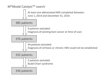
Gadoxetate enhanced abbreviated MRI (AMRI) is a simple, rapid acquisition protocol aimed at reducing the cost and increasing the throughput of MRI-based HCC surveillance. Here we analyze 330 consecutive patients with cirrhosis or chronic HBV who underwent at least one screening AMRI. The rate of HCC detected at cross sectional analysis (3.3%) was in line with published incidence of HCC, while the technical failure rate was low (5.8%) despite high prevalence of cirrhosis and ascites. Longitudinal analysis demonstrated high sensitivity, specificity, and negative predictive value in HCC detection, using a composite reference standard.
|
09:15
|
0077.
 |
Volumetric Apparent Diffusion Coefficient Histogram Analysis in Differentiating Intrahepatic Cholangiocarcinoma from Hepatocellular Carcinoma
Xianlun Zou, Yaqi Shen, Zhen Li, Daoyu Hu
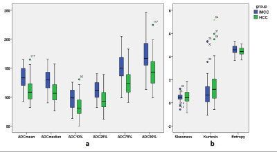
Accurate differentiation between intrahepatic cholangiocarcinoma (IHCC) and hepatocellular carcinoma (HCC) is essential for adequate treatment planning. In the present study, non-contrast volumetric ADC histogram analysis was employed to differentiate IHCC (n=33) and HCC (n=98). The results suggested that except the kurtosis and skewness, all the volumetric ADC histogram parameters, were helpful in distinguishing IHCC from HCC. Among all the parameters, 75th percentile ADC was most helpful to distinguish the two diseases. This non-contrast method provides useful information in differentiating IHCC from HCC, it benefits patients who are contraindicate to contrast agents.
|
09:27
|
0078.
 |
Motion Correction of Diffusion-weighted imaging in the analysis of Apparent Diffusion Coefficient for preoperative staging of hepatocellular carcinoma
Wu Zhou, Qiyao Wang, Guoxi Xie, Fei Yan, Yaoqin Xie, Guangyi Wang, Zaiyi Liu, Changhong Liang, Hairong Zheng, Lijuan Zhang
Preoperative tumor staging of hepatocellular carcinoma (HCC) is a critical issue that influences tumor recurrence and patient survival in clinical practice. One of the challenges encountered in DWI of the liver is cardiac motion that can affect the accuracy of ADC measurements, which may inevitably influence the performance of DWI for HCC staging. However, the impact of motion for ADC and HCC staging has not been thoroughly investigated. In this work, we quantitatively investigate the relationship of motion correction, ADC and staging of HCC in order to widen the understanding of applications in DWI for tumor assessment.
|
09:39
|
0079.
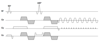 |
Partial velocity-compensated diffusion encoding for combined motion compensation and residual vessel signal suppression in liver DWI
Anh Van, Barbara Cervantes, Tetsuo Ogino, Johannes Peeters, Andreas Hock, Ernst Rummeny, Rickmer Baren, Dimitrios Karampinos
Despite its strong clinical significance in lesion detection and tumor staging, liver DWI remains challenged by its strong sensitivity to motion effects. Motion-compensated diffusion encoding schemes have been recently proposed to improve DW liver signal homogeneity especially in the left liver lobe, a region typically affected by cardiac motion. However, motion-compensated diffusion encoding is associated with hyperintense vessel signal even at high b-values, which can obscure lesion detection. The present work proposes a partial velocity-compensated diffusion encoding for combined motion compensation and residual vessel signal suppression in liver DWI.
|
09:51
 |
0080.
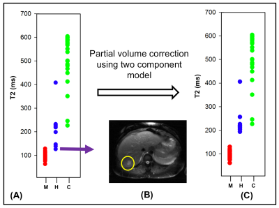 |
Characterization of Abdominal Neoplasms using a Fast T2 Mapping Radial TSE Technique
Mahesh Bharath Keerthivasan, Diego Blew, Jean-Philippe Galons, Diego Martin, Ali Bilgin, Maria Altbach
Radial turbo spin-echo (RADTSE) based methods have been proposed for quantitative T2 mapping. RADTSE yields high spatio-temporal resolution and allows the reconstruction of co-registered images at multiple TE times from a short acquisition (i.e. a breath hold). We investigate the clinical utility of RADTSE for the quantitative characterization of abdominal neoplasms.
|
10:03
|
0081.
 |
Using Tumor Stiffness as a Potential Biomarker for Predicting Hepatocellular Carcinoma Recurrence
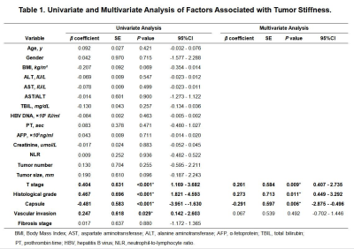
Jin Wang, Hao Yang, Yong Liu, Sichi Kuang, Bingjun He, Yao Zhang, Qungang Shan, Jingbiao Chen, TianHui Zhang, Kevin Glaser, Cairong Zhu, Jun Chen, Meng Yin, Bogdan Dzyubak, Sudhakar Venkatesh, Richard Ehman
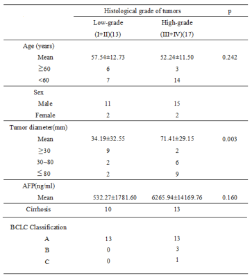
Hepatocellular carcinoma (HCC) is a highly aggressive cancer and one of the leading causes of cancer-related deaths around the world. Our preliminary analysis of 78HCCsshowed 3D MRE is a promising, noninvasive technique for predicting the early recurrence of HCCs after hepatic resection. MRE-assessed tumor stiffness correlates with features such as encapsulation, macrovascular invasion, and histological grade. In the future, larger studies will improve our understanding of the relationship between HCC stiffness, invasiveness, and outcome for better allocation of treatment strategies and surveillance follow-up.
|
|
