Hepatobiliary: Diffuse Liver Disease, Part 1
Oral
Body: Breast, Chest, Abdomen, Pelvis
Tuesday, 19 June 2018
| S06 |
13:45 - 15:45 |
Moderators: Reena Jha, Jeong Hee Yoon |
13:45
 |
0513.
 |
 Noninvasive Staging of Liver Fibrosis using Magnetic Resonance, Ultrasound, and Transient Elastography: a Comparison with Liver Biopsy Noninvasive Staging of Liver Fibrosis using Magnetic Resonance, Ultrasound, and Transient Elastography: a Comparison with Liver Biopsy
Thierry Lefebvre, André Ilinca, Claire Wartelle-Bladou, Giada Sebastiani, Hélène Castel, Bich Nguyen, Jessica Murphy-Lavallée, Damien Olivié, Guillaume Gilbert, An Tang
Elastographic techniques measure liver stiffness as surrogate biomarker of liver fibrosis. We performed paired comparisons of MRE, pSWE, and TE for staging liver fibrosis. For classification of dichotomized fibrosis stages F0 vs. ≥ F1, ≤ F1 vs. ≥ F2, ≤ F2 vs. ≥ F3, and ≤ F3 vs. F4, the AUCs were respectively 0.92, 0.84, 0.89, 0.89 for MRE; 0.76, 0.83, 0.80, 0.75 for pSWE; and 0.75, 0.70, 0.77, 0.84 for TE. Overall, MRE provided a diagnostic accuracy similar or higher than ultrasound-based elastographic techniques.
|
13:57
|
0514.
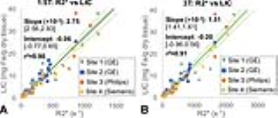 |
Liver R2* as a Biomarker of Liver Iron Concentration: Interim Results from a Multi-Center, Multi-Vendor Reproducibility Study at 1.5T and 3T
Diego Hernando, Ruiyang Zhao, Valentina Taviani, Mounes Aliyari Ghasabeh, Li Pan, Qing Yuan, Stefan Ruschke, Dimitrios Karampinos, Xiaodong Zhong, Ryan Mattison, Ihab Kamel, Ivan Pedrosa, Shreyas Vasanawala, Takeshi Yokoo, Scott Reeder
R2* is a promising biomarker of liver iron concentration (LIC), with application in the assessment of iron overload. Previous works have demonstrated the high correlation of liver R2* with biopsy-determined LIC. Although R2* measurements may be affected by multiple confounding factors, including the presence of fat and noise bias, confounder-corrected R2* mapping has been shown to be highly insensitive to the presence of these confounding factors. However, the multi-center reproducibility of confounder-corrected R2* for liver iron quantification remains unknown. This abstract demonstrates excellent reproducibility of R2* for liver iron quantification in a multi-center, multi-vendor study at both 1.5T and 3T.
|
14:09
|
0515.
 |
Estimating Liver Water and Fat T1 and T2, and PDFF using Flip Angle Corrected Multi-TR, Multi-TE 1H MRS
Gavin Hamilton, Alexandra Schlein, Rohit Loomba, Claude Sirlin
Multi-TR, multi-TE (MTRTE) 1H MRS estimates T1 and T2 of fat and water and liver proton density fat fraction (PDFF) in a single breath-hold by assuming a perfect 90° pulse, which is not guaranteed in vivo and may introduce bias. We introduce a flip angle corrected multi-TR, multi-TE (FAC MTRTE) 1H MRS sequence based on a non-steady state approach and compare the T1, T2, and PDFF given by MTRTE and FAC MTRTE MRS. T1 estimates given by MTRTE MRS and FAC MTRTE MRS were significantly different, due to the MTRTE sequence not being acquired with a perfect 90° flip.
|
14:21
|
0516.
 |
Rapid multi-slice abdominal T1 mapping with high spatial and temporal resolution using an inversion recovery radial steady-state free precession (IR-radSSFP) technique
Zhitao Li, Ali Bilgin, Kevin Johnson, Jean-Philippe Galons, Manoj Saranathan, Diego Martin, Maria Altbach
A radial IR steady-state free precession technique combined with a principal component based iterative algorithm are demonstrated for rapid high-resolution abdominal T1 mapping. This method yields high quality T1 maps for 10 slices with spatial resolution as high as of 0.83x0.83x3mm from data acquired in a breath-hold or a short free-breathing scan. The method is a significant improvement over existing abdominal T1 mapping techniques.
|
14:33
|
0517.
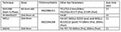 |
Evaluation of Four T1 Mapping Sequences for Obtaining the Extracellular Volume Fraction in Abdominal Imaging
Temel Tirkes, Chen Lin, Xuandong Zhao, Dominik Nickel, Kelvin Chow, Alex Stuckey, Robert Grimm, Shivraman Giri
Many different T1 mapping sequences and extracellular volume (ECV) fraction have proven to be useful tools for evaluation of tissue fibrosis, however their potential has not been explored in abdominal imaging. We evaluated 4 different T1 mapping techniques; Dual-flip angle VIBE (DFA VIBE), MOLLI, SASHA and IR-SNAPSHOT and obtained similar ECV fractions of the liver and pancreas. DFA VIBE has the highest spatial coverage in 1 breath hold but suffers from inhomogeneous T1 in the aortic blood. IR-SNAPSHOT has advantage of not requiring cardiac gating and provides the most homogenous T1 of the blood within the aorta.
|
14:45
|
0518.
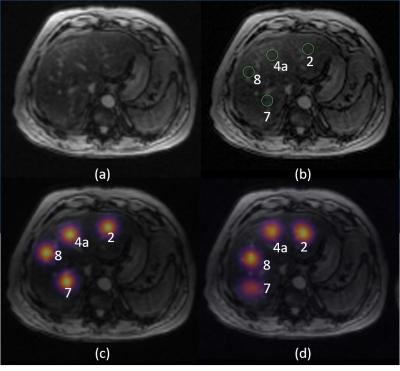 |
Automated liver-segment localization and segmental hepatic proton-density fat fraction measurement
Kang Wang, Adrija Mamidipalli, Kevin Blansit, Jonathan Hooker, Claude Sirlin, Albert Hsiao
Hepatic proton-density fat fraction (PDFF) has emerged as an imaging biomarker for liver fat content in non-alcoholic fatty liver disease. To account for the spatial heterogeneity of liver fat, one approach is to manually place a region of interest (ROI) and estimate the PDFF in each hepatic Couinaud segment separately. We trained a convolutional neural network to automatically locate each liver segment and calculate segmental PDFF values. We show that segmental PDFF measurement based on our automated approach closely matches those based on manual placement by a trained image analyst, demonstrating the feasibility of utilizing CNNs to automatically extract clinically valuable quantitative information from source images.
|
14:57
|
0519.
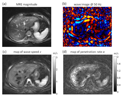 |
Assessment of hepatic fibrosis and steatosis in pediatric non-alcoholic fatty liver disease using multifrequency MR elastography
Jing Guo, Christian Hudert, Heiko Tzschätzsch, Jürgen Braun, Ingolf Sack
Multifrequency MR elastography MRE was applied to detect fibrosis and steatosis, two pathological features associated with pediatric non-alcoholic fatty liver disease (NAFLD). Shear wave speed (c) obtained from MRE is a measure of tissue stiffness and can differentiate moderate from advanced fibrosis. Penetration rate (a) as another MRE parameter is able to detect moderate steatosis and is negatively correlated with hepatic fat fraction. Both MRE derived mechanical parameters c and a are independently responsive to fibrosis and steatosis and could be used in the future as imaging markers for the noninvasive assessment of prediatric NAFLD.
|
15:09
|
0520.
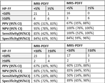 |
Can Negligible Hepatic Steatosis Determined by MRI-Proton Density Fat Fraction Obviate the Need for Liver Biopsy in Potential Liver Donors?
Kartik Jhaveri, Janakan Satkunasingham, Hooman Hosseini Nik, Sandra Fischer, Ravi Menezes, Nazia Selzner, Mark Cattral, David Grant
Hepatic steatosis in potential liver donor candidates has important implications towards outcomes of liver transplant recipients and donor safety. Hepatic steatosis in potential donors in excess of established but varying institutional thresholds are considered as grounds for donor ineligibility. However, negiblible/absent hepatic steatosis (<5%) is considered acceptable universally. Currently Liver biopsy is regarded as reference standard. We show in our study that MR-PDFF has very high NPV for excluding significant hepatic steatosis (>10%). Thus MRI-PDFF can be utilized for liver donor screening and obviates the need for liver biopsy when MRI-PDFF values are <5%
|
15:21
|
0521.
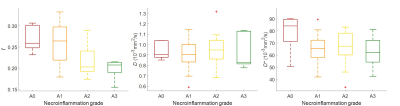 |
Intravoxel Incoherent Motion Diffusion-weighted MRI for Assessing Necroinflammation in Patients with Chronic Liver Disease
Thierry Lefebvre, Guillaume Gilbert, Claire Wartelle-Bladou, Giada Sebastiani, Hélène Castel, Bich Nguyen, Damien Olivié, An Tang
Necroinflammation is a hallmark feature in several causes of chronic liver disease. Because it has multiple tissue contrast mechanisms, MRI is ideally suited for characterization of histopathological changes (i.e. inflammation, fat, iron, and fibrosis) that may occur concomitantly in chronic liver disease. We evaluated intravoxel incoherent motion (IVIM) diffusion-weighted imaging (DWI) MRI for assessment of necroinflammation. Perfusion fractions were significantly correlated with necroinflammation grades (ρ = 0.49, P < 0.0001) and could discriminate grades ≤ A1 vs. ≥ A2 and ≤ A2 vs. A3 with good accuracy (AUC: 0.81 and 0.80, respectively). Our results suggest that perfusion fraction may be used for assessing liver necroinflammation.
|
15:33
|
0522.
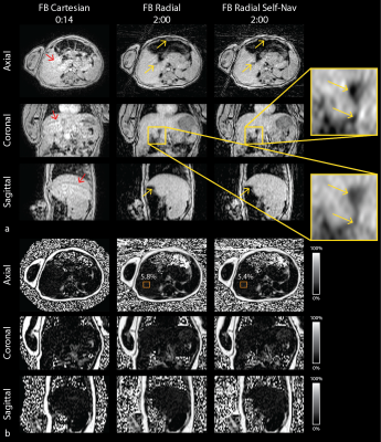 |
Free-Breathing Hepatic Fat Quantification in Children and Infants Using a 3D Stack-Of-Radial Technique: Assessment of Accuracy and Repeatability
T TeArmstrong, Karrie Ly, Yu Wang, Thomas Martin, Shahnaz Ghahremani, Kyunghyun Sung, Kara Calkins, Holden Wu
Non-alcoholic fatty liver disease (NAFLD) has increasing prevalence in children and early risk factors for NAFLD may be present during infancy. MRI can non-invasively quantify hepatic fat, but current techniques require breath-holding (BH), which is not possible in many children and infants. In this study, a novel free-breathing (FB) 3D stack-of-radial fat quantification technique was developed and evaluated in children and infants. The proposed FB technique achieved accurate and repeatable hepatic fat quantification and improved image quality compared to conventional BH techniques. This FB technique can potentially improve the diagnosis and monitoring of NAFLD in children and infants.
|
|
