Pancreas/GI
Oral
Body: Breast, Chest, Abdomen, Pelvis
Wednesday, 20 June 2018
| S05 |
13:45 - 15:45 |
Moderators: T.B.A. |
13:45
|
0823.
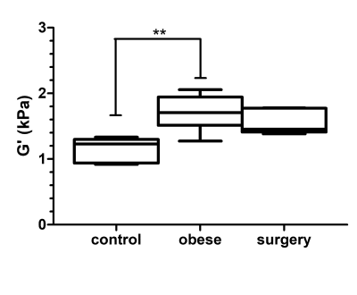 |
 MR elastography and multiparametric MRI of chronic pancreatitis reversal after bariatric surgery in obese rats MR elastography and multiparametric MRI of chronic pancreatitis reversal after bariatric surgery in obese rats
Philippe Garteiser, Vinciane Rebours, Sabrina Doblas, Gwenael Pagé, André Bado, Valérie Paradis, Maude Le Gall, Anne Couvelard, Bernard Van Beers
In obesity, pancreas is affected by fatty infiltration and fibrosis. Bariatric surgery is one of the only therapies which demonstrably improves pancreas status in obesity. In the present work we investigated the mechanical properties at several frequencies, the PDFF and the T2* of pancreatic explants in an obese rat model of bariatric surgery. Bariatric surgery reversed the MRI parameters to values not significantly different than controls. MRI parameters closely matched the reference histology observations. MRE and multiparametric MRI may be used to monitor pancreatic status and treatment response in an obese rat model of bariatric surgery.
|
13:57
|
0824.
 |
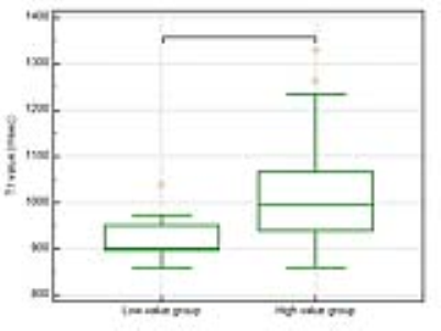
T1 Mapping and MR Elastography (MRE) for the Diagnosis of Mild Chronic Pancreatitis
Yu Shi, Min Wang, Xiaoqi Wang, Yanqing Liu, Ruoyun Ji, Lizhuo Cang, Qiyong Guo
We have investigated the value of both MR elastography (MRE) and T1 mapping for early detection of Mild Chronic pancreatitis(CP). Our study found that both MRE and T1 mapping had good diagnostic performance for detecting mild CP, with MRE slightly outperforming T1 mapping.
|
14:09
|
0825.
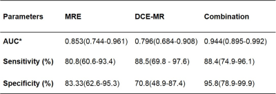 |
Differentiation of Mass-forming Focal Pancreatitis (MFFP) from Pancreatic Cancer (CP): Added Value of Magnetic Resonance Elastography (MRE) to Dynamic Contrast-enhanced Magnetic Resonance Imaging (DCE-MRI)
Yu Shi, Yanqing Liu, Xiaoqi Wang, Min Wang, Ruoyun Ji, Lizhuo Cang, Qiyong Guo
The differential diagnosis of mass-forming focal pancreatitis (MFFP) and pancreatic cancer (PC) is clinically important. Our study shows that combined assessment of magnetic resonance elastography (MRE) with dynamic contrast-enhanced magnetic resonance imaging (DCE-MRI) is a promising technique with improved specificity of diagnosing PC from MFFP cases, when comparing with DCE-MRI.
|
14:21
|
0826.
 |
T1 mapping of the pancreas: correlation with HbA1c values
Yoshifumi Noda, Satoshi Goshima, Yusuke Tsuji, Kimihiro Kajta, Yuta Akamine, Tomoyuki Okuaki, Masatoshi Honda, Hiroshi Kadohara, Hiroshi Kawada, Nobuyuki Kawai, Yukichi Tanahashi, Masayuki Matsuo
The presence of pancreatic fibrosis is a representative feature of the pancreas in patients with impaired glucose tolerance (IGT). The T1 signal intensity of the pancreas is reported to be associated with pancreatic fibrosis and HbA1c values. In this study, we evaluated the feasibility of T1 mapping of the pancreas for the assessment of HbA1c values. Our results showed that increased T1 values of the pancreas was significantly correlated with HbA1c values, so the T1 values of the pancreas could serve as a potential imaging biomarker for the assessment of patients with IGT.
|
14:33
|
0827.
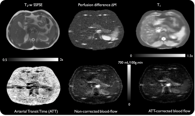 |
Pancreas perfusion and transit-time measurement using pseudo-continuous ASL
Manuel Taso, Arnaud Guidon, Li Zhao, Koenraad Mortele, David Alsop
While there are strong interests in non-contrast pancreatic perfusion measurement, reports of ASL use for this purpose are rare due to several challenges. In this work, we investigate the robustness of background-suppressed-pCASL for pancreatic perfusion, with an emphasis on quantifying blood-flow (PBF) and transit-time (ATT) using multiple-delays ASL. Robust ASL signal was obtained in all volunteers, and ATT was measured to be 1029±89ms. Measured ATT-corrected PBF was 162±12mL/100g.min. Evidence of heterogeneity in ATT and PBF was found, potentially linked to the complex vascular supply and different exocrine/endocrine functions. Hence, pCASL PBF measurement is feasible and holds promise for clinical studies.
|
14:45
|
0828.
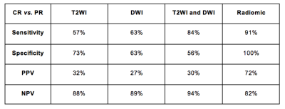 |
Comparison of T2WI and DWI qualitative assessment and T2W-based radiomic features for predicting complete response in patients with rectal cancer after neoadjuvant chemoradiotherapy
Video Permission Withheld
Natally Horvat, Harini Veeraraghavan, Monika Khan, Ivana Blazic, Junting Zheng, Marinela Capanu, Evis Sala, Julio Garcia-Aguilar, Marc Gollub, Iva Petkovska
Radiomics is a novel science that encompasses a computer-based extraction of quantitative features from images. Some studies have demonstrated that radiomics may help distinguishing malignant from benign diseases. We hypothesize that radiomics extracted from T2WI may improve qualitative MRI assessment in the evaluating of complete response in patient with locally advanced rectal cancer after neoadjuvant chemoradiotherapy.
|
14:57
|
0829.
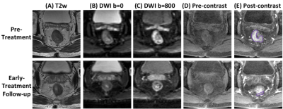 |
Machine Learning for Prediction of Chemoradiation Therapy Response in Patients with Locally-Advanced Rectal Cancer (LARC) Using Pre- and Early-Treatment Follow-up Multiparametric MRI
Yang Zhang, Liming Shi, Xiaonan Sun, Tianye Niu, Ning Yue, Tiffany Kwong, Peter Chang, Melissa Khy, Daniel Chow, Min-Ying Su, Ke Nie
A convolutional neural network (CNN) was implemented to predict the response of LARC patients receiving neoadjuvant chemoradiation therapy. The pre-treatment MRI, and the early-treatment follow-up MRI done at 2-3 weeks after the initiation of radiation were used. The MRI protocol included T2, DWI and DCE. A total of 41 patients were studied, with 8 pCR, 27 Tumor Regression Grade 1, and 9 TRG 2+3. The prediction accuracy was 0.71-0.89 for pCR vs. non-pCR; 0.70-0.77 for TRG(0+1) vs. TRG(2+3), not very good due to the limitations of a relatively small dataset. Using manually extracted tumor features in conjunction with neural network classifiers may achieve a higher accuracy.
|
15:09
|
0830.
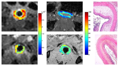 |
Multiparametric evaluation of inflammation and fibrosis in a radiation-induced murine model of colitis
Sabrina Doblas, Magaly Zappa, Dominique Cazals-Hatem, Fabien Milliat, Philippe Garteiser, Eric Ogier-Denis, Bernard Van Beers
The evaluation of fibrosis severity in Crohn’s disease is essential for patient management and prognosis, albeit seldom investigated. We validated a MR approach including diffusion-weighted imaging, magnetization transfer and FAIR perfusion to distinguish between moderate and severe forms of fibrosis in a radiation-induced murine model of colitis. The presence of fibrotic tissue and its accompanying vascular alterations induced a decrease in apparent diffusion coefficient and in perfusion, and an increase in the magnetization transfer ratio. This approach could be applied for diagnosis and assessment of intestinal fibrosis in patients.
|
15:21
|
0831.
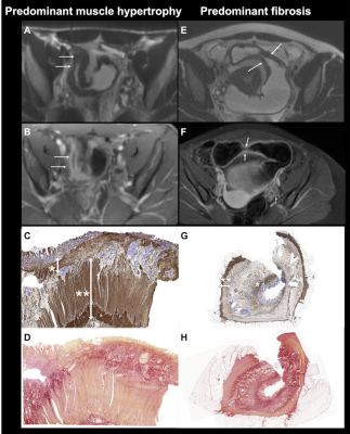 |
MRI predicts histopathologic composition of ileal Crohn's disease.
Mathilde Wagner, Mabel Ko, Manjil Chatterji, Cecilia Besa, Joana Torres, Xiaofei Zhang, Hinaben Panchal, Judy Cho, Jean-Frederic Colombel, Noam Harpaz, Bachir Taouli
The aim of our study was to assess the value of MRI for the characterization of histopathologic tissue composition of small bowel Crohn’s disease (CD), including assessment of inflammation, fibrosis and smooth muscle hypertrophy. Our results showed that MRI-based parameters including ADC, MaRIA score and layered pattern of enhancement can predict the degree of bowel inflammation. We also showed that wall thickness measured on T2WI can distinguish prominent muscle hypertrophy from prominent fibrosis in ileal CD.
|
15:33
|
0832.
 |
MRI of whole gut transit time in newly diagnosed coeliac disease.
Video Permission Withheld
Carolyn Costigan, Nina Lewis, Robin Spiller, Paul Morgan, Jennifer Price, Carolina Ciacci, Paola Iovino, Caroline Hoad, Penny Gowland, Luca Marciani
Coeliac disease (CD) is an autoimmune disease which affects 1 in 100 people. There is no cure and the only treatment is a lifelong gluten free diet. This study aims to improve our understanding of the functional motility disorder associated with CD using MRI. Whole gut transit time (WGTT), measured using MRI transit markers, was significantly delayed in the coeliac patients compared to helathy controls (p<0.04). The MRI gut transit test is quick and is acceptable to patients and could help long term monitoring and follow up, complementing existing more invasive techniques.
|
|
