Loose Cartilage
Oral
Musculoskeletal
Monday, 18 June 2018
| W03/04 |
13:45 - 15:45 |
Moderators: Jeff Dunn, Konstantin Momot |
13:45
|
0197.
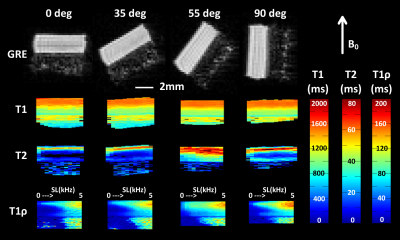 |
 Correlations of T1? with properties of articular cartilage depend on the spin-lock amplitude and orientation of the sample Correlations of T1? with properties of articular cartilage depend on the spin-lock amplitude and orientation of the sample
Mikko Nissi, Isabel Stavenuiter, Nina Hänninen
Several studies have reported different findings on the correlations of the CW-T1ρ relaxation time in articular cartilage with its different properties. Most studies agree on the sensitivity of CW-T1ρ to cartilage proteoglycans, although reports specifically against this also exist. Furthermore, CW-T1ρ has been connected to the collagen network properties and also correlated with T2 relaxation time. Orientation dependence of CW-T1ρ has been reported, as well as its dependence on the spin-locking amplitude. This study aims to combine all of these aspects in a single study.
|
13:57
|
0198.
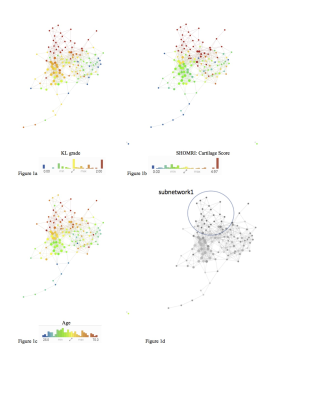 |
Using Multidimensional Data Analysis to Identify Traits of Hip OA
Jasmine Rossi-deVries, Valentina Pedoia, Michael Samaan, Adam Ferguson, Richard Souza, Sharmila Majumdar
This study aims to use big data analytics and imaging to simultaneously analyze all the combined variables in order to identify biomarkers able to classify the different disease progression of hip OA. 102 subjects and their 184 variables were examined. Big data analytics tool, Topological Data Analysis (TDA), was used to generate hypotheses. Three main groups were identified: healthy control subjects, subjects with radiographic and morphological evidence of OA, and subjects who progressed inconsistently were separated by knee biomechanics. The analysis obtained with TDA proposes new phenotypes of these subjects also shows the potential for further examination.
|
14:09
 |
0199.
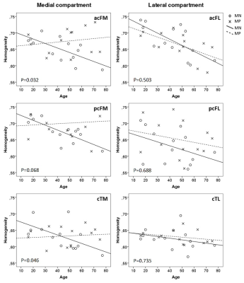 |
T2 Texture Analysis Reveals Potential Cartilage-preserving Effect in Presence of Heterozygous WNT1 Mutation in Human
Sami Lehtovirta, Riikka Mäkitie, Victor Casula, Marianne Haapea, Jaakko Niinimäki, Tuukka Niinimäki, Arttu Peuna, Eveliina Lammentausta, Outi Mäkitie, Miika Nieminen
Quantitative MRI (qMRI) assessment of tibiofemoral articular cartilage was performed in 13 WNT1 mutation-positive (MP) subjects and 13 mutation-negative (MN) controls. Cartilage thickness, T2 and T1r relaxation times, and texture features contrast,homogeneity and dissimilarity of T2 maps were determined in six regions of interests. Texture features demonstrated an opposing trend with age between the two groups in medial tibiofemoral cartilage, suggesting a possible age-related cartilage preservation in MP subjects. Similar differences were not observed in the other qMRI parameters, suggesting that texture analysis is a more sensitive and accurate tool for quantitative cartilage assessment than mere mean relaxation time measurements.
|
14:21
|
0200.
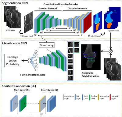 |
Can A Machine Diagnose Knee MR Images? Fully-automated Cartilage Lesion Detection by using Deep Learning
Fang Liu, Zhaoye Zhou, Kevin Lian, Shivhumar Kambhampati, Richard Kijowski
This study evaluated a fully-automated cartilage lesion detection system utilizing a deep convolutional neural network (CNN) to segment bone and cartilage followed by a second CNN classification network to detect structural abnormalities within the segmented tissues. The CNN network was trained to detect cartilage lesions within the knee joint using sagittal fat-suppressed T2-weighted fast spin-echo images in 125 subjects. The proposed CNN model achieved high diagnostic accuracy for detecting cartilage lesions with a 0.914 area under curve on receiver operation characteristics analysis. The optimal threshold for sensitivity and specificity of the CNN model was 84.3% and 84.6% respectively.
|
14:33
|
0201.
 |
Automated Knee Cartilage Segmentation with Very Limited Training Data: Combining Convolutional Neural Networks with Transfer Learning
Alexander Toews, Zhongnan Fan, Marianne Black, Jin Lee, Garry Gold, Brian Hargreaves, Akshay Chaudhari
Magnetic resonance imaging is commonly used to study osteoarthritis. In most cases, manual cartilage segmentation is required. Recent advances in deep-learning methods have shown promise for automating cartilage segmentation, but they rely on the availability of large training datasets that rarely represent the exact nature or extent of data practically available in routine research studies. The goal of this study was to automate cartilage segmentation in studies with very few training datasets available by creating baseline segmentation knowledge from larger training datasets, followed by creating transfer learning models to adapt this knowledge to the limited datasets utilized in typical study.
|
14:45
|
0202.
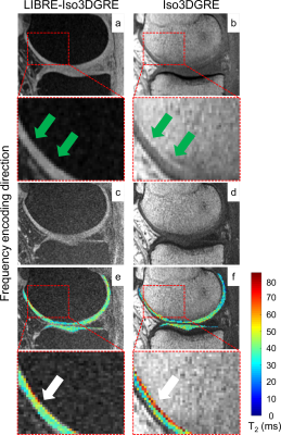 |
Isotropic 3D T2 mapping of knee cartilage with a novel water excitation technique
Roberto Colotti, Jessica Bastiaansen, Patrick Omoumi, Ruud van Heeswijk
The goal of this study was to develop an isotropic 3D lipid-insensitive T2 mapping technique of knee cartilage. Therefore we combined an existing isotropic 3D T2-prepared gradient-echo T2 mapping technique (Iso3DGRE) with the novel lipid-insensitive binomial off-resonant RF excitation (LIBRE) pulse. LIBRE pulse optimization was performed through numerical simulations and verified in phantom experiments, yielding complete fat signal nulling using a LIBRE pulse as short as 1 ms. T2 mapping of knee cartilage performed in five healthy volunteers with LIBRE excitation allowed for improved cartilage delineation and precise T2 values compared with normal excitation.
|
14:57
|
0203.
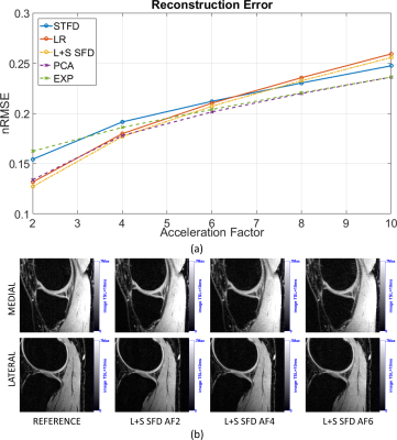 |
Accelerating 3D-Biexponential T1? Mapping of Cartilage using Compressed Sensing with Different Regularizations
Marcelo Zibetti, Azadeh Sharafi, Ricardo Otazo, Ravinder Regatte
Quantitative T1ρ imaging usually requires multiple spin-lock times to obtain T1ρ maps, which makes the acquisition time demanding especially for biexponential models. Compressed Sensing has demonstrated significant acquisition time reduction in MRI. Similar improvements are expected for T1ρ relaxation mapping, given the extensive correlations in the series of images. However, it is not clear which combination of sparsifying transform and regularization function performs best for biexponential T1ρ mapping. Here, we compare five CS approaches: l1-norm of principal component analysis, spatio-temporal finite differences, exponential dictionaries, low rank, and low rank plus sparse. Our preliminary results, with three datasets, suggest that L+S is the most suitable method with least T1ρ estimation error.
|
15:09
|
0204.
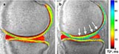 |
UTE-T2* Shows Deep Cartilage Subsurface Matrix Changes 2 Years After ACL Reconstruction
Ashley Williams, Matthew Titchenal, Aditi Guha, Bao Do, Constance Chu
The purpose of this study is to compare 2-D and 3-D assessments of UTE-T2* maps for evidence of alterations to the subsurface cartilage matrix suggestive of cartilage at risk for early OA 2 years after ACL reconstruction. UTE-T2* values from small 2-D, single slice ROIs correlated to 3-D ROI values that encompassed a larger degree of weight-bearing cartilage. Results indicate that single slice 2-D UTE-T2* mapping may be an efficient means to assess the medial femoral cartilage as an imaging marker of pre-osteoarthritis while 3-D assessments provide additional sensitivity to changes in the tibial plateau.
|
15:21
|
0205.
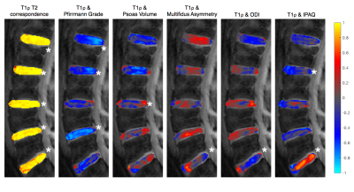 |
Local associations between intervertebral disc T1rho/T2, muscle health, physical activity, and clinical disability using voxel-based relaxometry
Claudia Iriondo, Valentina Pedoia, Jason Talbott, William Dillon, Sharmila Majumdar
Region of interest based analysis of intervertebral disc composition in low back pain populations is (1) time-consuming and (2) limited in reproducibility, even more so in patients with advanced degeneration. This study applies voxel-based relaxometry (VBR) to investigate the spatial distributions of T1ρ and T2 in lumbar intervertebral discs, and their association to patient reported outcomes and spinal muscle health. Our results demonstrate the potential to use VBR as a tool to more effectively measure biochemical differences in the intervertebral discs across low back pain subgroups and monitor changes over time.
|
15:33
|
0206.
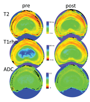 |
Quantitative MRI in early intervertebral disc degeneration: T1rho correlates better than T2 and ADC with biomechanics and matrix content
Video Permission Withheld
Cornelis Paul, Theodoor Smit, Magda de Graaf, Roderick Holewijn, Arno Bischop, Peter van de Ven, Margriet Mullender, Marco Helder, Gustav Strijkers
We correlated quantitative T2, T1rho and Apparent Diffusion Coefficient (ADC) values to disc mechanical behavior and gold standard early DDD markers in a graded degenerated lumbar IVD caprine model to assess their potential for early DDD detection. T1rho nucleus values correlate better than T2 and ADC with biomechanical, histological, and GAG changes.
|
|
