Little Bones
Oral
Musculoskeletal
Thursday, 21 June 2018
| S05 |
15:30 - 17:30 |
Moderators: Mikko Nissi |
15:30
 |
1241.
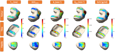 |
 Simultaneous PET-MRI for Investigating Associations Between Bone and Cartilage Biomarkers of Knee Osteoarthritis Simultaneous PET-MRI for Investigating Associations Between Bone and Cartilage Biomarkers of Knee Osteoarthritis
Hatef Mehrabian, Valentina Pedoia, Jasmine Rossi-Devries, Emma Bahroos, Dragana Savic, Sharmila Majumdar
Knee osteoarthritis (OA) is caused by cartilage degeneration which has been shown to be accompanied by the changes in adjacent subchondral bone. Simultaneous PET-MRI enables investigating the biomarkers of cartilage degeneration in OA with MRI (i.e. T1ρ, T2 and Gadolinium uptake) as well as biomarkers of bone remodeling in OA with PET (uptake of NaF tracer in bone) at the same time. PET-MRI also allows for probing the associations between different bone and cartilage markers. Voxel-wise assessment of these associations provides insight into the local correlations between different OA markers and yields comprehensive assessment of knee OA progression.
|
15:42
|
1242.
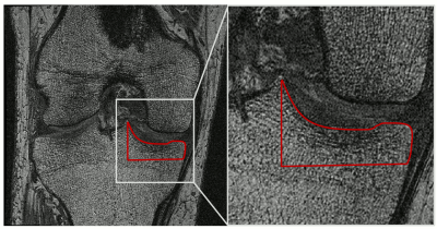 |
Automated Textural Classification of Osteoarthritis Magnetic Resonance Images
Joshua Kaggie, Rob Tovey, James MacKay, Fiona Gilbert, Ferdia Gallagher, Andrew McCaskie, Martin Graves
Osteoarthritis (OA) is the most common cause of disability in the United Kingdom and United States. Identifying the rate of OA progression remains an important clinical and research challenge for early disease monitoring. Texture analysis of tibial subchondral bone using magnetic resonance imaging (MRI) has demonstrated the ability to discriminate between different stages of OA. This work combines texture analysis with machine learning methods (Lasso, Decision Tree, and Neural Network) to predict radiographic disease progression over 3 years, trained using data from the Osteoarthritis Initiative. We achieved high sensitivity (86%), specificity (64%) and accuracy (74%) for predictions of OA progression.
|
15:54
 |
1243.
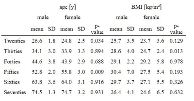 |
Anatomical variation of age-related changes in vertebral bone marrow composition using chemical shift encoding-based water-fat MRI
Thomas Baum, Alexander Rohrmeier, Jan Syväri, Maximilian Diefenbach, Daniela Franz, Michael Dieckmeyer, Andreas Scharr, Hans Hauner, Stefan Ruschke, Jan Kirschke, Dimitrios Karampinos
The assessment of vertebral bone marrow water-fat composition is attracting growing interest for applications in osteoporosis and bone metabolism. Chemical shift encoding-based water-fat MRI allows spatially resolved assessment of proton density fat fraction (PDFF) at the spine. This study demonstrated that males started with greater PDFF values in the Twenties compared to females. However, females showed an accelerated bone marrow fatty conversion until the Seventies on. This finding can be explained by the (patho-)physiological process of menopause. Interestingly, the relative age-related PDFF changes from the Twenties to the Seventies were dependent on the anatomical location and were most pronounced at lower lumbar vertebral levels in both genders.
|
16:06
|
1244.
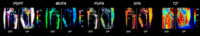 |
Unravelling bone marrow adipose tissue composition in proximal femur sub-regions through 3T Chemical Shift Encoded-MRI: differences between osteoporosis and glucocorticoid-induced osteoporosis
Dimitri MARTEL, Benjamin LEPORQ, Amit SAXENA, H.Michael BELMONT, Gabrielle TURYAN, Stephen HONIG, Ravinder R. REGATTE, Gregory CHANG
Osteoporosis (OP) is due to weak bone and can ultimately lead to fracture. Recent findings shows link between bone marrow adipose tissue (bMAT) composition and amount and OP. OP can be induced by drugs such as glucocorticoids resulting in glucocorticoid-induced osteoporosis (GIO) and can affect energy metabolism pathways, induce changes in bone including increased total marrow adiposity and changes in bMAT composition. The composition of bMAT in GIO has not been previously investigated. Our aim was to assess the bMAT composition of a GIO population and compare it to OP patients using 3T Chemical Shift Encoded- MRI (CSE-MRI).
|
16:18
|
1245.
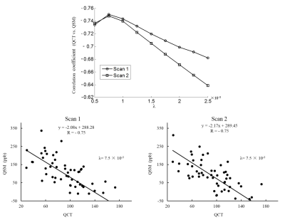 |
Quantitative Susceptibility Mapping of Human Spine: Correlation with Quantitative Computed Tomography and Reproducibility
Yihao Guo, Yingjie Mei, Xintao Zhang, Yanjun Chen, Xiaodong Zhang, Yanqiu Feng
This work aim to investigate the correlation between quantitative susceptibility mapping (QSM) and quantitative computed tomography (QCT) values, and the reproducibility of QSM value in human spine. Sixty-one subjects underwent QCT measurement for once and QSM measurement for twice in spine. The results showed that QSM value has strong correlation with QCT value, and has good reproducibility between two scans. These indicated that QSM has the potential to be an effective and stable measurement of BMD in clinical application.
|
16:30
|
1246.
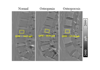 |
Preliminary Application Researches of Quantitative Susceptibility Mapping in Evaluating the Osteoporosis
Xintao Zhang, Yihao Guo, Yanjun Chen, Yingjie Mei, Yanqiu Feng, Xiaodong Zhang
The purpose of this work was to explore the clinical application of quantitative susceptibility mapping(QSM)using an ultrashort echo time (UTE) gradient echo (GRE) sequence in evaluating the bone mineral density(BMD)of lumbar vertebra by analyzing the correlation between QSM and quantitative computed tomography (QCT). Our results showed that there was strong correlation between QSM and QCT. QSM has the potential to be a biomarker providing new insights into osteoporosis.
|
16:42
|
1247.
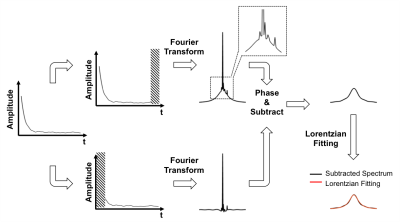 |
In Vivo Bone 31P Relaxation Measurement and Its Implications on Mineral Quantification
Xia Zhao, Hee Kwon Song, Felix Wehrli
In MRI-based bone mineral assessment, pixel intensity of the bone is compared against that of the sample to estimate the 31P density after correcting for their relaxation properties. Knowledge of bone 31P relaxation times is therefore crucial. Using saturation-recovery spectroscopy and ZTE-PETRA, T1, T2* and density of bone 31P in healthy subjects (26 to 76 y/o) were measured yielding 36.8±1.4s, 196.9±10.1μs and 6.10±0.62 mol/L. Measured T1 and T2* errors are expected to be within 6% and 15%, resulting in an error of quantified [31P] of ≤ 6.3%. The small inter-subject variations may therefore obviate the need for individual T1 measurements.
|
16:54
|
1248.
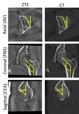 |
Zero TE Hip MRI: Osseous Measurements Assessing Femoroacetabular Impingement
Ryan Breighner, Eric Bogner, Brian Kelly, Matthew Koff, Hollis Potter
Due to short tissue relaxation times and signal scarcity, routine MRI does not provide direct visualization of bone. This study investigates the use of proton density zero echo time (ZTE) MRI of bone in the hip, with emphasis on femoroacetabular impingement (FAI). Hip CT and ZTE images were acquired for 24 hips (14 patients). Ten measures of osseous morphology were compared between modalities. 'Moderate' to 'excellent' agreement was seen in all but one of the measurements. Zero Echo Time MRI may mitigate the need for CT in some cases.
|
17:06
|
1249.
 |
Three-Dimensional Ultrashort Echo Time Cones Actual Flip Angle Imaging with Variable Repetition Time (3D UTE-Cones AFI-VTR) for Accurate T1 mapping of Short T2 Tissues
Yajun Ma, Xing Lu, Michael Carl, Yanchun Zhu, Nikolaus Szeverenyi, Graeme Bydder, Eric Chang, Jiang Du
To overcome the challenges (i.e. fast signal decay and low excitation efficiency) which would other render T1 measurements inaccurate for short T2 tissues, we propose a new approach by combining 3D UTE-Cones the actual flip angle imaging (AFI) with UTE-Cones variable repetition time (VTR) (3D UTE-Cones-AFI-VTR) method, where the identical RF pulses and flip angles are used for signal excitation in both sequences.
|
17:18
|
1250.
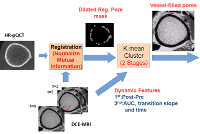 |
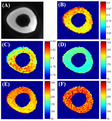
CORTICAL BONE VESSEL IDENTIFICATION ON CONTRAST-ENHANCED MR IMAGES
Po-hung Wu, Jing Liu, Julio Carballido-Gamio, Misung Han, Roland Krug, Galateia Kazakia
Pathological cortical bone porosity negatively impacts bone strength, but the mechanisms of pathological pore growth are not understood. We hypothesize that the contents of cortical bone pores (marrow or vessels) may be useful indicators of pore expansion mechanisms. In this study, we developed a technique using high resolution CT and DCE-MRI to visualize and identify pore contents. Dynamic features such as temporal intensity difference and transition slope within pore voxels were evaluated and clustered by a K-means clustering algorithm. The average intensity of segmented vessel-filled pores increased over time, demonstrating the ability of our technique to positively identify vessel-filled pores.
|
|
