MSK: Harder, Better, Faster, Stronger
Oral
Musculoskeletal
Thursday, 21 June 2018
| S02 |
08:00 - 10:00 |
Moderators: James MacKay, Erin Englund |
08:00
|
1037.
 |
 Fully Automated Deep Learning Pipeline for Meniscus Segmentation and Lesion Detection Fully Automated Deep Learning Pipeline for Meniscus Segmentation and Lesion Detection
Berk Norman, Valentina Pedoia, Thomas Link, Sharmila Majumdar
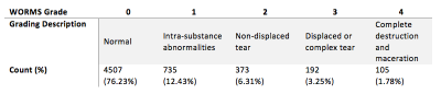
Damage to the meniscus is a physically limiting injury that can lead to further medical complications. Automatically classifying this type of meniscal damage poses the advantage for quicker and more accurate diagnosis at the time of an MRI scan. Using a fully automated deep learning pipeline we identify the region around the 4 meniscal horns and then classify if a lesion exists and if so, its severity based on WORMS grading. Lesion detection achieved 89.81% specificity and 81.98% sensitivity. This algorithm has the ability to quickly identify meniscal lesions from MRI and filter higher risk lesion subjects.
|
08:12
|
1038.
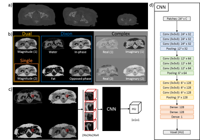 |
The influence of different MR contrasts in multi-channel convolutional neural networks on pseudo-CT generation for orthopedic purposes
Mateusz Florkow, Frank Zijlstra, Koen Willemsen, René Castelein, Harrie Weinans, Bart van der Wal, Max Viergever, Marijn van Stralen, Peter Seevinck
Conventional MR images and pseudo-CT’s (pCT’s) generated using state-of-the-art machine learning techniques poorly characterize bone anatomies, preventing applicability for orthopedic applications. We hypothesize that smart use of several specific MR contrasts will expose the information needed for diagnostic quality bone visualization. We designed a patch-based convolutional neural network taking groups of different MR contrasts -which were obtained from a single multi-gradient sequence- as inputs . It generated competitive pCT scans, capturing local anatomical variances present in the dataset. We show that Dixon reconstructed inputs appear to generate better soft-tissue visualization, while complex-valued data show promising results in bone reconstruction.
|
08:24
 |
1039.
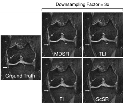 |
Super-Resolution Musculoskeletal MRI using Deep Learning
Akshay Chaudhari, Zhognan Fang, Feliks Kogan, Jeff Wood, Kathryn Stevens, Jin Hyung Lee, Garry Gold, Brian Hargreaves
Near-isotropic high-resolution magnetic resonance imaging (MRI) of the knee is beneficial for reducing partial volume effects and allowing multi-planar image analysis. However, previous methods exploring isotropic resolutions, typically compromised in-plane resolution for thin slices, due to intrinsic signal-to-noise ratio (SNR) limitations. Even computer-vision-based super-resolution methods have been rarely been used in medical imaging due to limited resolution improvements. In this study, we utilize deep-learning-based 3D super-resolution for rapidly generating high-resolution thin-slice knee MRI from slices originally 2-8 times thicker. Through quantitative image quality metrics and a reader study, we demonstrate superior performance to both conventionally utilized and state-of-the-art super-resolution methods.
|
08:36
|
1040.
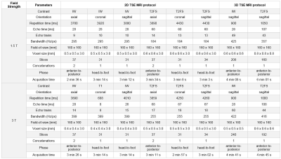 |
Fully-Automated One-Button-Push 10-min 3D CAIPIRINHA SPACE TSE MRI of the Knee: A Multi-Center Multi-Reader Multi-Field-Strength Validation Study
Video Permission Withheld
Filippo Del Grande, Marco Delcogliano, Riccardo Guglielmi, Esther Raithel, Derek Papp, Steven Stern, Christian Candrian, Jan Fritz
2D TSE MRI is widely used for the evaluation of internal knee derangement but is time-consuming. Recently introduced 3D TSE acceleration strategies, such as CAIPIRINHA allow for fast sampling, and together with AutoAlign technology enable now fully automated one-button-push 3D MRI protocols in 10 minutes total scan time. In a prospective study of 150 subjects, we analyzed the frequencies of structural abnormalities, inter-reader reliability, inter-method concordance, diagnostic definitiveness, and interchangeability of 10-min 3D CAIPIRINHA SPACE TSE protocols and 20-min 2D TSE standard-of-reference protocols. Our results indicated that that 10-min 3D TSE protocols are at least equivalent to 20-min 2D TSE protocols for the diagnosis of internal derangement of the knee.
|
08:48
|
1041.
 |
Reducing scan time for full MRI examination of the ankle by 45% while maintaining diagnostic value using combined compressed sensing and parallel imaging.
Onno Baur, Chiel Den Harder, Robert Hemke, Mario Maas, Farood Faridmojtahedi, Elwin De Weerdt, Maarten Versluis, Mascha Van Der Kwaak, Aart Nederveen
Dixon imaging is a well-known imaging technique that provides robust water-fat separation but requires relatively long acquisition times. We scanned twelve healthy volunteers with a standard ankle examination containing multiple Dixon sequences and an accelerated examination using CS-SENSE, a combination of Parallel Imaging and Compressed Sensing. A five-point Likert scale was used to compare reference images to CS-SENSE images. Although using CS-SENSE caused a slight SNR reduction, the diagnostic value was not impaired. Applying CS-SENSE on an ankle examination protocol resulted in a feasible reduction of the total acquisition time by 45% from 13’38” to 7’49”, without losing diagnostic value.
|
09:00
|
1042.
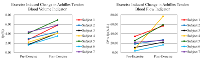 |
Intravoxel Incoherent Motion (IVIM) Imaging in Human Achilles Tendon
Kenneth Wengler, Dharmesh Tank, Mingqian Huang, Elaine Gould, Mark Schweitzer, Xiang He
Clinically, Achilles tendon (AT) rupture accounts for 40-60% of all operative tendon repairs. AT microcirculation plays a crucial role in the progression of tendinopathy and tendon repair. In this study a novel ste-RESOLVE IVIM protocol was developed to image AT microcirculation. Healthy participants were imaged pre- and post-exercise, and exercised induced increases in blood volume and blood flow were observed. AT tendinopathy patients exhibited greater baseline blood volume and blood flow when compared to healthy participants. For the first time, a robust MRI-based technique was developed to investigate the role of Achilles tendon microcirculation in tendinopathy.
|
09:12
|
1043.
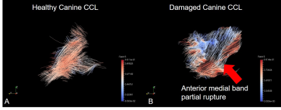 |
Visualisation and quantification of collagen fibers in a partially torn ligament using magic angle imaging
Karyn Chappell, Catherine Van Der Straeten, Donald McRobbie, Wladyslaw Gedroyc, Mihailo Ristic, Djordje Brujic, Richard Meeson
Human partial anterior cruciate ligament tears can be extremely difficult to diagnose with conventional MRI. Variations of signal intensity within the ligament are suggestive of injury but it is not possible to confirm damage or assess the collagen alignment within the ligaments. We have shown that magic angle imaging has the ability to visualise and quantify collagen fibers in a partially torn canine cruciate ligament. Furthermore it can delineate between damaged and healthy fiber bundles within the same ligament. This method has the potential to become a non-invasive alternative to arthroscopy for assessing and monitoring ligament damage and repair outcomes.
|
09:24
|
1044.
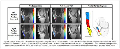 |
Longitudinal Changes in Quantitative MRI and Ultrasound Metrics of Patellar Tendon are Associated with Tendon Degeneration and Leg Dominance within of Collegiate Basketball Players over One Season of Play
Erin Argentieri, Matthew Koff, Bin Lin, Parina Shah, Hollis Potter, O. Nwawka
This study assessed correlations between quantitative T2* and Shear wave elastography (SWE) ultrasound metrics of the patellar tendon and qualitative morphologic grades patellar tendinosis (PT) within collegiate basketball players over one season of play. Within the current study, significant and strong correlations existed between T2* and SWE metrics, though morphologic PT grades were correlated with T2* metrics only. These findings support the notion that T2* relaxometry could benefit the clinical management of PT, as it is sensitive to changes in pathologic severity over time, and could therefore serve as a metric to guide treatment plans and evaluate intervention efficacy.
|
09:36
|
1045.
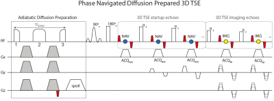 |
Isotropic resolution DTI of lower back nerves using a phase-corrected diffusion-prepared 3D TSE
Barbara Cervantes, Anh Van, Dominik Weidlich, Hendrik Kooijman, Andreas Hock, Ernst Rummeny, Alexandra Gersing, Jan Kirschke, Dimitrios Karampinos
Diffusion-prepared 3D TSE (dprep-3D TSE) has been applied for isotropic-resolution distortion-free proximal- and peripheral-nerve DWI. Dprep-3D TSE has been combined with magnitude stabilizers to reduce magnitude modulation induced by motion and eddy currents and has used velocity compensation to reduce motion-induced phase modulation in diffusion-weighted signals. However, due to the multi-shot nature of dprep-3D TSE, remaining motion-induced phase leads to image and DTI-parameter artifacts and requires phase navigation. The purpose of this work is to develop a phase-navigation and phase-correction scheme for dprep-3D TSE and to apply the developed method in vivo for isotropic-resolution DTI of lumbosacral and sciatic nerves.
|
09:48
|
1046.
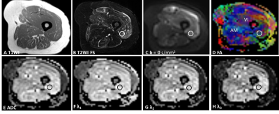 |
Simultaneous Multislice Accelerated Diffusion-Tensor Imaging of Thigh Muscles in Myositis
Fengdan Wang, Chanyuan Wu, Caiyuan Sun, Dong Liu, Yi Sun, Qian Wang, Zhengyu Jin
We investigated the clinical feasibility of using simultaneous multislice accelerated echo planar imaging diffusion-tensor imaging (SMS-EPI-DTI) to image thigh muscles of both 10 healthy control subjects and 20 dermatomyositis (DM)/ polymyositis (PM) patients. This technique yielded a reduced scan time to only about five minutes. The results showed that the tractographic imaging and DTI-derived parameters of edematous muscles differed among affected and unaffected muscles of the DM/PM patients and normal muscles of the control subjects. In conclusion, SMS-EPI-DTI is clinically feasible for imaging thigh muscles and quantitatively evaluating edematous muscles of DM and PM patients.
|
|
