Novel Contrast & Targeted Molecular Imaging
Oral
Molecular Imaging
Tuesday, 19 June 2018
| S04 |
08:15 - 10:15 |
Moderators: Luisa Ciobanu, A. Dean Sherry |
08:15
|
0398.
 |
 Intracellular MEMRI signals reflect the frequency of action potentials in Aplysia neurons Intracellular MEMRI signals reflect the frequency of action potentials in Aplysia neurons
Pavel Svehla, Alexis Bedecarrats, Caroline Jahn, Romuald Nargeot, Luisa Ciobanu
We show a positive correlation between electrical activity and MEMRI signal intensity in identified neurons in Aplysia buccal ganglia and demonstrate that the MEMRI signal reflects mainly fast and high membrane depolarization processes such as action potentials, and it is not sensitive to slow and small membrane depolarization, such as post-synaptic potentials.
|
08:27
|
0399.
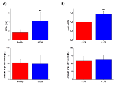 |
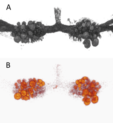
Active Targeting of human neutrophil granulocytes by non-invasive 19F MRI
Pascal Bouvain, Bodo Steckel, Wolfgang Krämer, Rolf Schubert, Sebastian Temme, Ulrich Flögel
The purpose of the present study was to target human neutrophils by PFCs to enable their visualization by 19F MRI. We coupled a neutrophil-binding peptide (NG2) to PFCs and showed the specific binding and internalization of NG2-PFCs by 19F MRI, microscopy and FACS. Interestingly, NG2-PFCs show an increased labelling of neutrophils from MI patients and these cells also showed an increased migration into an artificial circulation system which contained an IL-8 doped matrigel placed in a flow chamber. In conclusion, NG2-PFCs are suited to label neutrophil granulocytes after MI and enable their non-invasive visualization by 1H/19F MRI.
|
08:39
 |
0400.
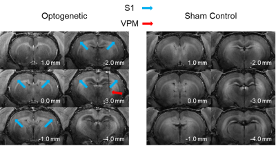 |
Layer specific neural interactions in the thalamo-cortical and cortico-cortical networks: An optogenetic manganese-enhanced MRI study
Karim El Hallaoui, Eddie Wong, Xunda Wang, Alex Leong, Russell Chan, Celia Dong, Ed Wu
The thalamo-cortical projections terminating in the primary somatosensory cortex are anatomically organized into layers. However, the functional layer-specificity of the axonal pathways which comprise the thalamo-cortical circuit remains to be studied. Combining optogenetic stimulation and manganese-enhanced MRI, the present study selectively induces neural activity in the ventral posteromedial nucleus to induce the deposit of manganese in its projections in-vivo. By identifying the cortical regions with increased contrast, using images acquired with the MDEFT MRI experiment, we infer the layer-specific connections from the thalamus to the primary somatosensory cortex.
|
08:51
|
0401.
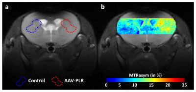 |
Noninvasive imaging of transgene expression using CEST-MRI
Julien Flament, Jérémy Pépin, Marianne Maugard, Mylène Gaudin, Julien Valette, Gilles Bonvento
Gene therapy often uses viral vectors to insert a therapeutic gene into cells. It is necessary to develop molecular imaging techniques to safely monitor gene expression. We designed a strategy to image expression of a transgene using a genetically engineered CEST reporter. An adeno associated vector encoding for 150 L-arginine residues fused to a fluorophore was produced and injected in the mouse brain. Specific signals were observed in both CEST and fluorescence modalities, demonstrating the possibility to use CEST-based reporter genes to monitor transgene distribution and expression. Such noninvasive imaging modality could be valuable for further developments of gene therapy.
|
09:03
|
0402.
 |
OATP1A1 reporter gene-enhanced magnetic resonance imaging of Triple Negative Breast Cancer in animal models at 3 Tesla
Nivin Nyström, Amanda Hamilton, Francisco Martinez, Timothy Scholl, John Ronald
Preclinical cancer models are invaluable for studying oncogenic pathways and assessing therapies. However, precise detection of small tumours with specificity, sensitivity and high resolution remains challenging. A member of the Organic Anion Transporting Polypeptide 1 (OATP1) family of proteins, called OATP1A1, can take up the clinically-approved, liver-specific paramagnetic agent Gd-EOB-DTPA. Significant increases in spin-lattice relaxation rates were exhibited at 3T by triple negative breast cancer (TNBC) cells engineered to express OATP1A1 and pilot data showed enhancement of OATP1A1-expressing orthotopic TNBC tumours in mice. Our data supports the utility of OATP1A1 for improved MR detection of TNBC tumours in animal models.
|
09:15
|
0403.
 |
MR/ fluorescence dual-modality image-guided multifunctional albumin nanoassembies for sonodynamic therapy of glioma
Qian Wan, Chao Zou, Mengjie Chen, Zonghai Sheng, Jun Zhou, Yanwen Zhu, Hairong Zheng, Xin Liu
Sonodynamic therapy (SDT) is a non-invasive and effective therapeutic modality for solid cancerous tumors. In this study, to improve the effectiveness of SDT, we fabricated a high-performance multifunctional albumin nanoassembies (HSA-Ce6-Mn NCs) having higher cell uptake efficiency and the tumor accumulation. It also exhibited an excellent ability of MR/ fluorescence (FL) dual-modality imaging so that glioma margin and HSA-Ce6-Mn NCs metabolism could be clearly visualized. The SDT has been demonstrated for suppressing the tumor growth in-vivo under a MRI-guided FUS system and it is highly expected to have a great potential in clinical translation.
|
09:27
|
0404.
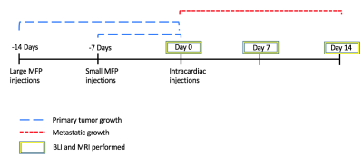 |
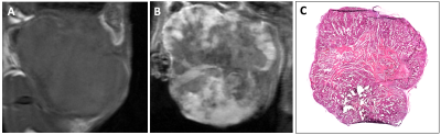
Multimodality cellular and molecular imaging of the impact of a primary tumor on metastatic growth in a syngeneic mouse model of breast cancer brain metastasis
Katie Parkins, Veronica Dubois, Amanda Hamilton, Ashley Makela, John Ronald, Paula Foster
The mechanisms that influence metastatic growth rates are poorly understood. One mechanism of interest known as concomitant tumor resistance (CTR) can be defined as the inhibition of metastatic growth by existing tumor mass. Conversely, the presence of a primary tumor has also been shown to increase metastatic outgrowth, termed concomitant tumor enhancement (CTE). The goal of this research was to use conventional and cellular MRI , and bioluminescence imaging to study the impact of a primary tumor on the development of breast cancer brain metastases in a syngeneic mouse model.
|
09:39
|
0405.
 |
Europium(2+/3+) dual mode MRI contrast agents: combining paraCEST and T1w contrast in one oxygen-sensitive agent.
Alexander Funk, Veronica Clavijo-Jordan, Dean Sherry, James Ratnakar, Zoltan Kovacs
The ability to determine hypoxia in tumors in vivo could provide useful diagnostic contrast information. MRI contrast agents could provide this information. Eu2+ is isoelectronic with Gd3+, and produces T1w contrast on a similar level, in addition, it is not oxidatively stable. In an aerobic atmosphere, it oxidizes rapidly to Eu3+, which does not produce T1 contrast, but can belong to a conceptually different class of contrast agents: paraCEST (chemical exchange saturation transfer) agents. A Eu2+/3+ agent was tested in different tissues in-vivo to show the correlation between the rate of oxidation and the surrounding oxidative environment of the tissue.
|
09:51
|
0406.
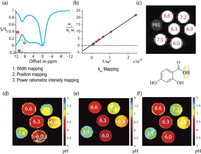 |
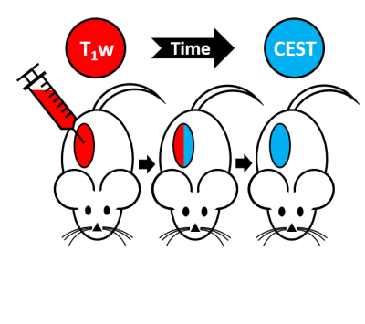
Renal pH Imaging Using Aspirin Analogs and CEST MRI
KowsalyaDevi Pavuluri, Michael T McMahon
Developing new tools that can be used to detect changes in renal tissue is of great interest for nephrologists. Indeed, standard measurements of blood-urea nitrogen and serum creatinine might not detect kidney injury until a 50% loss in function occurs. As kidneys play a preeminent role in controlling acid-base balance, pH mapping should enable early detection of changes in renal health. Here we propose administration of salicylate based diaCEST MRI agents to map renal pH. As we show, aspirin and three analogs: salicylic acid, 2,4-dihydroxy benzoic acid, 2,5-dihydroxy benzoic acid displayed promise in vitro and in vivo for pH mapping.
|
10:03
|
0407.
 |
Development and Proof of Principle for Free Radical Imaging with the Novel Field-cycling in vivo DNP-MRI
Hideo Utsumi, Toshiki Masumizu, Ryoma Kobayashi, Utaroh Motosugi, Tatsuya Shimizu, Tomoko Tahira, Atsushi Iikura, Hidenori Kajiwara
In vivo DNP-MRI was reported as a new imaging method for free radical species in vivo1, and the field-cycling was utilized to overcome the large difference between electron and nuclear spin resonances 2,3. Unfortunately the development of the clinical DNP-MRI has severe difficulties due to less applicability. Here, we developed the novel field-cycling DNP-MRI for clinical trial by rotating the magnets for MRI (0.3T) and ESR (4.7mT), and the free radicals placed under the palm of volunteers were visualized, superimposed on the anatomical MRI image, and demonstrated the proof of principle for free radical imaging with the newly developed field-cycling DNP-MRI.
|
|
