Methods & Applications for PET/MR
Oral
Molecular Imaging
Thursday, 21 June 2018
| S06 |
15:30 - 17:30 |
Moderators: Uwe Himmelreich, Tone Bathen |
15:30
 |
1251.
 |
ZTE MRI for attenuation correction in PET/MR: performance in a large cohort of cognitive disorder patients
Video Permission Withheld
Maya Khalifé, Brian Sgard, Arthur Bouchut, Brice Fernandez, Marine Soret, Gaspar Delso, Marie-Odile Habert, Aurélie Kas
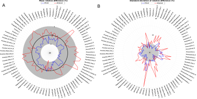
Several attenuation correction (AC) methods based on Zero Echo Time (ZTE) MRI are available in brain PET/MR. However, most of them were evaluated in healthy subjects or in patients without apparent brain disease. In this work, we investigated ZTE-AC in a 50-patient cohort imaged for cognitive disorders and we compared its performance with default atlas-AC and reference CT-based AC. The impact of the two AC methods (ZTE-AC and atlas-AC) was evaluated and compared to reference CT-AC, using a Volume of Interest (VOI) analysis and voxelwise group comparison between patients with normal cortical metabolism vs. metabolic pattern suggestive of Alzheimer’s disease.
|
15:42
|
1252.
 |
 UTE with Multi-Echo Radial Acquisition for PET/MRI Attenuation Correction UTE with Multi-Echo Radial Acquisition for PET/MRI Attenuation Correction
Zhengyang Ming, Paul Han, Debra Horng, Shuang Hu, Kui Ying, Chao Ma, Georges El Fakhri
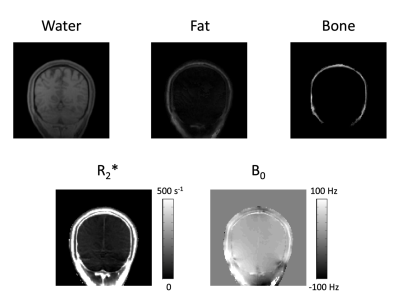
Reliable estimation of PET attenuation coefficient (AC) is a fundamental problem in PET/MR imaging. Accurate measurement of bone density variation is vital to generate reliable subject-specific AC maps using MR. Also, minimizing the data acquisition time of MR sequences for AC is essential to allow more MR scans in a single PET/MR scanning session. In this work, we propose a new acquisition scheme combining UTE and multi-echo radial acquisition to reliably estimate the composition of water, fat and bone in a minute scan. Results show that water, fat and bone signals can be estimated in using the proposed method, which can be potentially used to estimate subject-specific AC maps.
|
15:54
|
1253.
 |
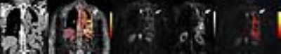
Deep Learning based pseudo-CT computation and its application for PET/MR attenuation correction and MR-guided radiation therapy planning
Video Permission Withheld
Sandeep Kaushik, Cristina Cozzini, Mikael Bylund , Jaewon Yang, Dattesh Shanbhag, Joakim Jonsson, Josef Lundman, Thomas Hope, Tufve Nyholm, Andrew Leynes, Peder Larson, Florian Wiesinger
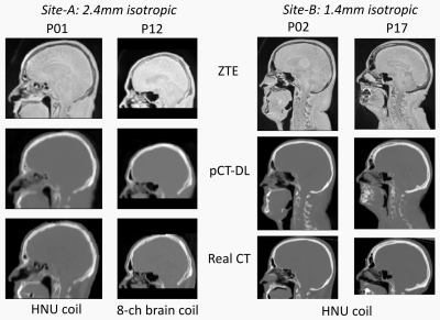
In this work, we demonstrate a generic deep learning (DL) model that computes pCT images (i.e. continuous density bone) using a single channel ZTE MRI data and is robust to protocol and coil variations (as dictated by application needs). The method was evaluated for PET/MR attenuation correction protocol (low resolution for speed) and MRgRTP dose planning protocol (higher resolution for spatial accuracy). The advantages include a single model for multiple protocols, pCT which are very much like real CT in appearance, as well as excellent quantitative accuracy of estimated bone values in the computed pCT.
|
16:06
|
1254.
 |
MR-Based Respiratory and Cardiac Motion Corrected PET/MR
Philip Robson, MariaGiovanna Trivieri, Nicolas Karakatsanis, Ronan Abgral, Marc Dweck, Jason Kovacic, Zahi Fayad
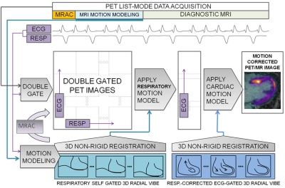
A major advantage of hybrid PET/MR systems is the radiation-free high spatial and temporal resolution of MR imaging that can be used to estimate cardio-respiratory motion present during PET data acquisition. This information can be incorporated into reconstruction algorithms to correct for motion in the PET data to reduce blurring and increase target-to-background ratios (TBR) of PET hotspots. This may be of particular importance in cardiac imaging where the heart is in constant motion. In this work, we demonstrate a method for cardio-respiratory motion correction and evaluate the effect on TBR in the myocardium in a cohort of patients.
|
16:18
|
1255.
 |
Simultaneous characterization of tumor cellularity and aerobic glycolysis with PET, MR and MRSI
Christian Hundshammer, Miriam Braeuer, Christoph Müller, Adam Hansen, Mathias Schillmaier, Stephan Düwel, Benedikt Feuerecker, Steffen Glaser, Axel Haase, Jorge Cabello, Franz Schilling, Jan Hövener, Andreas Kjaer, Stephan Nekolla, Schwaiger Markus
The new paradigm in oncology to tailor patient-specific therapies triggered the invention of powerful imaging techniques to non-invasively quantify tumor biology in depth. PET/MR is an emerging new hybrid imaging modality that allows the simultaneous acquisition of high resolution anatomical, functional and metabolic information.
We established a multimodal imaging workflow on a whole-body PET/MR system to quantify tumor cellularity, glucose uptake and aerobic glycolysis. We applied this workflow on a pre-clinical breast cancer model in a longitudinal study to analyze the effect of necrosis and tumor growth on metabolic parameters and measure the correlation of glucose uptake and LDH activity.
|
16:30
|
1256.
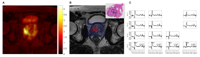 |
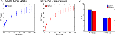
Simultaneous PET and MRSI of Prostate Cancer
Kirsten Selnæs, Morteza Esmaeili, Nassim Tayari, Tom Scheenen, Mattijs Elschot, Arend Heerschap, Tone Bathen
Both positron emission tomography (PET) and magnetic resonance spectroscopic imaging (MRSI) can give important information about cancer metabolism. With an integrated PET/MR scanner, MRSI and PET images can be acquired simultaneously. The aim of this study was to examine possible correlations between 18F-Fluciclovine PET and MRSI measurements and evaluate their diagnostic performance in prostate cancer. The results showed a significant moderate correlation between both SUVmean and SUVmax and the CCS/C ratio. A combination of the imaging outcomes derived from the integrated PET-MRSI modalities improved the localization accuracy of prostate cancer lesions.
|
16:42
|
1257.
 |
Comparison of Staging Capability among PET/MRI, MRI, PET/CT and Conventional Staging Method in Patients with Malignant Pleural Mesothelioma
Video Permission Withheld
Yoshiharu Ohno, Masao Yui, Yuji Kishida, Shinichiro Seki, Kota Aoyagi, Katsusuke Kyotani, Yoshimori Kassai, Takeshi Yoshikawa
No direct comparisons for TNM staging capability among whole-body PET/CT, whole-body PET/ MRI, whole-body MRI and conventional radiological examination in malignant pleural mesothelioma (MPM). We hypothesize that whole-body FDG-PET/MRI and MRI have better potential for TNM stage assessment than whole-body FDG-PET/CT and conventional staging method in MPM patients. The purpose of this study was to directly and prospectively compare the TNM staging capability among whole-body FDG-PET/MRI, MRI with DWI, FDG-PET/CT and conventional staging method in MPM patients
|
16:54
|
1258.
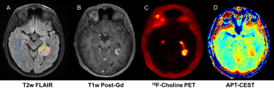 |
Correlation between APT-CEST and 18F-Choline PET in glioma at 3T
marilena rega, Francisco Torrealdea, Joe Hearle, Moritz Zaiss, Ana Carvalho, Asim Asaf, Shonit Punwani, Xavier Golay, John Dickson, Anath Shankar, Harpreet Hyare
Chemical exchange saturation transfer MRI is emerging as a powerful diagnostic tool and has been shown to correlate with glioma tumour grade and molecular genetics. In this study, we aim to investigate whether APT signal is a non-invasive biomarker of Teenage and Young Adult glioma cell proliferation through correlation with 18F-Cho PET SUV as the gold standard. The strong positive correlation found in APT and 18F-Cho PET SUV indirectly demonstrates that APT SI may be a marker of glioma cell proliferation and further demonstrates the potential of APT in the assessment of glioma burden.
|
17:06
|
1259.
 |
Separating BOLD into local and non-local components using a novel simultaneous fMRI-fPET dual analysis
Phillip Ward, Francesco Sforazzini, Sharna Jamadar, Jakub Baran, Shenpeng Li, Zhaolin Chen, Gary Egan
The objective was to separate the MRI BOLD response into local and non-local components using a simultaneously acquired functional FDG-PET (fPET) dataset. We used a visual task, with both high-frequency and low-frequency temporal components, to simultaneously evoke glucose and BOLD responses. Joint analysis of the fMRI and fPET identified two components, including a positively-correlated map of the visual cortex, and a negatively-correlated map of sub-regions of the visual cortex in fMRI and the draining vasculature in fPET. These findings provide preliminary evidence that we can deconstruct the fMRI BOLD signal into local (neuronal) and non-local (haemodynamic) components using simultaneous fMRI-fPET.
|
17:18
|
1260.
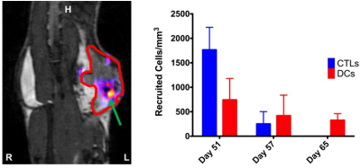 |
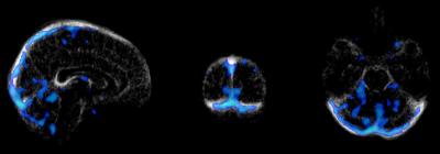
Using Simultaneous PET/MRI and Cell Tracking to Evaluate Immunotherapies in Breast and Ovarian Cancer Models
Brianna Kelly, Victoria Gonzalez, Marie-Laurence Tremblay, Andrea Nuschke, Christa Davis, Alecia MacKay, Andrea West, Barbara Vanderhyden, Genevieve Weir, Marianne Stanford, Kim Brewer
Epithelial ovarian cancer and triple negative breast cancer (TNBC) are aggressive cancers with poor survival outcomes. Simultaneous FDG-PET/MRI and quantitative cell tracking were used to monitor orthotopic ovarian cancer and TNBC models for longitudinal tumor growth and metabolism in response to therapy while tracking immune cell subsets labeled with superparamagnetic iron oxide (SPIO). PET/MRI enabled monitoring of tumor growth and internal physiological changes and allowed quantitative assessment of tumor volumes over time. MRI cell tracking results indicated changes in the recruitment rates of three unique immune cell types over time in the group level with significant individual-level variability.
|
|
