Magnetic Resonance Elastography: Applications & Methods
Oral
Contrast Mechanisms
Thursday, 21 June 2018
| S06 |
08:00 - 10:00 |
Moderators: Thomas Royston, Jeong Hee Yoon |
08:00
 |
1067.
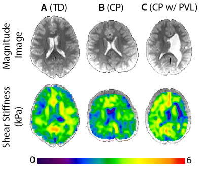 |
 Altered brain tissue stiffness in pediatric cerebral palsy measured with magnetic resonance elastography Altered brain tissue stiffness in pediatric cerebral palsy measured with magnetic resonance elastography
Charlotte Chaze, Daniel Smith, Grace McIlvain, Gabrielle Villermaux, Nicole Maguire, Freeman Miller, Jeremy Crenshaw, Curtis Johnson
Magnetic resonance elastography (MRE) measures the viscoelastic mechanical properties of tissues, which vary extensively between normal and disease states. In this study, we hypothesized that the mechanical integrity of brain tissue is reduced in children with cerebral palsy (CP). Through MRE, we found the stiffness of the cerebrum in children with CP ages 5-12 is significantly lower than in typically developing (TD) children. This finding indicates that there is a difference in brain tissue health in children with CP that is quantifiable through stiffness measured with MRE.
|
08:12
|
1068.
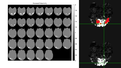 |
Imaging Visual Cortex Activity with Intrinsic Activation MRE
Reihaneh Forouhandehpour, Elijah Van Houten
Initial RD-MRE results from intrinsic activation during repeated ON-OFF visual stimulation cycles are presented. Processed probability values from independent t-tests show areas of isolated activity in the region of the visual cortex for certain mechanical parameters, including the real shear modulus and the imaginary bulk modulus. Overlays of these probability value images with co-registered BOLD images show complimentary regions of activation.
|
08:24
 |
1069.
 |
Assessing tumor mechanical properties and blood perfusion with MRI and correlation with tumor pressure at different compression levels in mice
Gwenaël Pagé, Marion Tardieu, Laurent Besret, Bernard Van-Beers, Philippe Garteiser
The purpose of this study was to evaluate the changes of total tumor pressure and mechanical properties as a function of increasing stress. MR elastography and perfusion measurements (FAIR method) were performed in mice with tumors xenografted subcutaneously. Tumor pressure was measured with a catheter-transducer system. Measurements were performed at different stress levels by externally compressing the tumor with an inflatable balloon. The results show that increasing the externally applied compression results in increased mechanical properties and tumor pressure and decreased perfusion. These results suggest that elevated tumor pressure can be explained by solid stress rather than fluid pressure.
|
08:36
|
1070.
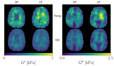 |
Multi-Excitation MRE in Aging Human Brain
Aaron Anderson, Curtis Johnson, Tracey Wszalek, Bradley Sutton, Elijah Van Houten, John Georgiadis
The adult aging process affects human brains in different ways and becomes more prone to neurodegenerative diseases. MRE has shown it’s sensitivity to both changes within healthy brains and identifying biomarkers in diseased brains. This study builds on previous MRE aging research and adds higher-resolution, full-coverage MRE imaging and the ability to identify tissue anisotropy, or lack thereof, with the multi-excitation experiment. We were able to identify important anisotropic differences in the loss modulus for some white matter (WM) regions within the young group and a loss of group-level anisotropy in the select WM regions in the older group.
|
08:48
|
1071.
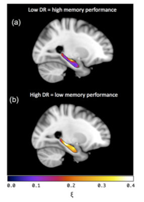 |
Hemispheric specialisation of hippocampal viscoelasticity for memory performance in healthy older adults
Lucy Hiscox, Curtis Johnson, Matthew McGarry, Hillary Schwarb, Edwin van Beek, Neil Roberts, John Starr
MR Elastography of the hippocampus has been associated with memory performance in young adults and thus may have potential as a novel imaging biomarker for Alzheimer’s disease (AD). In healthy older-adults, hippocampal damping ratio ξ was significantly associated with performance on a verbal memory task. Due to greater hippocampal atrophy present in older populations, the contributions of voxels containing CSF were analysed. Stronger correlations with memory were found once CSF voxels were excluded, and when the left hippocampus was analysed separately. MRE of the hippocampus may be a sensitive marker for detecting early pathological changes in patients with AD.
|
09:00
|
1072.
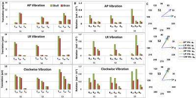 |
In Vivo Characterization of 3D Skull and Brain Motion using MR Elastography with Multi-Excitation Head Driver
Ziying Yin, Yi Sui, Joshua Trzasko, Phillip Rossman, Armando Manduca, Richard Ehman, John Huston III
Characterization of skull-brain interactions during applied motion is essential to understanding the mechanics of traumatic brain injury. In this study, MR elastography was performed on volunteers to study in vivo skull-brain motion responding to different vibrational directions using a multi-excitation driver. With novel dual-saturation imaging and dual-sensitivity motion encoding schemes, we directly measured relative skull-brain displacement on a voxel basis. Our results show that the skull-brain interface tends to significantly attenuate and delay rotational motion compared to translational motion. In slip interface imaging, the skull-brain slip interface is not completely evident, and the slip pattern is spatially heterogeneous.
|
09:12
|
1073.
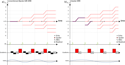 |
Ultra-Fast 4D MR Elastography using Down-Stream Echoes
Christian Guenthner, Sweta Sethi, Ayse Sila Dokumaci, Ralph Sinkus, Sebastian Kozerke
We propose the readout of the lowest order SSFP echo also referred to as down-stream echo of a phase-locked, spoiled SSFP sequence and utilize the unbalanced gradient moment as a highly efficient motion encoding gradient for ultra-fast MRE and demonstrate its feasibility for rapid breast MRE.
|
09:24
|
1074.
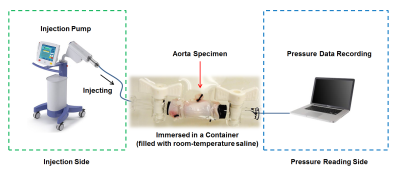 |
In Vivo MR Elastography in Abdominal Aortic Aneurysm Porcine Model: A Comparison to Burst Testing and Mechanical Testing
Huiming Dong, Matthew Joseph, Alan Litsky, Xiaokui Mo, Prateek Kalra, Richard White, Arunark Kolipaka
Abdominal aortic aneurysm (AAA) can result in death due to rupture. Aortic stiffness is an important biomechanical property that can potentially provide accurate rupture risk evaluation. MR elastography (MRE) is a non-invasive technique to estimate aortic stiffness and has not been validated. Therefore, the aim of this study is to use in vivo aortic MRE to estimate aortic stiffness in AAA-induced animal models, and compare it with mechanical testing as well as burst testing. Results demonstrated that aortic stiffness was significantly higher in AAA when compared to normal aorta, while bursting pressure and peak stress was significantly lower in AAA.
|
09:36
|
1075.
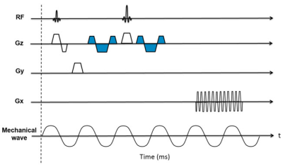 |
Respiratory-triggered (RT) spin-echo echo-planar imaging (SE-EPI) based MR Elastography (MRE)
Hui Wang, Jean Tkach, Tom Cull, Andrew Trout, Charles Dumoulin, Jonathan Dillman
In this work, we describe the development of respiratory-triggered (RT) SE-EPI MR elastography (MRE) and its validation with respect to breath-hold (BH) SE-EPI MRE in adult volunteer subjects.
|
09:48
|
1076.
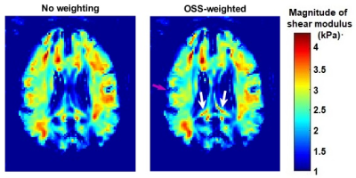 |
Usage of Octahedral Shear Strain Weights in the Inversion of Multifrequency MR Elastography
Cemre Ariyurek, Bilal Tasdelen, Eric Barnhill, Arif Ergun, Yusuf Ider, Ergin Atalar
Multifrequency MR elastography (MMRE) is useful for compensating the influence of amplitude nulls on the elastogram by combining computations at different frequencies by amplitude-weighted averaging or directly without using weights. Previously, it was shown that strain-SNR measures the quality of the data for reconstructing accurate elastography maps. Therefore, using octahedral shear strain (OSS) weights may lead to more accurate elastograms. In this study, k-MDEV and multifrequency Helmholtz inversion have been used. Including OSS-weights in the inversions yielded more reliable elastograms for simulation and experiment phantom. Furthermore, elastograms in higher resolution were obtained for the brain model and human brain data.
|
|
