Novel CEST Methods in Cancer & Beyond
Oral
Contrast Mechanisms
Tuesday, 19 June 2018
| S05 |
08:15 - 10:15 |
Moderators: Xiang Xu, Greg Stanisz |
08:15
 |
0408.
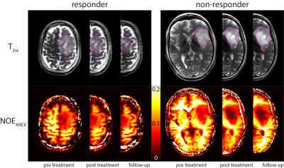 |
 NOE-mediated CEST Imaging of Glioma at 7 Tesla Aids Early Response Evaluation of Patients Undergoing Radio-Chemotherapy NOE-mediated CEST Imaging of Glioma at 7 Tesla Aids Early Response Evaluation of Patients Undergoing Radio-Chemotherapy
Jan-Eric Meissner, Sebastian Regnery, Andreas Korzowski, Steffen Goerke, Jürgen Debus, Heinz-Peter Schlemmer, Mark Ladd, Peter Bachert, Sebastian Adeberg, Daniel Paech
Reliable biomarkers for an early assessment of treatment response are urgently needed. In this study, we investigated the rNOE-mediated CEST signals in patients undergoing radio- and chemotherapy at three different time points including pre and post treatment. The results were compared to the standard clinical determination of responders and non-responders (RANO criteria) and the relaxation compensated CEST contrast NOEAREX showed a statistically significant difference between the two groups directly after therapy. Hence, NOEAREX might potentially help determining early tumor response to therapy.
|
08:27
 |
0409.
 |
CEST-Dixon for human breast cancer characterization at 3T: a preliminary study
Shu Zhang, Stephen Seiler, Xinzeng Wang, Ananth Madhuranthakam, Jochen Keupp, Emily Eads, Robert Lenkinski, Elena Vinogradov
Previous studies have demonstrated the application of CEST to breast malignancies and its potential to aid tumor characterization. However, artifacts can develop in breast CEST imaging due to strong lipid signals. In this work, CEST-Dixon sequence is used for fat free CEST imaging to characterize suspicious lesions in patients. The CEST effects are higher in the ER- IDC than the ER+ IDC, benign and normal groups. The results also indicate positive correlation of CEST with the Ki-67 level. Thus, CEST-Dixon has a potential for improved non-invasive characterization of breast lesions, potentially differentiating more aggressive from less aggressive tumors.
|
08:39
|
0410.
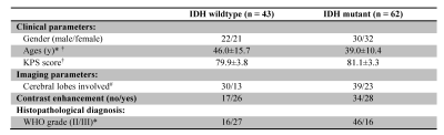 |
Decoding IDH Genotype in Grade-II and -III Gliomas with Protein-based Amide Proton Transfer-Weighted (APTw) MRI
Shanshan Jiang, Qihong Rui, Hao Yu, Yu Wang, Yi Zhang, Hye-Young Heo, Jinyuan Zhou, Zhibo Wen
We explored the possibility of using the APTw signal intensity as a surrogate marker to identify IDH mutation status in gliomas. 105 patients with newly diagnosed grade-II/III gliomas were included. APTw histogram data obtained from tumor burden was recorded. The Mean, Peak, 10th, 25th, 50th, 75th, 90th Percentiles of APTw signal histograms were significantly higher in the IDH-wildtype group than in the IDH-mutant group. The classification for 50th Percentile APTw to differentiate these two glioma groups was 78.1%. Preoperative APTW imaging may assist in predicting the IDH mutation status in gliomas.
|
08:51
|
0411.
 |
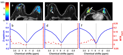
Quantifying the glucose CEST Effect in Patients with oropharyngeal squamous cell carcinoma at 3T in vivo using an oxygen challenge
Alex Smith, Tessa Greenhalgh, Nia Taylor, Benjamin Irving, Ketan Shah, Daniel Bulte, Martin Craig, Michael Chappell, Fergus Gleeson, Brian Burns
Characterizing tumor treatment response is an ongoing radiological challenge as both the anatomical and physiological characteristics of the tumor are changing as the treatment course progresses. GlucoCEST imaging may provide an avenue towards understanding the evolving tumor metabolic environment over the course of treatment. In this preliminary assessment, we show that an oxygen challenge can highlight changes in the vasculature of tumors post radiotherapy.
|
09:03
|
0412.
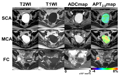 |
Amide proton transfer (APT) imaging of benign ovarian cystic lesions
Keisuke Ishimatsu, Akihiro Nishie, Yukihisa Takayama, Yoshiki Asayama, Kousei Ishigami, Yasuhiro Ushijima, Daisuke Kakihara, Nobuhiro Fujita, Koichiro Morita, Seiichiro Takao, Osamu Togao, Kenzo Sonoda, Jochen Keupp, Hiroshi Honda
It is important to diagnose benign ovarian cystic lesions as early and correctly as possible because some types of lesions have malignant potential. The objective of our study is to investigate whether amide proton transfer (APT) imaging is useful for evaluation of benign ovarian cystic lesions. We compared the APT signal in three different benign ovarian cystic lesions using three different durations of presaturation pulse.
|
09:15
|
0413.
 |
Separation of the contribution of protein concentration and pH to measured APT signal changes in a preclinical model of brain metastases
Kevin Ray, Manon Simard, James Larkin, Michael Chappell, Nicola Sibson
Amide proton transfer (APT) studies usually attribute the altered APT signal in tumours to an increased cytosolic protein concentration. However, other concomitant changes in pH, T1, or water content make absolute quantification of protein concentration or pH from APT signals challenging. In this study, we separate the contributions of protein concentration and pH to APT signal differences in a preclinical model of brain metastases by combining in vivo and ex vivo measurements. We show that 66% of the observed APT signal difference was caused by protein concentration alterations, with the remaining 34% signal change reflecting an increase in tumour pH.
|
09:27
|
0414.
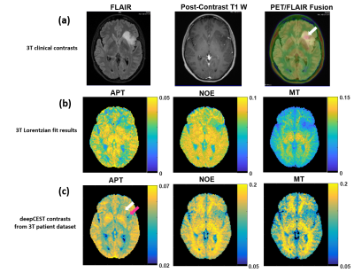 |
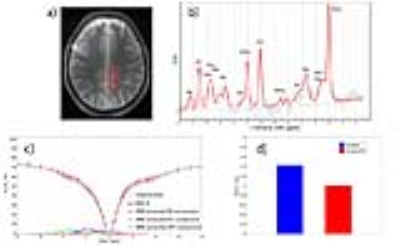
Deep CEST MRI – 9.4T spectral super-resolution from 3T CEST MRI data
Moritz Zaiss, Anagha Deshmane, Kai Herz, Max Braun, Benjamin Bender, Tobias Lindig, Klaus Scheffler
CEST peaks are easy to detect at ultra-high-field strengths due to high signal and spectral separation. However, spectral coalescence and line broadening makes modeling of CEST effects at clinical field strengths (<=3T) a challenge. In this proof-of-concept study of super-resolution CEST imaging, the underlying spectral features of 3T Z-spectra were predicted using a neural network trained on 9.4T data. Applying the neural network to untrained volunteer and patient data acquired at 3T resulted in the expected contrast in healthy gray and white matter and tumor tissue in Z-spectra and APT, NOE, and MT CEST maps.
|
09:39
|
0415.
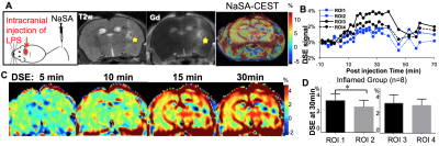 |
NaSA-CEST: MR monitoring of brain inflammation using an aspirin metabolite as contrast agent
Xiaolei Song, Yanrong Chen, Chenwang Jin, Chengyan Chu, Irina Shats, Yuguo Li, Yue Yuan, Xiaowei He, Piotr Walzcak, Jeff Bulte
Sodium salicylate (NaSA), a nonsteroidal anti-inflammatory drug and the main metabolite of aspirin, can accumulate specifically in inflamed tissue. We investigated the use of NaSA-enhanced CEST MRI for in vivo mapping of brain inflammation, induced by intrastriatal injection of lipopolysaccharide (LPS). Inflamed mice exhibited an increase of ~8% NaSA-CEST signal on the AUC10-30min images, with signals for the ipsilateral striatum significantly higher than those for the mirrored contralateral regions. While for the non-inflamed sham mice, which received saline instead of LPS, no such NaSA-CEST signal could be observed. Our NaSA-CEST approach could possibly lead to the development of inflammation-specific theranostics.
|
09:51
|
0416.
 |
Multicolor metabolic quantitative CEST (mmqCEST) imaging: possibility and limitations
Vitaliy Khlebnikov, Alex Bhogal, Olivier Mougin, Vincent Boer, Jannie Wijnen, Penny Gowland, Peter Luijten, Jeanine Prompers, Hans Hoogduin, Dennis Klomp
Multicolor metabolic CEST imaging: possibility and limitations
|
10:03
|
0417.
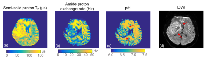 |
CEST Fingerprinting: Initial Study in Human Subjects
Zhengwei Zhou, Qi Yang, Zhaoyang Fan, Pei Han, Debiao Li
CEST fingerprinting was recently proposed to achieve efficient exchange rate quantification for pH mapping. Here we present the initial data of pH mapping in healthy subjects and one acute stroke patient at clinical scanners in an 8-min scan using CEST fingerprinting. The pH maps of the healthy volunteers are homogenous and the values fall in the normal physiological range. The pH map in the patient shows an inhomogenous acidic area. This study shows CEST fingerprinting has the potential to detect acidosis in human subjects.
|
|
