Magnetic Susceptibility Imaging
Oral
Contrast Mechanisms
Monday, 18 June 2018
| S06 |
13:45 - 15:45 |
Moderators: Jürgen Reichenbach, Masaki Fukunaga |
13:45
|
0187.
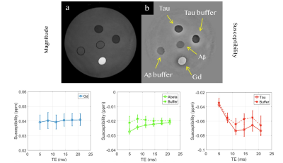 |
 Direct Imaging of Diamagnetic Susceptibility of Beta Amyloid Aggregates in Transgenic Mouse Models of Alzheimer’s Disease using Quantitative Susceptibility Mapping MRI Direct Imaging of Diamagnetic Susceptibility of Beta Amyloid Aggregates in Transgenic Mouse Models of Alzheimer’s Disease using Quantitative Susceptibility Mapping MRI
Nan-Jie Gong, Russell Dibb, Chunlei Liu
We demonstrated in a phantom that beta amyloid is diamagnetic and can generate strong contrast on susceptibility maps. Based on this, it is further shown both in vivo and ex vivo that magnetic susceptibility mapping could be used to monitor accumulation of amyloid plaques in AD mouse models. Most importantly, the diamagnetic susceptibility map and paramagnetic susceptibility map provided histology-like image contrast for identifying deposition of beta amyloid plaques and iron.
|
13:57
 |
0188.
 |
Iron-induced relaxation mechanisms in the human substantia nigra: towards quantifying iron load in dopaminergic neurons
Malte Brammerloh, Isabel Weigelt, Thomas Arendt, Filippos Gavriilidis, Nico Scherf, Steffen Jankuhn, Markus Morawski, Nikolaus Weiskopf, Evgeniya Kirilina
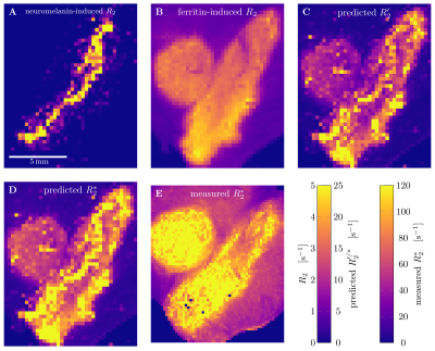
Pathological iron accumulation in the human brain is a biomarker for neurodegeneration. Several diagnostically promising MR-based methods for in vivo iron quantification were proposed, based on the empirical relationship between R2* and iron concentration. However, these do not account for different chemical forms and cellular distribution of iron. We combined post mortem MRI, advanced quantitative histology and biophysical modeling to develop a generative theory linking obtained iron concentrations to quantitative MR parameters. The impact of nanoscale molecular interaction of water with iron and of iron-rich dopaminergic neurons was quantified in substantia nigra.
|
14:09
 |
0189.
 |
Accurate and Efficient QSM Reconstruction using Deep Learning
Enhao Gong, Berkin Bilgic, Kawin Setsompop, Audrey Fan, Greg Zaharchuk, John Pauly
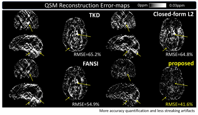
Quantitative Susceptibility Mapping (QSM) is a powerful MRI technique to quantify susceptibility changes and reveal pathology such as multiple sclerosis (MS) lesions and demyelination. QSM reconstruction is very challenging because it requires solving an ill-posed deconvolution and removing the effects of a dipole kernel on tissue phases to obtain susceptibility. To address the limitations of existing QSM reconstruction methods in accuracy, stability and efficiency, an iteration-free data-driven QSM reconstruction is proposed that trains a deep learning model to approximate COSMOS QSM quantification from acquired signals and pre-processed phases. Cross-validated on in-vivo datasets with 15 single direction QSM scans and 3 COSMOS QSM results from 3 healthy subjects, the proposed deep learning method achieves accurate QSM reconstruction, outperforming state-of-the-art methods across various metrics. The deep learning solution is also faster than iterative reconstruction by several orders of magnitude, which enables broader clinical applications.
|
14:21
|
0190.
 |
Quantitative susceptibility mapping using deep neural network
Jaeyeon Yoon, Jingyu Ko, Jingu Lee, Hosan Jung, Berkin Bilgic, Kawin Setsompop, Jongho Lee
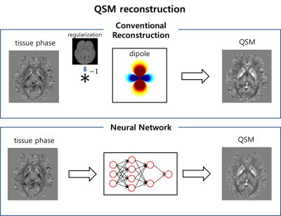
In this study, we designed a deep neural network that functions as dipole deconvolution in QSM reconstruction. For label data, COSMOS reconstructed QSM maps were used so that the network produces ground truth like COSMOS results without streaking artifacts. The performance of our network was superior to conventional QSM results with lower RMSE for multiple head orientation input data.
|
14:33
|
0191.
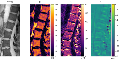 |
Vertebral Column Quantitative Susceptibility Mapping using Joint Background Field Removal and Dipole Inversion
Maximilian Diefenbach, Anh Van, Jakob Meineke, Andreas Scharr, Jan Kirschke, Alexandra Gersing, Thomas Baum, Benedikt Schwaiger, Dimitrios Karampinos
Quantitative susceptibility mapping (QSM) with joint background field removal and dipole inversion is applied in the spine of osteoporosis patients and healthy volunteers. Preliminary multi-MR-parametric patient results are compared to low-dose CT scans to investigate the feasibility of QSM to qualitatively and quantitatively detect features of diseased tissues and differentiate positive and negative susceptibility sources in comparison to R2*-mapping.
|
14:45
|
0192.
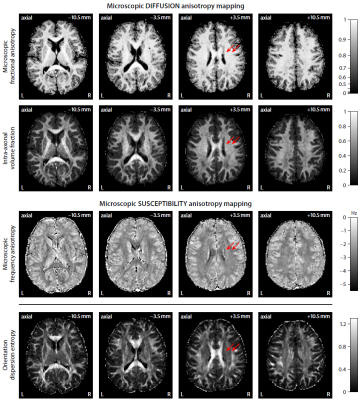 |
Microscopic susceptibility anisotropy imaging: A clinically viable gradient-echo MRI technique
Enrico Kaden, Umesh Rudrapatna, Irina Barskaya, Mark Does, Derek Jones, Daniel Alexander
The orientation dependence of the gradient-echo MR signal in brain white matter conflates two principal effects, (i) the susceptibility properties of tissue microenvironments, especially the myelin microstructure, and (ii) the axon orientation distribution with respect to the external magnetic field. This work introduces a clinically feasible MRI method based on gradient-echo and diffusion measurements, which we refer to as microscopic susceptibility anisotropy imaging, that disentangles both effects, hence enabling us to estimate microscopic susceptibility anisotropy unconfounded by fibre crossings and orientation dispersion as well as magnetic field direction.
|
14:57
|
0193.
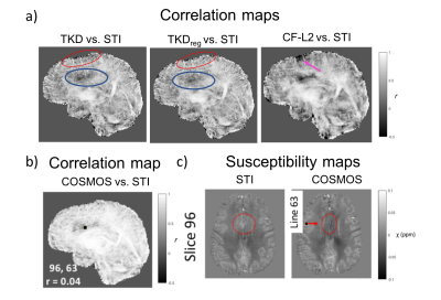 |
How should we compare QSM results? A correlation based analysis as an alternative to traditional error metrics
Jiaen Liu, Pinar Özbay
During the last QSM Workshop, the results of first “Quantitative Susceptibility Mapping (QSM) Reconstruction Challenge” were presented, which was performed to allow a systematic comparison of various QSM algorithms. One unresolved issue was the fact that the comparison metrics did not properly deal with the effect of smoothing. Here, we propose a comparison method based on pearson correlations, which are calculated over 1D lines throughout the QSM volumes, and show its robustness relative to other metrics under the influence of over-smoothing.
|
15:09
|
0194.
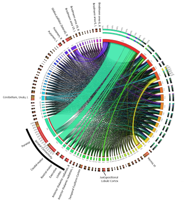 |
Age- and sex-related spatial patterns of variation in normal brain magnetic susceptibility (QSM) revealed by Blind Source Separation (BSS) and Supervised Machine Learning
Ferdinand Schweser, Balint Sule, Juliane Damm, Niels Bergsland, Michael Dwyer, Akshay Dhamankar, Bianca Weinstock-Guttman, Robert Zivadinov
Previous studies using QSM have demonstrated a relatively high inter-subject variation of brain susceptibility. In the present work, we combined a blind source separation technique with a machine learning strategy to disentangle spatial networks of independent variation of brain susceptibility. As a first step toward a better understanding of the underlying causes of variation, we studied their associations with age and sex. The analysis revealed several networks with distinct anatomical features, although the applied analysis technique did not involve any information about anatomy, age, or sex.
|
15:21
|
0195.
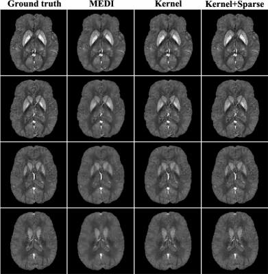 |
Constrained Dipole Inversion for Quantitative Susceptibility Mapping Using a "Kernel+Sparse" Model
Xi Peng, Yudu Li, Fan Lam, Rong Guo, Bryan Clifford, Zhi-Pei Liang
In quantitative susceptibility mapping (QSM), constrained dipole inversion is often necessary to overcome the ill-posedness of the underlying dipole deconvolution problem. Existing methods achieve this by the use of spatial regularization. In this work, we propose a novel "kernel+sparse" model for constrained dipole inversion. In this model, the kernel term absorbs the prior information by representing the susceptibility as a function of prior features while the sparse term accounts for the localized novel features. The proposed method has been evaluated using both simulated and in vivo data, producing impressive results. This method may prove to be useful for many QSM studies.
|
15:33
|
0196.
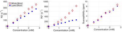 |
Sensitivity of Relaxometry and Quantitative Susceptibility Mapping to Microscopic Iron Distribution
Timothy Colgan, Gesine Knobloch, Scott Reeder, Diego Hernando
MRI-based iron quantification enables the non-invasive assessment of tissue iron concentration. MRI relaxation parameters such as R2 and R2* are sensitive to iron concentration, but may depend on the microscopic spatial distribution of iron. Quantitative Susceptibility Mapping (QSM) is a promising iron quantification technique, but its sensitivity to the spatial distribution of iron remains unknown. In this work, we performed simulations and in vitro experiments using whole versus lysed erythrocytes to investigate this sensitivity. Our results suggest that QSM, unlike R2 and R2* relaxometry, is independent of the microscopic distribution of iron.
|
|
