Techniques for Myelin & Microstructure Imaging
Oral
Contrast Mechanisms
Wednesday, 20 June 2018
| N03 |
13:45 - 15:45 |
Moderators: Alex MacKay, Se-Hong Oh |
13:45
 |
0783.
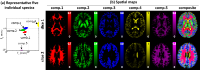 |
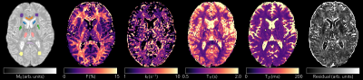
 Multidimensional T1 Relaxation-T2 Relaxation Correlation Spectroscopic Imaging (RR-CSI) for In Vivo Imaging of Microstructure Multidimensional T1 Relaxation-T2 Relaxation Correlation Spectroscopic Imaging (RR-CSI) for In Vivo Imaging of Microstructure
Daeun Kim, Jessica Wisnowski, Christopher Nguyen, Justin Haldar
We propose a new multidimensional MRI experiment called T1 Relaxation-T2 Relaxation Correlation Spectroscopic Imaging (RR-CSI) for probing microstructure. RR-CSI acquires imaging data with two-dimensional relaxation contrast encoding and estimates a high-dimensional spectroscopic image by using spatially-constrained reconstruction. The spectroscopic image comprises a full 2D T1-T2 spectrum at every voxel. The distinct peaks in these spectra correspond to different microscopic tissue compartments, which enables spatial mapping of microstructure. Compared to conventional methods, RR-CSI has improved capabilities for resolving tissue microenvironments with similar relaxation parameters. RR-CSI is demonstrated with real MRI data, including the first in vivo human brain results.
|
13:57
 |
0784.
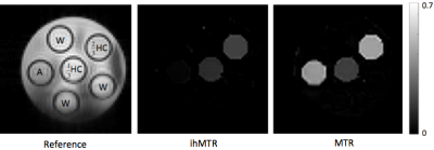 |
Comparison of Inhomogeneous Magnetization Transfer and Myelin Water Fraction Ex-Vivo at 7T
Michelle Lam, Andrew Yung, Alan Manning, Cornelia Laule, G.R. Wayne Moore, Anastasia Smolina, Irene Vavasour, Erin MacMillan, Carl Michal, Alex Mackay, Piotr Kozlowski
Inhomogeneous magnetization transfer (ihMT) shows promise as a myelin specific MR technique. This specificity is thought to emerge from ihMT’s sensitivity to dipolar relaxation times, which can differ dramatically between lipids (which are the main component of myelin) and other brain constituents. We compared both ihMT and conventional MT to myelin water imaging using ex-vivo normal and multiple sclerosis brain tissue, to determine how the white matter and grey matter signal intensities vary across these three proposed myelin specifictechniques.
|
14:09
|
0785.
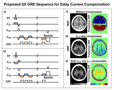 |
A Sequence for High Quality Gradient Echo Myelin Water Imaging (GRE-MWI) at 3T and 7T
Hyeong-Geol Shin, Se-Hong Oh, Masaki Fukunaga, Doohee Lee, Yoonho Nam, Sooyeon Ji, Woojin Jung, Jongho Lee
In this study, we propose an eddy current compensated 2D GRE-MWI sequence and demonstrate that the sequence is robust to physiological noises. The resulting myelin water fraction maps at 3T and 7T shows high quality images at high resolution (2 × 2 × 2 mm3 at 3T and 1.5 × 1.5 × 2 mm3 at 7T).
|
14:21
|
0786.
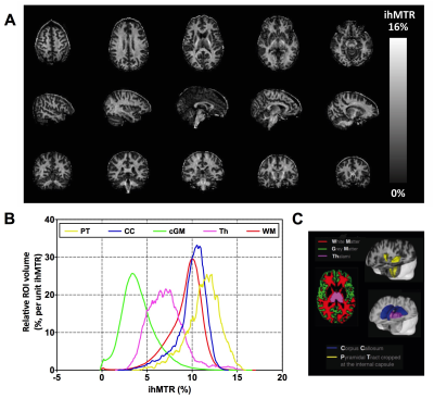 |
Whole brain inhomogeneous Magnetization Transfer (ihMT) imaging at 3T: concentrating RF power to mitigate RF inhomogeneities effects
Samira Mchinda, Gopal Varma, Robin Draveny, Arnaud Le Troter, Victor Carvalho, Valentin Prevost, Maxime Guye, Jean Pelletier, Jean-philippe Ranjeva, David Alsop, Guillaume Duhamel, Olivier Girard
Inhomogeneous magnetization transfer (ihMT) is a new MRI modality that provides strong sensitivity to myelinated tissues. Previously, a 3D whole brain ihMT sequence, based on GRE readouts interleaved with bursts of MT saturation pulses, was developed and optimized at 1.5T. In this work we demonstrate that concentrating the MT pulse energy can mitigate the sensitivity of ihMT to RF inhomogeneities encountered in high field systems. Overall, this allows for 1.5mm3 resolution ihMTR maps to be acquired at 3T, with a reduced B1-induced bias and strong signal within the whole brain, allowing robust clinical applications of ihMT at 3T.
|
14:33
|
0787.
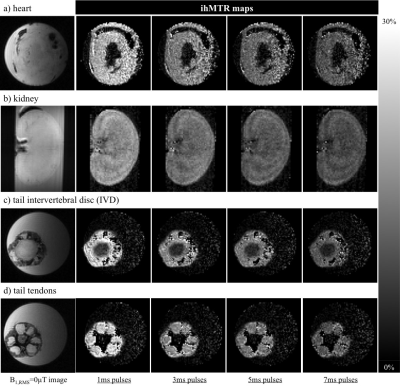 |
Towards short dipolar relaxation time, T1D, MRI
Gopal Varma, Patricia Coutinho de Souza, Valentin Prevost, Olivier Girard, Victor Carvalho, Samira Mchinda, Leo Tsai, Guillaume Duhamel, Aaron Grant, David Alsop
The inhomogeneous magnetization transfer (ihMT) technique has shown myelin sensitivity, and is understood to be dependent on power and the dipolar relaxation time parameter, T1D, which is longer in myelinated tissues. Implementation of ihMT can be adapted to provide a smaller, but non-negligible signal from other, relatively short T1D tissues. Simulations showed a measurable ihMT signal, achieved from fixed low duty cycle MT preparations with high B1 pulses, decayed with pulse width at a rate dependent on T1D. Thus short, high B1 pulses were implemented to acquire ihMT data from ex-vivo samples of rat heart, kidney, and tail tendon, demonstrating the feasibility of short T1D imaging.
|
14:45
|
0788.
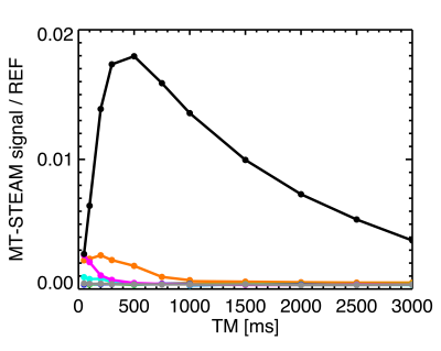 |
MT-Based Detection of Semi-Solids without Background Water Signal.
Peter van Gelderen, Jacco de Zwart, Jeff Duyn
Brain tissue can be modeled as a system with a water pool and an invisible pool of semi-solids. The exchange between these pools can be measured by saturating the semi-solids and observing the decrease in water signal after exchange. This requires making a differential measurement, comparing data with and without saturation. Here we present a variation of the STEAM sequence, adding water suppression, which allows for observation of exchanging spins in a single acquisition without a water background signal. The method was demonstrated in both a phantom and human subject at 3 T.
|
14:57
|
0789.
 |
Elliptical Magnetization Transfer: Calculating MT Parameters from the bSSFP Signal Ellipse
Tobias Wood, Anna Combes, Shaihan Malik
Balanced Steady-State Free-Precession images are sensitive to T1, T2, off-resonance and Magnetization Transfer effects. Previously, Gloor et al extracted MT parameters from SSFP data, but required external T1 & B1 maps and did not account for off-resonance effects. Here we show that by incorporating the elliptical method of Shcherbakova et al, B0 can be measured and the need for an external T1 map removed. We present results covering the whole brain at 1.5mm isotropic voxel size acquired in 20 minutes. We also discuss an interesting asymmetry seen in the SSFP ellipse at low flip-angles.
|
15:09
|
0790.
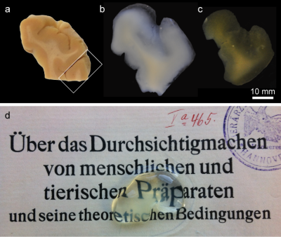 |
3D microscopy with CLARITY on human brain tissue: a tool for informing and validating MRI-based histology
Markus Morawski, Evgeniya Kirilina, Nico Scherf, Carsten Jäger, Katja Reimann, Robert Trampel, Filippos Gavriilidis, Stefan Geyer, Bernd Biedermann, Thomas Arendt, Nikolaus Weiskopf
Recent developments of methods for mapping tissue microstructure with MRI require histological 3D data for validation. Here, CLARITY on post mortem human brain was adapted for this purpose. We demonstrated clearing of up to 8 mm thick samples, 3D microscopy of up to 5 mm thick samples, and the application of multiple stains including markers for neurons, glia, and fibers. The result is a detailed histology-based characterization of cyto- and myelo-architectonics within a volume corresponding to a typical MRI voxel. This approach promises to help integrate MRI-based histology and optical microscopy in 3D, and enable the further development and validation of in vivo histology using MRI.
|
15:21
|
0791.
 |
Disentangling the contributions of brain tissue fraction and composition to quantitative MRI
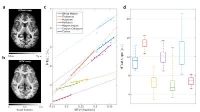
Shir Filo, Oshrat Shtangel, Aviv Mezer
In-vivo quantitative MRI (qMRI) aims at characterizing the biological properties of brain tissue. However, qMRI parameters are sensitive both to the molecular tissue properties and to the water content within each voxel. We introduce a novel approach that disentangles these two contributions to qMRI parameters and provides tissue-specific measurements. This is achieved by evaluating the dependency of qMRI parameters on the non-water fraction. Using phantoms, we show that this dependency changes as a function of molecular composition. In the human brain, our method reveals unique tissue signatures for different brain regions, along with region-specific age-related changes.
|
15:33
|
0792.
 |
Longitudinal assessment of adolescent brain myelination using a pulsed magnetization transfer approach
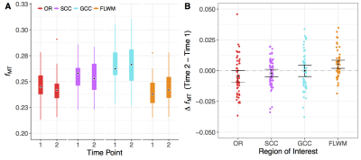
Erika Raven, Peter van Gelderen, Diana Fishbein, John VanMeter, Jeff Duyn
The spatiotemporal growth trajectories of white matter, and in particular myelin, are an important part of cognitive development during adolescence. Quantitative magnetization transfer (qMT) imaging can be used to measure the fraction of non-water protons (fMT) as an estimate of myelin in vivo. Here we used a recently developed, time-efficient pulsed MT approach to extract fMT from white matter regions at different stages of development in a community-based cohort of adolescents. We tested the sensitivity of this approach for detecting region-specific change in fMT in repeated scans that covered a period of 18 months.
|
|
