Measuring & Correcting Imperfections
Oral
Acquisition, Reconstruction & Analysis
Monday, 18 June 2018
| S03 |
13:45 - 15:45 |
Moderator: S. Johanna Vannesjo |
13:45
 |
0167.
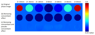 |
 On pseudo-concomitant fields caused by gradient coil vibrations On pseudo-concomitant fields caused by gradient coil vibrations
Yi-Cheng Hsu, Ying-Hua Chu, Fa-Hsuan Lin, Maxim Zaitsev
We demonstrate that a negative quadratic phase accrual, which we term a pseudo-concomitant field, is likely to be caused by gradient vibration and has a nearly constant spatial distribution. Accounting for this effect may be of importance for phase-sensitive MR applications utilizing strong gradients, such as flow imaging.
|
13:57
|
0168.
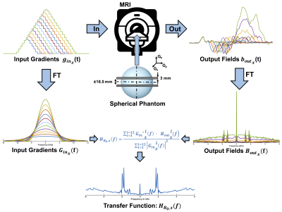 |
$$$B_{0}$$$-component determination of the gradient system transfer function using standard MR scanner hardware
Manuel Stich, Tobias Wech, Anne Slawig, Gudrun Ruyters, Andrew Dewdney, Ralf Ringler, Thorsten Bley, Herbert Köstler
As a linear and time-invariant (LTI) system, the dynamic gradient system can be described by the system transfer function. While special measurement equipment like field cameras can be used to precisely determine even higher orders of the transfer function, phantom-based approaches were introduced for alternative determination without additional hardware needed. This study reports on phantom-based measurements of B0-components, which resulted in transfer functions with sufficiently high resolution for the characterization of mechanical resonances.
|
14:09
|
0169.
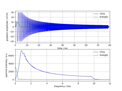 |
GIRF measurement using a combination of triangular and chirp waveform input functions
Video Permission Withheld
Peter Mazurkewitz, Jürgen Rahmer, Peter Börnert
Gradient-impulse-response-function (GIRF) measurement is a well-established method for MRI gradient-system characterization. Typical GIRF input-functions are triangles or chirps. For triangles, measurements have to be performed with different pulse lengths to get a continuous frequency spectrum due to blind spots in the spectrum, requiring long scan times. In contrast, the spectrum of the chirp waveform covers a large frequency range without blind spots. However, at low frequencies the chirp fails due to a diverging intensity in its spectrum. We interleaved both waveforms and obtained a continuous gradient modulation transfer function (GMTF) spectrum down to low frequencies in short measurement time.
|
14:21
|
0170.
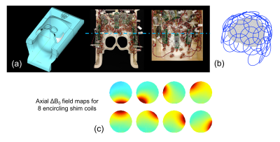 |
Spatially-selective excitation using a tailored nonlinear ?B0 pattern generated by an integrated multi-coil ?B0/Rx array
Jason Stockmann, Nicolas Arango, Benedikt Poser, Thomas Witzel, Jacob White, Lawrence Wald, Jonathan Polimeni
Multi-coil ΔB0 shim arrays have recently been used for zoomed imaging of target ROIs in mice, providing improved acquisition efficiency without the high SAR, long RF pulses, and other limitations of conventional selective excitation methods that rely solely on linear gradients. We extend this work to zoomed 3D EPI in humans using an integrated ΔB0/Rx array coil to dynamically switch-on a static, spatially-tailored nonlinear ΔB0 pattern during RF excitation. Proof-of-concept selective excitations of the occipital visual cortex and the peripheral cerebrum are shown, with strong correspondence to the target patterns. Four-fold in-plane undersampled EPI of occipital visual cortex demonstrates the method’s potential for efficient high-resolution neuroimaging.
|
14:33
|
0171.
 |
Improved multi-band RF performance using GRATER-based predistortion
Vanessa Landes, Krishna Nayak
The Gradient Reversal Approach to Evaluate RF (GRATER) has been shown to accurately measure small-tip RF pulses in phantoms, without the need for added hardware or sophisticated processing. Imperfect RF production can be measured with GRATER, and RF waveforms can be appropriately predistorted. We demonstrate substantial improvements in multi-band RF performance using this approach. For example, a 0.648 ms, 30º FA, 4- band RF pulse with 2 cm center-to-center spacing and 5 mm slice thickness (desirable for SMS cardiac imaging) excites spurious side-lobes with 10.4% and 3.4% max signal before and after GRATER-based RF predistortion, respectively.
|
14:45
|
0172.
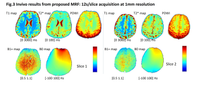 |
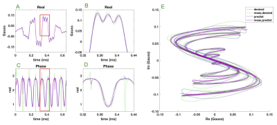
MR Fingerprinting (MRF) incorporating simultaneous detection of RF transmit field and B0 inhomogeneity
Huihui Ye, Qing Li, Xiaozhi Cao, Qiuping Ding, Hongjian He, Huafeng Liu, Jianghui Zhong
MR fingerprinting with B1+ and B0 field inhomogeneity detection is proposed with an IR-spoiled GRE based sequence. With this sequence, a 12s/slice acquisition simultaneously provide unbiased T1, T2* maps and B1+, B0 information at isotropic 1mm resolution.
|
14:57
|
0173.
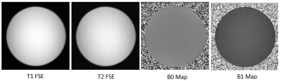 |
Deep Learning Based Approach for Main and RF Field Maps Estimation in MRI
Kavitha Manickam, Jaganathan Vellagoundar
In MRI, automatic estimation of main (B0) and RF (B1) field maps from the scanned images will help daily quality assurance and field corrected reconstruction. In this paper, a novel approach based on deep learning technique is presented to estimate B0 and B1 maps from the scanned images. A modified version of stacked convolutional encoder with random skip connections deep learning network is constructed. Two separate networks are used to estimate B0 and B1 maps individually. The networks are trained and tested with phantom images. The results show that the estimated maps are comparable to the actual field maps. Automatic map estimation based on deep learning approach is the first step towards achieving daily quality assurance and field correction from the regular scanned images.
|
15:09
|
0174.
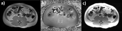 |
An empirical $$$B^-_1$$$ Non-uniformity Correction of Phased-Array Coil Images without Measuring Coil Sensitivity
Frederick Damen, Kejia Cai
Radio Frequency (RF) receiving coil arrays improve the signal-to-noise ratio (SNR), and enable partial parallel imaging. However, these benefits often come at the cost of image non-uniformity. $$$B^-_1$$$ non-uniformity correction techniques are confounded by signal that not only varies due to coil induced $$$B^-_1$$$ sensitivity, but also due to true signal variations in proton density, susceptibility, and relaxation rates. Herein, we propose an empirical method that produces a $$$B^-_1$$$ non-uniformity-corrected complex image from the phased-array coil images themselves using minimal assumptions and without measuring the coil sensitivities. This method is validated using MRI of the abdomen, brain, and a homogeneous phantom.
|
15:21
|
0175.
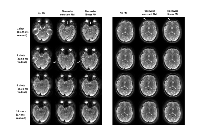 |
Improved image quality for long readout imaging using piecewise linear field map model
Giang-Chau Ngo, Alex Cerjanic, Bradley Sutton
Trajectories such as spiral or EPI enable fast acquisition compared to spin warp imaging. While a spin warp acquisition has a readout of 2-3 ms, a spiral trajectory readout can be as long as 60 ms. The efficiency of the spiral trajectory comes at a cost of image quality through magnetic field inhomogeneity and T2* decay during long readouts. In this work, a signal model with a piecewise linear field map is used to correct for the magnetic field inhomogeneity effects during long imaging readouts . The proposed method shows the ability to correct for resulting image artifacts.
|
15:33
|
0176.
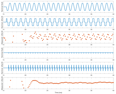 |
Measuring MRI Gradient Trajectory Dynamics using Simultaneous EEG-FMRI
Mark Chiew, Jostein Holmgren, Dean Fido, Catherine Warnaby, S Vannesjo
Simultaneous EEG-FMRI data acquisition provides an opportunity for characterization of magnetic field dynamics during imaging, by leveraging the information contained in the EEG induced gradient artefacts. Using a simple effective loop model, we reduce the complex EEG electrode geometry to small effective loops located at off-isocentre positions, which we use to fit first order (linear in space) models of the magnetic field rate of change. From these, estimates of the actual field dynamics, and trajectory/encoding information can be derived at no cost. In this proof-of-principle, we demonstrate estimation of gradient dynamics during a conventional EPI acquisition using simultaneous EEG recordings.
|
|
