Motion Correction: Beyond Gating
Oral
Acquisition, Reconstruction & Analysis
Tuesday, 19 June 2018
| S02 |
13:45 - 15:45 |
Moderators: Freddy Odille, Giulia Ginami |
13:45
|
0473.
 |
 Assessment of respiratory motion-resolved and nonrigid motion-corrected 3D Cartesian coronary MRA Assessment of respiratory motion-resolved and nonrigid motion-corrected 3D Cartesian coronary MRA
Teresa Correia, Gastao Cruz, Giulia Ginami, Imran Rashid, Radhouene Neji, Rene Botnar, Claudia Prieto
Slow acquisitions and susceptibility to respiratory motion artifacts are major challenges in free-breathing 3D whole-heart coronary MR angiography (CMRA). Recently, a respiratory-resolved approach has been proposed to improve scan efficiency and reduce motion artifacts using non-Cartesian acquisitions. However, irregular respirations compromise its suitability for Cartesian imaging. Here, sparsity in a motion-corrected domain is exploited to generate high-quality respiratory-resolved Cartesian images, used to estimate nonrigid motion fields. These are incorporated into a motion-corrected generalized matrix reconstruction, to further improve coronary vessel sharpness. Thus, this approach provides high-quality respiratory-resolved Cartesian CMRA images and a motion-corrected CMRA image at a given respiratory phase.
|
13:57
 |
0474.
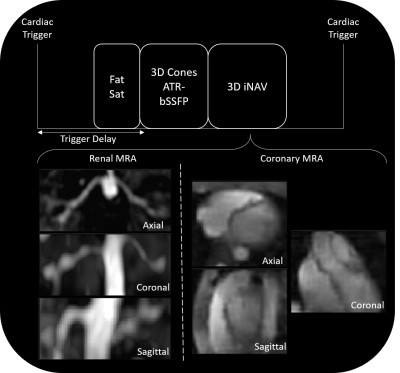 |
Nonrigid Motion Correction using 3D iNAVs with Generalized Motion Compensated Reconstruction and Autofocusing
Srivathsan Koundinyan, Corey Baron, Nicholas Dwork, Joseph Cheng, Dwight Nishimura
We present a novel framework to combine two well-known methods for motion correction: generalized motion compensated reconstruction (GMCR) and autofocusing. In this hybrid technique, 3D image-based navigators (3D iNAVs) are utilized for motion tracking. The beat-to-beat and voxel-by-voxel motion information within the 3D iNAVs are directly inputted into GMCR to mitigate motion artifacts. To reduce computation time, an autofocusing step is incorporated. The overall correction scheme is evaluated in free-breathing coronary magnetic resonance angiography and renal magnetic resonance angiography exams. In all six in vivo studies, images reconstructed with the proposed strategy outperform those generated with beat-to-beat 3D translational correction.
|
14:09
|
0475.
 |
Effects of reconstruction with deformable motion correction on abdominal DCE-MRI images
Adam Johansson, James Balter, Yue Cao
Image reconstruction with deformable motion correction for dynamic contrast-enhanced (DCE) MRI can counteract artifacts in dynamic images and estimated perfusion maps. In this study, we present the results of applying a reconstruction method with deformable motion correction to 54 DCE-MRI examinations of 31 patients. Deformable motion correction is found to compensate for up to 15 mm of residual motion after rigid-body motion correction. Motion-correction also reduces liver-wide bias caused by motion-distorted input functions and removes localized artifacts at liver edges. Finally, for several cases, motion-correction makes lesion edges sharper in reconstructed images.
|
14:21
|
0476.
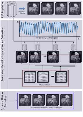 |
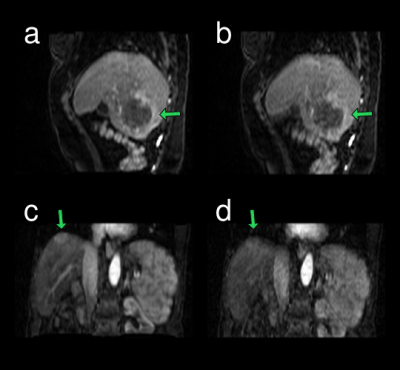
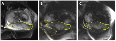
3D non-rigid motion-corrected dynamic contrast enhanced MRI of the liver with high isotropic resolution
Matteo Ippoliti, Marcus Makowski, Tobias Schaeffter, Christoph Kolbitsch
Dynamic Contrast Enhanced (DCE) MRI of the liver is an important diagnostic tool. The main challenge is complex respiratory motion of the abdomen. Here we present a motion correction approach for DCE-MRI. Golden Radial Phase Encoding data was acquired continuously in a patient after contrast injection during free-breathing. In a first step, non-rigid respiratory motion was estimated from the data. The motion information was then used to reconstruct 3D motion corrected dynamic images with a temporal resolution of 13s and an isotropic spatial resolution of 1.5mm3. The proposed technique strongly reduced motion artefacts and ensured high image quality.
|
14:33
|
0477.
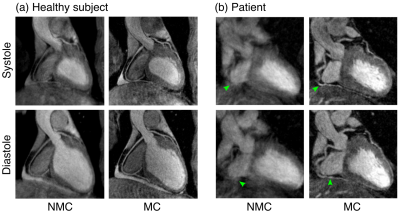 |
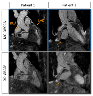
Cardiac and respiratory motion-corrected whole-heart PET-MR imaging for simultaneous assessment of coronary anatomy, cardiac function and myocardial integrity
Camila Munoz, Radhouene Neji, Karl Kunze, Imran Rashid, Christoph Rischpler, Stephan Nekolla, René Botnar, Claudia Prieto
Image degradation due to cardiac and respiratory motion remains a challenge for cardiac PET-MR imaging. Here we propose a simultaneous dual-phase PET and Coronary MR angiography (CMRA) acquisition and reconstruction framework that allows for the visualisation of coronary anatomy, estimation of ventricular function and motion-corrected myocardial PET in a single efficient examination. A validation study in healthy subjects shows that left ventricular ejection fraction can be estimated from dual-phase CMRA images at 3T. Results from patients with cardiovascular disease show improvements in respiratory motion corrected dual-phase CMRA and in PET image quality when applying both cardiac and respiratory motion correction.
|
14:45
|
0478.
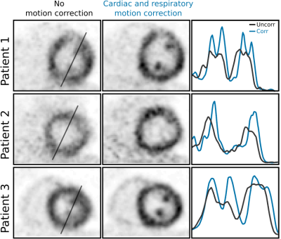 |
Joint PET-MR image registration for cardiac and respiratory motion correction of simultaneous cardiac PET-MR
Christoph Kolbitsch, Radhouene Neji, Matthias Fenchel, Andreas Schuh, Andrew Mallia, Paul Marsden, Tobias Schaeffter
Cardiac Positron Emission Tomography (PET) can provide diagnostic information about myocardial perfusion and metabolism with excellent sensitivity. The main challenge is physiological motion of the heart due to breathing and heartbeat. Here we present a joint PET-MR image registration approach which provides non-rigid cardiac and respiratory motion information utilising image data from a simultaneous PET-MR scan of less than 4min. In addition, attenuation correction information is also derived from the MR scan. The motion information is utilised in a motion-corrected PET image reconstruction which improves PET image quality, enhances visualisation of small features and increases measured uptake values by 22±7%.
|
14:57
|
0479.
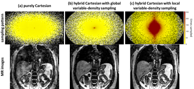 |
Improved subsampling trajectory for reliable assessment of cardiac and respiratory motion in 5D MRI for body motion correction
Thomas Küstner, Martin Schwartz, Petros Martirosian, Konstantin Nikolaou, Sergios Gatidis, Bin Yang, Fritz Schick
Respiratory and cardiac motion may cause artifacts in body trunk imaging if patients cannot hold their breath or triggered acquisitions are not practical. Retrospective correction strategies cope with motion by fast sequences under free-movement conditions. In the acquisition, a random subsampling with global variable-density scaling is usually applied. Improved sampling efficiency in terms of acquired SNR per sample can be achieved if the underlying energy distribution is considered. An image upscaling via dictionary learning from the localizer with extraction of the energy distribution and local shaping of the hybrid Cartesian subsampling improves the overall image quality and SNR by 45%.
|
15:09
|
0480.
 |
Estimation of a signal-appropriate reference volume to improve registration of high b-value diffusion volumes in the human placenta
Conrad Rockel, Barbra de Vrijer, Charles McKenzie
A technique is presented to aid in better motion correction of high b-value diffusion images of the human placenta in utero for the purposes of quantification using intra-voxel incoherent motion (IVIM). A registration reference volume is calculated with signal intensities and tissue features with similarity to volumes with high diffusion weighting, and produce more successful registrations than when using the b=0 volume as a reference.
|
15:21
|
0481.
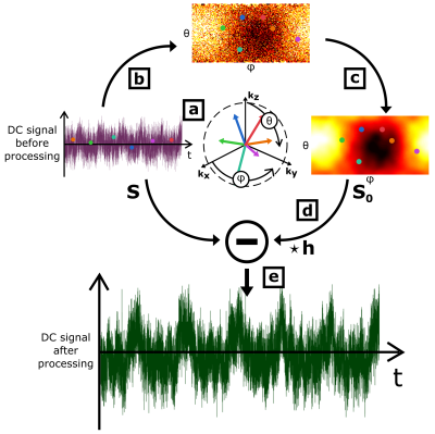 |
Off k-space center and spin-history artifacts Self-Navigator Intelligent Filter (SNIF) for quasi-random 3D-radial acquisitions
Tanguy Boucneau, Brice Fernandez, Peder Larson, Luc Darrasse, Xavier Maître
For 3D radial acquisitions featuring a quasi-random order of spoke orientations in k-space, implementable in UTE pulse sequences, respiratory and cardiac self-navigators are usually extracted from the assumed k-space center thanks to standard bandpass filters to perform self-gating. To remove more efficiently the background noise, SNIF, Self-Navigator Intelligent Filter, takes into account the underlying physics of the MR acquisition to filter out of the DC signal both off k-space center and spin-history effects. SNIF systematically enhances both respiratory and cardiac self-navigators. It even sometimes reveals their spectral first harmonics, which remained otherwise buried into the background noise.
|
15:33
|
0482.
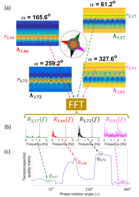 |
CAPTURE: Consistently-Acquired Projections for Tuned and Robust Estimation - A Self-Navigated Respiratory Motion Correction Approach
Cihat Eldeniz, Tyler Fraum, Amber Salter, Yasheng Chen, H. Michael Gach, Parag Parikh, Kathryn Fowler, Hongyu An
In this study, we present a fully-automated and robust self-navigated approach to obtain 4D motion-resolved images during free breathing. A 1D navigator was acquired consistently in order to reduce the signal contamination due to system imperfections. The resulting projections were then 'tuned' using complex phase rotation for adapting to scan-to-scan variations, followed by the detection of the respiratory curve. CAPTURE successfully detected respiratory motion in all subjects. A blind radiological review by two radiologists shows the significant quality improvement in motion-corrected images.
|
|
