Novel Pulse Sequences
Oral
Acquisition, Reconstruction & Analysis
Monday, 18 June 2018
| N04 |
08:15 - 10:15 |
Moderators: Gigi Galiana, Christian Guenthner |
08:15
|
0056.
 |
 High Spatial Resolution BOLD fMRI Using Simultaneous Multislice Excitation with Echo-Shifting Gradient Echo at 7 Tesla High Spatial Resolution BOLD fMRI Using Simultaneous Multislice Excitation with Echo-Shifting Gradient Echo at 7 Tesla
Shi Su, Na Lu, Xiaojing Long, Chunxiang Jiang, Hang Zhang, Ye Li, Rong Xue, Haifeng Wang, Lijuan Zhang, Liang Dong, Xin Liu, Guoxi Xie
Signal to noise gain at ultra-high field has pushed blood oxygen level dependent functional MRI towards high spatial resolution with the benefit of improved accuracy in functional mapping. However, the techniques available for high spatial resolution fMRI are mainly based on echo-planar imaging technique, which faces geometric distortion. In this work, we proposed a technique combining simultaneous multislice excitation with echo-shifting, which can be virtually free from distortion artifacts, for high spatial resolution fMRI. Significant activation was identified in visual and motor experiments with in-plane resolution of 1.0×1.0 mm2 and an acceleration factor of 10 at 7 Tesla.
|
08:27
|
0057.
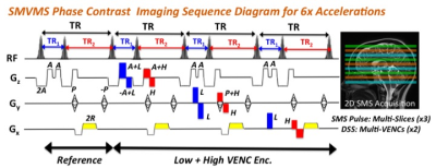 |
Simultaneous Multi-VENC and Multi-Slice (SMVMS) Phase Contrast Imaging Using Dual Steady-State Sequence
Suhyung Park, Liyong Chen, David Feinberg
Phase contrast (PC) MRI has been successfully applied to quantify flow velocity over the cardiac cycle by adding bipolar gradients with opposite polarity to cine gradient echo (GRE) sequences with prospective cardiac gating. However, PC-MRI suffers from a couple of disadvantages: 1) velocity aliasing which requires multiple acquisition without prior knowledge of the highest potential velocity and 2) excessive lengthy scan with whole brain coverage. In this work, we propose the Simultaneous Multi-VENC and Multi-Slice (SMVMS) technique to eliminate both issues. Instead of sequentially acquiring multiple slices with low and high velocity encoding (VENC) schemes, this interleaves an echo-shift technique in a Dual TR Steady-State (DSS) sequence using SMS excitation, which allows fast acquisition by sharing low- and high-VENC acquisitions with multiple slices in a single measurement.
|
08:39
|
0058.
 |
Diffusion interleaved and slice-shuffled (DiSS) imaging for joint diffusion-relaxometry studies
Jana Hutter, Daan Christiaens, Thomas Roberts, Paddy Slator, Anthony Price, Joseph Hajnal
Imaging protocols that allow diverse contrast mechanisms to be sampled with different mixes open up exciting opportunities for joint modelling of tissue properties. However joint sampling with conventional sequence structures can be extremely inefficient and so prohibitively time consuming. Here we explore the joint inversion recovery-diffusion sampling challenge and create an efficient capability by breaking the “one-volume – one encoding” paradigm to interleave the diffusion encoding not per volume but for every slice. Flexible sampling during inversion recovery allowing sufficient samples for a joint fit to be acquired in a much shorter time. The approach has been tested in normal volunteer brain examinations.
|
08:51
|
0059.
 |
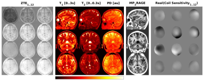
High Contrast and Resolution Simultaneous T1 and T2 MPRAGE at 7T
Chan Hong Moon, Hoby Hetherington, Jullie Pan
T2W MRI is useful for lesion detection in neurological disorders. At 7T, while SNR can be excellent for high-resolution imaging, T2W imaging is known to be difficult due to problems with B1 amplitude and homogeneity, as well as low T2 contrast between WM vs. GM. To address these, we developed new simultaneous T1W/T2W MP2RAGE sequence. We simulated the new sequence to optimize brain contrast and implemented on 7T combined with B1-shimmed pTx multi-transceiver and high-order B0 shim. The results show homogeneous contrast of WM vs. GM over whole brain with excellent SAR efficiency, giving high resolution detection of hippocampal sub-structures.
|
09:03
|
0060.
 |
A 2D multi-shot inversion recovery EPI (MS-IR-EPI) sequence for high spatial resolution T1-mapping at 7T
Video Permission Withheld
Rosa Sanchez Panchuelo, Robert Turner, Olivier Mougin, Susan Francis
The present study uses an efficient 2D, multi-shot, inversion-recovery EPI (MS-IR-EPI) acquisition that combines separately excited k-space segments after each inversion pulse together with steps in slice ordering to generate T1-maps with high SNR per unit time. We show that although the inversion times systematically vary across slices, consistent T1-maps can be generated across the whole brain. Such T1-maps provide high spatial resolution and SNR, with little image distortion and can be collected in a short acquisition time.
|
09:15
|
0061.
 |
Silent, 3D MR Parameter Mapping using Magnetization Prepared Zero TE
Florian Wiesinger, Martin Janich, Emil Ljungberg, Gareth Barker, Ana Beatriz Solana
Here we describe a novel method for 3D, quantitative, silent MR parameter mapping based on 1) combined T1 and T2 magnetization preparation, 2) Zero TE image encoding and 3) least-squares dictionary matching.
|
09:27
|
0062.
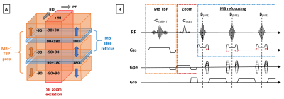 |
Multiband zoom TSE imaging: increasing efficiency with multiband tip-back preparation pulses
Anthony Price, Lucilio Cordero-Grande, Shaihan Malik, Joseph Hajnal
Inner volume imaging can be appealing as it negates the need to encode a large field of view when the region of interest resides within a larger tissue structure. However, conventional zoom approaches with precisely limited field of view produce strong saturation throughout the image volume, placing a restrictive lower limit on the minimum TR, in order to avoid reduced signal and contrast. Here we present a new multiband tip-back preparation pulse in combination with a zoom multiband TSE sequence, which realises the benefits of reduced field of view encoding without the penalty of saturation in the intermediate slice areas.
|
09:39
|
0063.
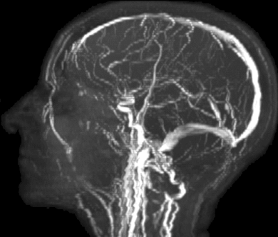 |
Simultaneous Magnetic Resonance Angiography and Multiparametric Mapping in the Transient-state
Pedro Gomez, Miguel Molina-Romero, Guido Buonincontri, Bjoern Menze, Marion Menzel
Quantitative Transient-state Imaging (QTI) is a non-random, dictionary-less MR Fingerprinting alternative. Through iterative reconstructions, QTI recovers a series of contrast-weighted images from transient-state acquisitions and subsequently estimates the parameters that best describe the resulting dynamic signal evolutions. Here, we extend the QTI framework by incorporating a simple velocity model that accounts for blood flowing into and out of the imaging slice. The model, however wrong, can be very useful: it predicts signal hyperintensities in the presence of flow, allowing for the simultaneous reconstruction of MR Angiography images, hundreds of dynamic contrast-weighted images, and their corresponding parametric maps.
|
09:51
|
0064.
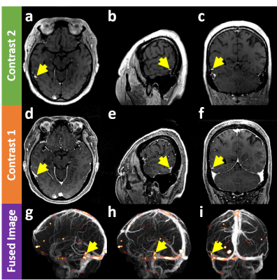 |
Novel Tumor-Selective Dual-Contrast 3D MRI Toward Zero False-Positiveness in Brain Metastases: A Feasibility Study
Hoonjae Lee, Seong-gi Kim, Jaeseok Park
The purpose of this work is to develop a novel, tumor-selective dual-contrast 3D MRI technique that can clearly differentiate small brain metastases from contrast-enhanced vessels while potentially eliminating false-positiveness in the corresponding diagnosis. After injecting contrast agents, the proposed pulse sequence employs a pair of mixed encodings in each TR, yielding highly tumor-selective, blood-suppressed images from the latter to increase the sensitivity of metastases detection while producing blood-enhanced signals from the former to evaluate the false-positiveness of the detected metastases. It is expected that the proposed method enhances detection sensitivity to brain metastases while substantially reducing false-positiveness.
|
10:03
 |
0065.
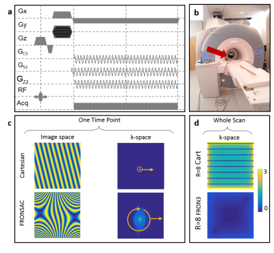 |
Clinical Imaging Potential of FRONSAC
Nadine Dispenza, Sebastian Littin, Maxim Zaitsev, R. Constable, Gigi Galiana
Despite potential for more flexible and efficient encoding that better complements receiver geometry, the past decade of work with nonlinear gradients (NLGs) has shown relatively modest improvements on accelerated image quality. In this work we present the first experimental evidence that the previously introduced ROtary Nonlinear Spatial ACquisition (FRONSAC) can notably improve accelerated image quality, both in vitro and in humans. Furthermore, this work introduces and demonstrates a number of robust and flexible attributes of this method, which are crucial to reducing scan times in a clinical setting.
|
|
