Parameter Quantification
Oral
Acquisition, Reconstruction & Analysis
Monday, 18 June 2018
| S03 |
16:15 - 18:15 |
Moderators: Tobias Kober, Jürgen Reichenbach |
16:15
 |
0262.
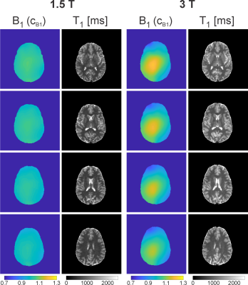 |
 Simultaneous B1 and T1 Mapping Using Spiral Variable-Flip-Angle Acquisitions for Whole-Brain Coverage in Less Than One Minute Simultaneous B1 and T1 Mapping Using Spiral Variable-Flip-Angle Acquisitions for Whole-Brain Coverage in Less Than One Minute
Rahel Heule, Josef Pfeuffer, Craig Meyer, Oliver Bieri
Rapid variable-flip-angle T1 mapping techniques are frequently applied in clinical settings, but their accuracy is often impaired by incomplete spoiling or flip angle miscalibrations. To eliminate these two error sources simultaneously, a combined B1 and T1 mapping method is proposed based on spiral 2D multislice spoiled gradient echo imaging with high spoiling efficiency. The transition to steady state is minimized by an optimized single preparation pulse. A single-shot spiral readout during the preparation module enables ultrafast B1 mapping and as a result reproducible bias-free T1 mapping with whole-brain coverage at clinically relevant resolution in less than one minute.
|
16:27
|
0263.
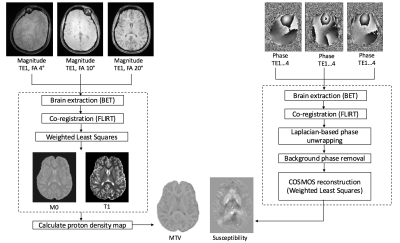 |
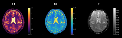
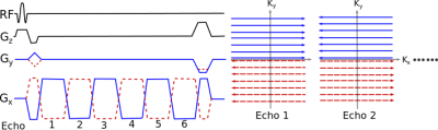
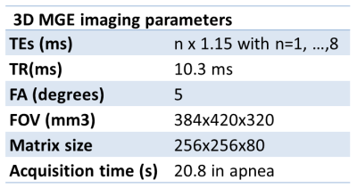
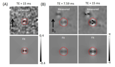
Rapid simultaneous acquisition of QSM and MTV
Fang Yu, Susie Huang, Tanguy Duval, Julien Cohen-Adad, Berkin Bilgic
Quantitative susceptibility mapping (QSM) and macromolecular tissue volume (MTV) represent quantitative methods that improve characterization of neurodegenerative diseases. MTV involves acquisition of 3D-Gradient Echo (GRE) at multiple flip angles with long TR and short TE. High-quality QSM requires 3D-GRE at multiple head orientations with long TE. We exploit (i) unused time due to long TR in MTV to collect additional late echoes that allow QSM processing, (ii) acquire each of the multiple flip angles at a different head orientation. These permit simultaneous acquisition of QSM and MTV, whereby two maps are obtained at the scan time of a single contrast.
|
16:39
|
0264.
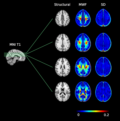 |
Myelin water atlas: a template for myelin distribution in the brain
Hanwen Liu, Cristina Rubino, Mike Jarrett, Emil Ljungberg, Irene Vavasour, Shannon Kolind, Erin MacMillan, Tony Traboulsee, Donna Lang, Alex Rauscher, David Li, Alex MacKay, Lara Boyd, John Kramer, Cornelia Laule
In-vivo information about myelin content in the brain is desirable for studying brain diseases and injuries. Normative information is key for determining what is abnormal when assessing neurological conditions that affect myelin. We used myelin water imaging to create a template specific to myelin, the myelin water atlas, for healthy brains. The resulting atlas shows strong agreement with well-known anatomical features that have demonstrated that different brain regions have distinct amounts of myelin. Our work shows one of the potential applications of using the myelin water atlas as a reference to visualize demyelination in the brain of individual subjects.
|
16:51
|
0265.
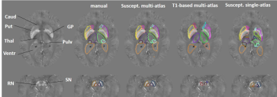 |
Multi-atlas tool for automated magnetic susceptibility quantification in brain nuclei
Andreia Faria, Lin Chen, Kwame Kutten, Can Ceritoglu, Ningdong Kang, Li Pan, Ye Qiao, Michael Miller, Susumu Mori, David Yousem, Peter van Zijl, Xu Li
Quantitative magnetic susceptibility offers a non-invasive measure of important brain tissue molecules, such as iron complexes and myelin, potentially providing significant information about normal and pathological conditions during aging. We developed an automated process to quantify tissue susceptibility in a biologically meaningful set of structures, thereby generating universal and sharable quantitative susceptibility measures. Our susceptibility-based multi-atlas outperformed the single atlas and the T1-weighted multi-atlases. Our tool and normalization algorithm offered consistent measures of magnetic susceptibility over different image protocols and platforms. Automatic and reliable quantitative susceptibility mapping measures will facilitate individual analyses and studies on aging and neurodegeneration.
|
17:03
|
0266.
 |
High-resolution in-vivo multi-parametric MRI using MR-STAT with a highly parallelized, limited-memory reconstruction algorithm
Oscar van der Heide, Alessandro Sbrizzi, Peter Luijten, Cornelis van den Berg
MR-STAT is a framework for obtaining multiple quantitative parameter maps from a very short scan. The parameter maps are obtained by fitting a Bloch-based signal model directly to the time domain data. No Fourier transform is needed. In this work we demonstrate that MR-STAT can obtain excellent high-resolution in-vivo quantitative maps using very short Cartesian acquisitions standardly available on clinical MR systems. The solution of the large-scale reconstruction problem is made possible by a highly parallelized, limited-memory algorithm.
|
17:15
|
0267.
 |
Increased Measurement Efficiency for Fat Quantification Using a Mixed Polarity Bipolar Acquisition Scheme
Ruitian Song, Nathan Artz, Ralf Loeffler, Claudia Hillenbrand
A new mixed polarity bipolar GRE sequence is proposed for fat quantification by mixing positive and negative readout polarity in one echo acquisition. Accurate fat fraction maps can be obtained using the proposed method with up to 50% scan time savings compared to a dual shot unipolar GRE method.
|
17:27
|
0268.
 |
Comparison of 3D spoiled-gradient multiple echo with STEAM for proton density fat fraction and fatty acid composition estimation
Angéline Nemeth, Hélène Ratiney, Benjamin Leporq, Kévin Seyssel, Bérénice Segrestin, Pierre-Jean Valette, Martine Laville, Olivier Beuf
A total of 39 volunteers underwent an imaging and spectroscopy protocol on a 3T Ingenia Philips system with an axial 3D spoiled-gradient multiple echo sequence on abdominal region and a set of three STEAM sequences acquired on subcutaneous adipose tissue, visceral adipose tissue and liver. The quantification of Proton density fat fraction (PDFF), proportion of saturated (SFA), monounsaturated (MUFA) or polyunsaturated (PUFA) fatty acids from both MRI and MRS methods were compared. Good correlation with a little bias was found for the liver PDFF. Values of PUFA, MUFA and SFA from both techniques were poorly correlated.
|
17:39
|
0269.
 |
Quantitative Measurements of Deep Medullary Vein Caliber and Oxygenation Level Using MRI Phase and Complex Images
Xiaopeng Zong, Weili Lin
Collagenosis-induced narrowing of deep medullary vein (DMV) caliber has been implicated as one of the main causes of small vessel disease. However, a non-invasive imaging method for monitoring the DMV narrowing is still lacking. We present an MRI method for non-invasive measurement of DMV caliber and oxygenation level base on MRI phase and complex images acquired using a double echo gradient echo sequence at 7 T. The measured DMV caliber distribution agreed well with earlier report. Our approach can serve as an invaluable tool for studying the role of venous lumen narrowing in the pathogenesis of small vessel disease.
|
17:51
|
0270.
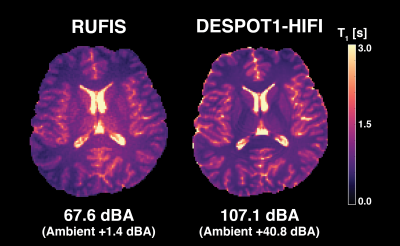 |
Silent T1-Mapping Using the Variable Flip Angle Method with Zero Echo Time
Emil Ljungberg, Ana Beatriz Solana Sanchez, Tobias Wood, Shannon Kolind, Florian Wiesinger, Gareth Barker
In this work we present a silent whole brain T1-mapping technique with zero echo time using a variable flip angle (VFA) scheme with a 3D radial sequence (RUFIS). The technique is compared to a conventional Cartesian gradient-echo based sequence (DESPOT1-HIFI) in a quantitative T1 phantom as well as in vivo in a single subject. Phantom measurements showed good agreement between techniques with average difference of 28 ms. In vivo T1 maps showed good correspondence between RUFIS and DESPOT1-HIFI.
|
18:03
|
0271.
 |
Explicit mathematical expression for the Cramér-Rao lower bound for experimental design and parameter estimation in parallel imaging
Video Permission Withheld
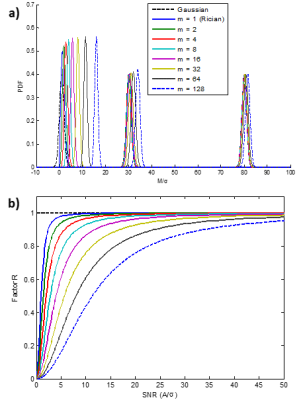
Mustapha Bouhrara, Richard Spencer
The Cramér-Rao lower bound (CRLB) is widely used in the design of magnetic resonance (MR) experiments for parameter estimation. Previous work has considered only Gaussian or Rician noise distributions in this calculation. However, the noise distribution for multiple-coil acquisitions, such as in parallel imaging, obeys the noncentral χ-distribution under many circumstances. Here, we present the general mathematical expression for the CRLB calculation for parameter estimation from multiple-coil acquisitions. Our results indicate that the CRLB calculation must account for the noncentral χ-distribution of noise in multi-coil acquisitions, especially in the low-to-moderate signal-to-noise ratio (SNR) regime.
|
|
