Approaches to Quantitative Mapping
Oral
Acquisition, Reconstruction & Analysis
Thursday, 21 June 2018
| N03 |
08:00 - 10:00 |
Moderators: Valentina Taviani, Jakob Assländer |
08:00
 |
1017.
 |
 Fast 3D MR fingerprinting with spiral projection acquisition for whole brain quantification imaging Fast 3D MR fingerprinting with spiral projection acquisition for whole brain quantification imaging
Xiaozhi Cao, Congyu Liao, Qing Li, Huihui Ye, Hongjian He, Jianhui Zhong
A spiral projection acquisition scheme was used for 3D MR fingerprinting to achieve isotropic resolution of 1.2x1.2x1.2 mm3 with FOV of 240x240x240 mm3 for whole brain T1 and T2 mapping within 4.3 minutes.
|
08:12
|
1018.
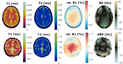 |
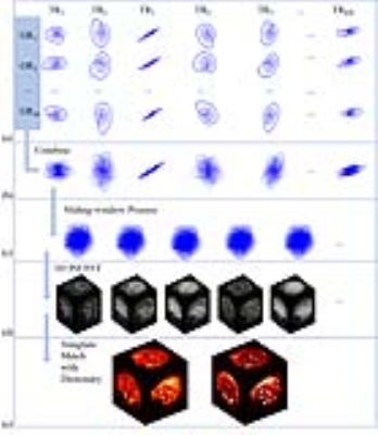
Magnetic Field Fingerprinting (MFF)
Gregor Körzdörfer, Yun Jiang, Peter Speier, Josef Pfeuffer, Dan Ma, Bartosz Guzek, Bernhard Hensel, Vikas Gulani, Mark Griswold, Mathias Nittka
Parameter maps obtained from Magnetic Resonance Fingerprinting (MRF) are sensitive to magnetic field inhomogeneities. Most MRF publications are based on FISP to minimize the dependency on B0. In FISP however, the magnetization is spoiled at the end of every TR resulting in lower T2 differentiation capability and SNR compared to TrueFISP. We propose an MRF variant that applies TrueFISP, FISP and FLASH in a continuous acquisition, and additionally incorporates B1 and B0 in the dictionary, which are resolved by a pattern match. This implementation enables increased spiral resolution and spiral deblurring with the derived high-resolution B0 maps.
|
08:24
|
1019.
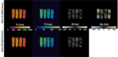 |
Improving uniqueness of Magnetic Resonance Fingerprinting (MRF) signal evolutions using spatio-temporal variation of non-linear ?B0 shim coil.
Bhairav Mehta, Michael Twieg, Mingrui Yang, Dan Ma, Yun Jiang, Simone Coppo, Haoqin Zhu, Shinya Handa, Labros Petropoulos, Hiroyuki Fujita, Mark Griswold
Magnetic Resonance Fingerprinting (MRF) framework uses variation in acquisition parameters to generate unique signal evolutions, which can be treated as “fingerprints”. The iPRES coil concept provides independent and dynamic variations of multiple magnetic fields, which can be used to improve the uniqueness of signal evolutions through spatio-temporal variations of multiple fields. In this study, we present a proof-of-concept implementation illustrating spatio-temporal variations of local non-linear ΔB0 fields improve the uniqueness of signal evolutions for MRF. Our phantom results show reduction in variation of estimated tissue properties for data with 500frames, thereby illustrating the capability of acceleration using these field variations.
|
08:36
|
1020.
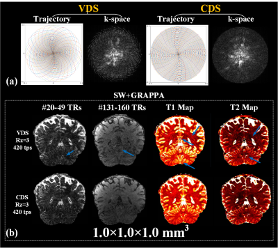 |
Optimized 3D Stack-of-Spirals MR Fingerprinting with Hybrid Sliding-Window and GRAPPA Reconstruction
Congyu Liao, Berkin Bilgic, Mary Manhard, Xiaozhi Cao, Jianhui Zhong, Lawrence Wald, Kawin Setsompop
We demonstrate 3D stack-of-spiral MRF acquisition with hybrid sliding-window and GRAPPA (SW+GRAPPA) reconstruction using a constant density spiral (CDS) rather than standard variable density spiral (VDS). The CDS was found to mitigates high-frequency artifacts after in-plane sliding-window (SW) combination and improve the subsequent GRAPPA and dictionary matching reconstruction. The proposed 3D constant density stack-of-spiral MRF allows whole-brain (240×240×192 mm3) parametric mapping at 1 mm isotropic resolution with high quality in 8 minutes.
|
08:48
|
1021.
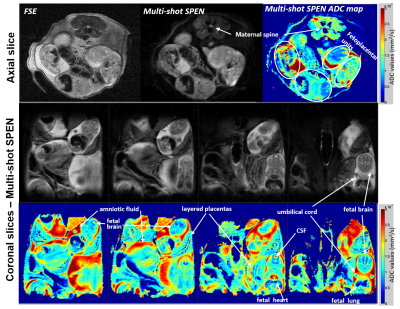 |
Diffusion-weighted in vivo imaging with =100 µm resolution: Principles and applications to ADC mapping of pregnant mice
Qingjia Bao, Gilad Liberman, Eddy Solomon, Miki Lustig, Lucio Fydman
Although a major advantage of SPatiotemporal ENcoding (SPEN) vs EPI is a higher immunity to artifacts, it suffers –as all single-shot experiments– from resolution and SNR limitations. These can be dealt by multi-scan interleaving, which unlike EPI counterparts leads to independent low-resolution images free from aliasing artifacts. We present an acquisition and processing protocol that reconstructs from these data a composite image free from hardware or motional imperfections, possessing unprecedented resolution. The power of this self-referenced method is demonstrated with in vivo ADC maps of brains, kidneys and fetal organs in pregnant mice, possessing resolutions in the 78-230 µm range.
|
09:00
|
1022.
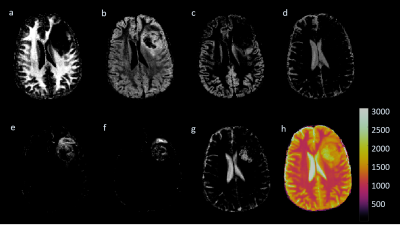 |
Segmentation of Brain Tissues using a Bayesian Estimation of Multicomponent Relaxation Values in Magnetic Resonance Fingerprinting
Debra McGivney, Yun Jiang, Dan Ma, Chaitra Badve, Vikas Gulani, Mark Griswold
A Bayesian methodology has been previously applied to MRF reconstructions to analyze subvoxel T1 and T2 distributions. The multidimensional results from this algorithm are difficult to visualize. We propose to apply this Bayesian approach in the brain to create T1 and T2Gaussian distributions to represent various tissue types. Using these distributions as prior information, the Bayesian methodology is applied over the brain with a smaller dictionary. Results from this Bayesian approach with a smaller dictionary are weighted by the Gaussian probabilities and summed to create tissue probability maps in normal volunteers and a brain tumor patient.
|
09:12
|
1023.
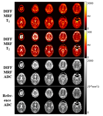 |
Simultaneous quantification of T1, T2 and Apparent Diffusion Coefficient using Magnetic Resonance Fingerprinting based on Echo Planar Imaging
Benedikt Rieger, Mehmet Akçakaya, Lothar Schad, Sebastian Weingärtner
In this study we propose to integrate diffusion imaging into magnetic resonance fingerprinting, a method that has shown promise for time-efficient simultaneous quantification of multiple tissue parameters. The proposed sequence for quantitative T1, T2 and the apparent diffusion coefficient (ADC) is based on using Cartesian EPI readout. The contrast is generated using spin-echo EPI and gradient spoiling with diffusion gradients of varying moment and varying TR and TE. Joint T1, T2 and ADC parameter-maps acquired in phantoms are in good agreement with reference measurements and demonstrate high quality in-vivo maps, within a scan time of 28 seconds per slice.
|
09:24
|
1024.
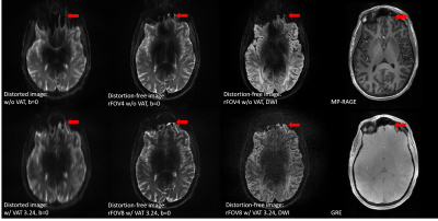 |
Accelerated Distortion-Free Diffusion Imaging at 7T – by Fusing PSF and VAT
Yi-Hang Tung, Myung-Ho In, Sinyeob Ahn, Alessandro Sciarra, Oliver Speck
In this study, combining view angle tilting (VAT) with the novel PSF diffusion EPI sequence demonstrates the feasibility to further accelerate distortion-free diffusion weighted imaging at high field.
|
09:36
 |
1025.
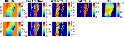 |
Musculoskeletal MR Fingerprinting with dictionary-based fat and water separation
Matteo Cencini, Laura Biagi, Joshua Kaggie, Rolf Schulte, Michela Tosetti, Guido Buonincontri
The purpose of this work is to obtain quantitative MRI maps of fat and water with a fast acquisition using MR Fingerprinting (MRF). The major advantage of MRF is its intrinsic multicomponent quantification capability, which overcomes the limitations of traditional separation techniques. Previous methods using MRF have repeated the same acquisitions for multiple echo times in order to obtain fat fraction maps. Here, variable echo times within a single MRF acquisition are used, in order to estimate T1 and PD of water and fat. We demonstrate quantitative maps in 16s per slice and improved delineation of anatomy in human knees.
|
09:48
|
1026.
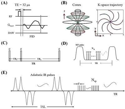 |
Quantitative Ultrashort Echo Time (UTE) Magnetic Resonance Imaging of Knee Osteoarthritis (OA)
Yajun Ma, Lidi Wan, Xin Cheng, Yinghua Zhao, Eric Chang, Jiang Du
In this study we aimed to develop a comprehensive quantitative UTE imaging package including UTE-Cones actual flip angle imaging (UTE-Cones-AFI) for accurate B1 mapping, UTE-Cones variable flip angle (UTE-Cones-VFA) for T1 mapping, 3D UTE-Cones-MT for MT modeling, UTE-Cones-AdiabT1rho for T1r mapping, and multi-echo UTE-Cones for T2* mapping. The techniques were evaluated on cadaveric human knee joints.
|
|
