Diffusion MRI: From Fiber Orientation to Connectivity
Oral
Diffusion
Monday, 18 June 2018
| N02 |
08:15 - 10:15 |
Moderators: Stamatios Sotiropoulos, Robert Smith |
08:15
 |
0036.
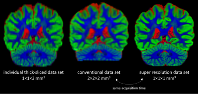 |
 Super-resolution for spherical deconvolution of multi-shell diffusion MRI data Super-resolution for spherical deconvolution of multi-shell diffusion MRI data
Ben Jeurissen, Gabriel Ramos-Llordén, Floris Vanhevel, Paul Parizel, Jan Sijbers
Multi-tissue constrained spherical deconvolution (MT-CSD) can simultaneously estimate the full white matter fiber orientation distribution function (fODF) and the apparent densities of cerebrospinal fluid and grey matter from multi-shell diffusion MRI data, making it an attractive option for clinical and neuroscientific studies. Unfortunately, MT-CSD at high spatial resolution is challenging due to scan time and signal-to-noise ratio constraints. We propose a new MT-CSD approach that enables super-resolution estimation from multiple thick-sliced data sets with varying slice orientation. Using data acquired on a clinical scanner, we demonstrate high-quality tissue density maps and fODFs at 1×1×1mm3 spatial resolution in under 10 minutes.
|
08:27
|
0037.
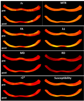 |
Differential Sensitivity of Various Microstructural Metrics to Training-Induced White Matter Dynamics
Debbie Anaby, Benjamin Tendler, Chantal Tax, Greg Parker, Yaniv Assaf, Derek Jones
Previous in-vivo MRI studies on training-induced WM microstructural dynamics were mostly based on DTI measurements showing changes in FA. The non-specificity of FA as a WM marker stimulated us to obtain a more specific characterization of WM microstructural changes. Using multi-parametric MR with the ‘Tractometry’ approach we show significant changes in the fornix post a navigation working memory task. Critically, we report here for the first time significant microstructural changes with susceptibility, shown in vast areas of the fornix. Fr, MD, RD and λ1 also show significant changes but in limited areas of the fornix.
|
08:39
|
0038.
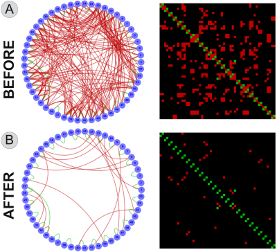 |
Reducing false positives in tractography with microstructural and anatomical priors
Alessandro Daducci, Muhamed Barakovic, Gabriel Girard, Maxime Descoteaux, Jean-Philippe Thiran
Tractography has proven particularly effective for studying noninvasively the neuronal architecture of the brain but recent studies have showed that the high incidence of false positives can significantly bias any connectivity analysis. We present a novel processing framework that can dramatically reduce these false positives, i.e. improving specificity, without affecting the sensitivity, by considering two very basic observations about white-matter anatomy. Our results may have profound implications for the use of tractography to study brain connectivity.
|
08:51
|
0039.
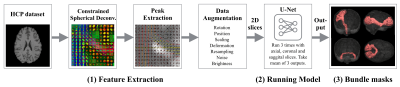 |
Fast and accurate white matter bundle segmentation
Jakob Wasserthal, Peter Neher, Klaus Maier-Hein
Automatic white matter fiber bundle segmentation in diffusion-weighted MRI brain scans enables detailed studies of white matter characteristics in healthy and diseased brains. Existing approaches combine processing steps such as tractography, atlas registration and cortical parcellation, resulting in pipelines that are computationally intensive and tedious to set up. We present a novel convolutional neural network-based approach that incorporates or circumvents most of the usually required processing steps (no registration, no tracking, no parcellation). We demonstrate in 105 subjects from the Human Connectome Project that the proposed approach is much faster than existing methods while providing more accurate results.
|
09:03
|
0040.
 |
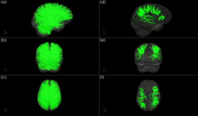
A data-driven groupwise fiber clustering atlas for consistent white matter parcellation and anatomical tract identification of subjects across the lifespan
Fan Zhang, Ye Wu, Isaiah Norton, Yogesh Rathi, Nikos Makris, Lauren O'Donnell
We propose an anatomically curated white matter parcellation atlas generated from a large population of 100 healthy adult subjects, leveraging a well-established data-driven groupwise fiber clustering pipeline and expert neuroanatomy knowledge. We demonstrate the ability of the proposed method to parcellate a total of 541 subjects ranging in age from 1 day to 82 years. The results suggest that our parcellation algorithm provides high generalization and consistency of white matter parcellation and tract identification for subjects across the lifespan.
|
09:15
|
0041.
 |
Bundle-Wise Deep Tracker: Learning to track bundle-specific streamline paths
Philippe Poulin, Francois Rheault, Etienne St-Onge, Pierre-Marc Jodoin, Maxime Descoteaux
We propose a novel bundle-wise tracking algorithm based on deep learning and recurrent neural networks. This allows bundle-specific features to be learned directly from the diffusion signal without the need to reconstruct a fiber orientation distribution. With a high amount of examples, the proposed method improves classic algorithms for several quantitative measures such as tracking efficiency, number of valid streamlines, and volume coverage.
|
09:27
|
0042.
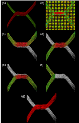 |
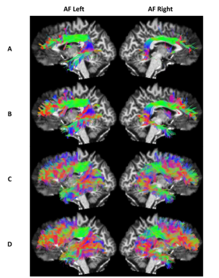
Anchor tracts: a novel concept for reducing false positives in fiber tractography
Peter Neher, Klaus Maier-Hein
Numerous reports have shown that fiber tractography suffers from a difficult sensitivity-specificity tradeoff. We present an approach that leverages knowledge about certain well studied tracts (anchor tracts) in a tractogram to quantitatively assess and score the remaining tracts (candidate tracts) according to their plausibility in conjunction with this context information. We show that our approach has the potential for greatly reducing the number of false positive tracts in fiber tractography while maintaining high sensitivities.
|
09:39
 |
0043.
 |
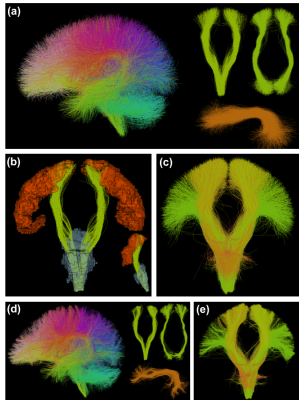
Investigating U-Shape Fibers from Data-Driven Clustering of White Matter Tractography
Jason Kai, Loxlan Kasa, Terry Peters, Ali Khan
Studies of white matter tractography typically investigate fibers that make up long association tracts, such as the arcuate fasciculus. As a result, the local structural connections comprising short association (cortico-cortical), U-shaped fibers, or U-fibers, are poorly understood. Previous work suggests these fibers play an important role in communication between adjacent cortical regions. We introduce a flexible, data-driven tool incorporating multimodal MRI techniques to aid differentiation of fiber tracts via clustering techniques, including a filter to identify and extract U-fibers. Using this tool, we present a preliminary investigation of U-fibers between controls (N=20) and patients diagnosed with temporal lobe epilepsy (N=19).
|
09:51
|
0044.
 |
Tractography-based parcellations show limited sensitivity to internal structure
Jonathan Clayden, David Thomas, Alexander Kraskov
Connectivity-based parcellation of subcortical structures using diffusion tractography is a common paradigm. Typically, these analyses imply voxel-level specificity of connectivity, and spatial coherence is taken as imaging-based evidence for anatomically distinct subnuclei. However, by spatially permuting diffusion parameters and repeating the parcellation, we demonstrate that internal structure in diffusion anisotropy is not necessary for a plausible parcellation to be obtained. This suggests that such parcellations should be interpreted with some caution.
|
10:03
|
0045.
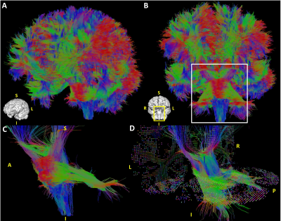 |
Ultra-high resolution multi-shell dMRI and tractography of the ex vivo human brain using kT-dSTEAM at 9.4T
Francisco Fritz, Shubharthi Sengupta, Robbert Harms, Benedikt Poser, Alard Roebroeck
Here we explore the high resolution acquisition of multi-shell and undersampled diffusion data with 9.4T kT-dSTEAM and analysis of such data for crossing fiber tractography. This permits effective usage of both high SNR and diffusion-weighting inherent to data with multiple b-values and shows superior definition of white matter tracks at ultra-high resolution for tractography. In addition, 3D undersampled acquisition allows more room in the trade-off between acquisition time, resolution, b-values and directions.
|
|
