Diffusion: Validation
Oral
Diffusion
Wednesday, 20 June 2018
| S06 |
08:15 - 10:15 |
Moderators: Els Fieremans Fieremans, Andrada Ianus |
08:15
 |
0733.
 |
 SYNCHROTRON X-RAY PHASE-CONTRAST IMAGING TO SIMULATE DIFFUSION TENSOR MRI: APPLICATION TO TRACTOGRAPHY SYNCHROTRON X-RAY PHASE-CONTRAST IMAGING TO SIMULATE DIFFUSION TENSOR MRI: APPLICATION TO TRACTOGRAPHY
Timothée Jacquesson, Justine Bosc, Hugo Rositi, Marlène Wiart, Fabien Chauveau, Françoise Peyrin, David Rousseau, Carole Frindel
As it provides the only method for mapping neural tracts in vivo, diffusion MRI tractography is gaining importance in clinical and neuroscience research. However, the precision of tractography results is influenced by many factors. In this study, we propose a highly realistic simulator based on real data acquired by synchrotron x-ray phase-contrast imaging. This imaging technique with histology-like resolution is demonstrated to reveal adequately the mouse brain in 3D by comparison with classical histology, which loses continuity along the sectioning axis. We expect that our simulator may serve as a tool for the validation and optimization of tractography algorithms.
|
08:27
|
0734.
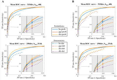 |
Validation of diffusion MRI models and tractography algorithms using chemical tracing
Giorgia Grisot, Suzanne Haber, Anastasia Yendiki
Brain circuitry is still poorly understood, posing a challenge to the validation of diffusion MRI (dMRI) tractography. Many aspects of tractography algorithms, such as their choice of diffusion model or deterministic vs. probabilistic approach, can impact on their performance. Therefore, beyond a qualitative validation, we need quantitative metrics for comparing and optimizing these algorithms. In this work we perform a systematic evaluation of different diffusion models and tractography algorithms by assessing their accuracy with respect to chemical tracing in macaques. We find that the combination of probabilistic tractography and GQI/DSI model yields the best results, and that accuracy does not always improve with higher angular resolution.
|
08:39
|
0735.
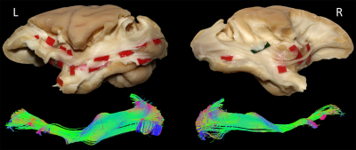 |
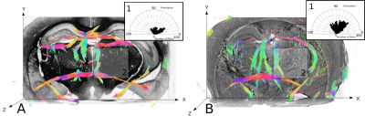
The existence of the inferior fronto-occipital fasciculus (IFOF) revealed in the non-human primate by ex-vivo diffusion-weighted tractography and blunt dissection.
Laurent Petit, Silvio Sarubbo, Alessandro De Benedictis, Franco Chioffi, Maurice Ptito, Tim Dyrby
The existence of a ventral fronto-occipital association pathway in non-human primates similar to the inferior fronto-occipital fasciculus (IFOF) in humans, is nowadays still largely debated. In this study, we elucidate the existence, course and terminations of such a pathway using in the same non-human primate (vervet monkey) both ex-vivo diffusion-weighted tractography and blunt microdissection. From a methodological point of view, it allows an unprecedented anatomical validation of advanced tractography with microdissection for the first time in the same specimen.
|
08:51
|
0736.
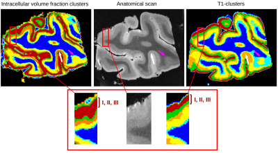 |
Post-mortem mapping of cortical layers using combined multicompartmental relaxometry and diffusometry at ultra-high field (7T and 11.7T)
Justine Beaujoin, Christophe Destrieux, Fabrice Poupon, Ilyess ZEMMOURA, Jean-François Mangin, Cyril Poupon
The human cerebral cortex has a laminar structure presenting distinct cell organization and myelination. In this work, we compare the lamination observed from multicompartmental quantitative relaxometry- or diffusometry-based microstructural ultra-high field MRI on a post-mortem human brain visual cortex sample. This study reveals that the quantitative intra-neuritic volume fraction map provides a better delineation of the line of Gennari in the primary visual cortex whereas multicompartment T1 relaxometry provides a better segmentation of the superfical layers. Thus, this study strongly plays in favour of a combination of relaxometry and diffusometry methods to robustly map the cortex lamination in humans.
|
09:03
|
0737.
 |
A tunable permeability phantom for validating time-dependent diffusion models
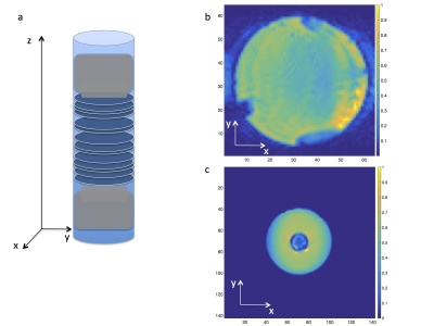
Antonios Papaioannou, Dmitry Novikov, Els Fieremans
Well characterized phantoms are essential for time-dependent diffusion model validations. Here we introduce a phantom made of permeable barriers with highly tunable micro-structural characteristics such as pore size, pore density and permeability and perform time-dependent diffusion measurements using three NMR and MRI systems. Our experimental results agree with the theory of time-dependent diffusion in disordered systems, making the phantom suitable for model validations in clinical and preclinical systems.
|
09:15
|
0738.
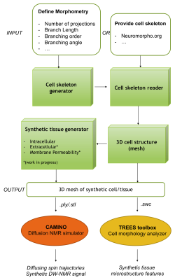 |
A new computational framework for complex numerical simulation of diffusion-weighted NMR signal in brain tissue
Marco Palombo, Daniel Alexander, Hui Zhang
In this work, we introduce a new framework that we developed to use numerical simulations to investigate the complexity of brain tissue at a microscopic level with a detail never realised before. Directly inspired by the advances in computational neuroscience for modelling brain cells, the proposed toolbox enables numerical simulation of molecular diffusion within realistic digitalised brain cells, such as neurons and glia. Here we show a select set of examples offered by this new toolbox to demonstrate its versatility and potentiality. Further development is ongoing, which will support even more realistic conditions and fully digitalised tissues.
|
09:27
|
0739.
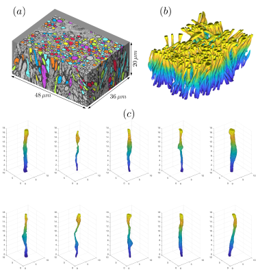 |
Monte Carlo simulations of diffusion in mouse corpus callosum reconstructed from 3-d electron microscopy validate the time-dependence along axons
Hong-Hsi Lee, Dmitry Novikov, Els Fieremans
We present a numerical framework for validation of diffusion models applied to biological tissues. By performing Monte Carlo simulations in a geometry created by automated segmentation of 3-dimensional mouse brain electron-microscopy images, we study the time-dependence of the diffusion coefficient and kurtosis along individual white matter axons, with or without orientation dispersion. Simulation results, together with the analysis of spatial correlations of axonal cross-section variations along axons, point at the short-range disorder in restrictions to diffusion, and agree with theoretical predictions. Our results are consistent with diffusion time-dependence observed in in vivo human brain data.
|
09:39
|
0740.
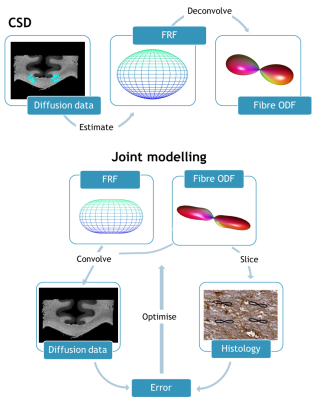 |
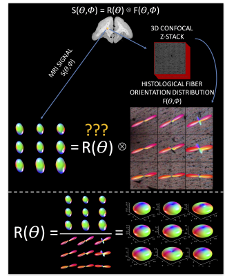
Joint modelling of diffusion MRI and histology
Amy Howard, Jeroen Mollink, Michiel Kleinnijenhuis, Menuka Pallebage Gamarallage, Matteo Bastiani, Karla Miller, Saad Jbabdi
Constrained spherical deconvolution determines the orientation of white matter fibres from the diffusion MRI signal. To do so, the diffusion profile of a single fibre is estimated and assumed constant across the sample. However, the diffusion signal is dependent on microstructural properties such as axonal diameter, packing and myelination, which questions the assumption of a single-fibre response function. This multimodal study combines diffusion MRI with histology from the same tissue sample, to test the validity of a ‘brain-wide’ fibre response function. Preliminary results indicate that in practice the fibre response function is indeed dependent on local anatomy.
|
09:51
|
0741.
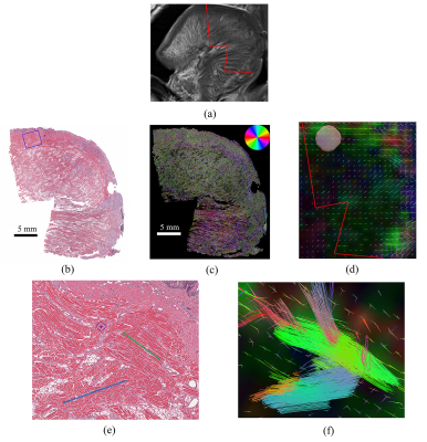 |
Validation of Diffusion Spectrum Imaging of the Human Tongue with Histology
Nahla Elsaid, Adam Puche, Maureen Stone, Jerry Prince, Steven Roys, Rao Gullapalli, Jiachen Zhuo
In this study provides validation procedure for DSI anisotropy measures versus the histological analysis of the aligned tongue muscles. A fresh ex-vivo sample of the tongue is scanned using DSI multi-shell acquisition then the sample is fixed for further histological analysis and comparison with the fiber tracts using DSI reconstruction. The DSI delineation of muscle fibers is matching the histology images of the same region.
|
10:03
 |
0742.
 |
Histologically-derived fiber response functions for diffusion MRI data reveal systematic differences from model-based deconvolution kernels
Kurt Schilling, Vaibhav Janve, Yurui Gao, Iwona Stepniewska, Bennett Landman, Adam Anderson
Spherical deconvolution for diffusion MRI requires a response function in order to accurately reconstruct the underlying voxel-wise fiber orientation distribution (FOD). Here, using 3D histologically-defined fiber orientation distributions and the corresponding diffusion signal, we derive the ground-truth fiber response functions. We show that there is significant variation in these response functions across the brain. We find that the current methods to estimate this function do not match the histological results, which leads to differences in fiber volume fractions. This is important because the wrong response function can amplify spurious peaks in the FOD and lead to inaccurate tractography.
|
|
